Original Article
MiR-124-3p inhibits PDGF-BB-induced vascular smooth
muscle cell proliferation and migration
through targeting STAT3
Lijie Yan, Haitao Yang, Hongyan Duan, Jintao Wu, Peng Qian, Xianwei Fan, Shanling Wang
Department of Cardiology, Henan Provincial People’s Hospital, Zhengzhou, China
Received December 1, 2015; Accepted March 25, 2016; Epub June 15, 2017; Published June 30, 2017
Abstract: Aberrant vascular smooth muscle cells (VSMCs) proliferation and migration contribute significantly to
the development of vascular pathologies, such as atherosclerosis. MicroRNAs (miR) have been reported to act as important gene regulators and play essential roles in the proliferation and migration of VSMCs in a cardiovascular disease. The aim of this study was to investigate the role of miR-124-3p in VSMCs. In the present study, we showed that platelet-derived growth factor (PDGF)-BB, as a stimulant, promoted VSMCs proliferation and suppressed the expression of miR-124-3p. Moreover, overexpression of miR-124-3p inhibited proliferation and also suppressed the proliferating cell nuclear antigen (PCNA) expression in PDGF-BB-induced VSMCs. In addition, we demonstrated that
ectopic expression of miR-124-3p repressed the VSMCs migration. Furthermore, dual-luciferase assays identified that STAT3 is a direct target gene of miR-124-3p in VSMCs, and overexpression of miR-124-3p significantly
down-regulated the mRNA and protein levels of STAT3. In addition, restored the expression of STAT3 in VSMCs could partially abolish the suppressive effects of miR-124-3p on the proliferation and migration. Collectively, these data suggest that miR-124-3p is a key molecule in regulating human VSMCs proliferation and migration by targeting
STAT3 and suggest that specific modulation of miR-124-3p in human VSMCs may represent an attractive approach
for the treatment of proliferative vascular diseases.
Keywords: miR-124-3p, proliferation, migration, STAT3, vascular smooth muscle cells (VSMCs)
Introduction
Cardiovascular disease is one of the leading causes of death worldwide. Despite technologi-cal advances in vascular angioplasty and stent-ing, the outcomes of these procedures are poorer in elderly patients than in younger pa- tients [1]. Atherosclerotic plaques are complex lesions in which repair of tissue injury is as- sociated with vascular smooth muscle cells (VSMCs) proliferation and migration, connec-tive tissue formation and calcium deposition [2]. VSMCs are an important component of vas-cular walls and their proliferation plays a crucial role in atherosclerotic lesion formation [3]. MicroRNAs (miRNAs) are a class of endogen- ous and small (18-24 nucleotides) non-coding RNAs that negatively modulate gene expres-sion through binding to the 3’-untranslated regions (3’-UTR) of target mRNAs to lead to
In the current study, we performed miRNA microarray analysis to identify the miRNAs involved in regulating human VSMCs
prolifera-tion. We, for the first time, demonstrated that
miR-124-3p, which is abundantly expressed in
VSMCs, was significantly down-regulated in
proliferative VSMCs. Furthermore, we found that miR-124-3p inhibits proliferation and mig- ration in PDGF-BB-induced VSMCs via directly targeting STAT3, which is a critical regulator implicated in proliferative vascular diseases. Materials and methods
Cells culture and oligonucleotide transfection
The VSMCs cell line was purchased from Cascade Biologics (Portland, OR) and kept in
DMEM/F12 medium (Dulbecco’s modified
Eagle’s medium; Invitrogen, Carlsbad, CA), sup-plemented with 10% FBS and 1% antibiotics (Gibco, MD, USA). Cells were cultured at 37°C in
a humidified incubator under a 5% CO2
atmo-sphere. VSMCs were seeded in complete medi-um for 24 h. Then, the cells were replaced with fresh serum-free media containing the 25 ng/ mL of PDGF-BB for 24 h. miR-124-3p and scramble oligonucleotide was purchased from GenePharma (Shanghai, China) and transfect-ed into the VSMCs ustransfect-ed Lipofectamine 2000 (Invitrogen, Carlsbad, CA) according to the man-ufacturer’s information.
Quantitative real-time PCR
Total RNAs were isolated with the use of TRIZol reagent (Invitrogen, Grand Island, NY). miRNAs were assayed by real-time PCR. The cDNAs
were produced from 100 ng purified total RNA
with Taqman1 MicroRNA Reverse Transcriptase Kit (Applied Biosystems, Foster City, CA) in com-bination with Taqman1 MicroRNA Assays for
quantification of specific miR-124-3p, accord -ing to the manufacturer’s conditions. U6 was used as an endogenous control for data
nor-malization. Real-time PCR analyses for amplifi
-cation and detection of specific miRNAs were
performed in a Light Cycler 480 II (Roche). The relative differences in expression levels of miRNA in VSMCs were calculated and present-ed as fold induction (2DDCt) after normaliza-tion to control U6.
Western blot analysis
starved in serum-free medium for 24 h. The cells were lysed in radio immunoprecipitation assay (RIPA) buffer with protease and phospha-tase cocktails. Equal amounts of protein were separated by 10% sodium dodecyl sulfate-po- lyacrylamide gel electrophoresis (SDS-PAGE) and electro-transferred onto PVDF membranes (Millipore, Billerica, MA, USA). The membranes were blocked, and then incubated with various antibodies overnight, and then with the horse-radish peroxidase-conjugated secondary anti-body (Beijing TDY Biotech Co., Ltd.) (1:5,000)
for 2 h. Specific protein expression levels were
normalized to GAPDH for total protein analyses or to total proteins for phosphorylated protein measurements. The blots were analyzed using the ChemiDoc™ MP imaging system (Bio-Rad Laboratories, Inc., Hercules, CA, USA). The ex- periments were replicated a number of times. Plasmid construction and luciferase reporter assays
The cells were seeded in triplicate in 24-well plates and allowed to settle for 12 h. The whole 3’-UTR of STAT3 gene was cloned and
ampli-fied. Mutation in 3’-UTR of STAT3 gene with
miR-124-3p putative target binding site deleted was generated with the Quick Change Site-Directed Mutagenesis kit (Stratagene, CA, USA). Both the wild and mutant STAT3 genes were cloned into the pGL-3-vector (Promega, Wisconsin, USA) immediately downstream of the Renilla luciferase gene. A luciferase report-er construct containing the miR-124-3p con-sensus target sequence served as the positive control (PC) and the pRL-TK vector was used as positive and internal controls, respectively.
Cells were co-transfected with pGL-3 firefly
luciferase reporter (50 ng), pRL-TK Renilla lucif-erase reporter (10 ng) and miR-124-3p or nega-tive control with Lipofectamine 2000 (Invi- trogen, Carlsbad, CA, USA). Cell lysates were prepared using Passive Lysis Buffer (Promega, Wisconsin, USA) 48 h upon transfection, and luciferase activity was measured using the Dual-Luciferase Reporter Assay (Promega, Wisconsin, USA). Results were normalized to the Renilla luciferase.
Cell proliferation assay
medium. After synchronization by DMEM with 0.1% FBS overnight, cells were stimulated with 20 ng/mL PDGF-BB and incubated for 24 h.
After experimental treatment, 20 μl of the Cell
Counting Kit-8 (Dojindo, Kumamoto, Japan) was added to each well. The absorbance value
was measured at 450 nm using a microplate reader (Perkin Elmer, Waltham, MA). Cell via- bility was assessed by trypan blue exclusion microscopy.
Cell migration assay
Cell migration activity was investigated with Transwell chamber system (Corning, Corning, NY) following the manufacturer’s instruction.
Briefly, cells were trypsinized, washed with PBS
and suspension (2×10⁴ cells) was added to the
upper chamber. The lower chamber was filled
with DMEM supplemented with PDGF-BB (20 ng/mL). The chamber was incubated at 37°C for 24 h. The cells on the upper surface of the
filters were carefully removed while the cells on the lower surface were fixed with 4% parafor -maldehyde and stained with 0.1% crystal violet.
Migrant cells were quantified by blind counting
under a microscope (Nikon, Tokyo, Japan). The number of transmigrated cells were counted and averaged in eight random areas.
Statistics analysis
Results were displayed as mean ± SD. The
sig-nificance between two groups was used Stu-dent’s t test. The significance between more
than two groups was used One-way ANOVA. A value of P<0.05 considered to indicate a
statis-tically significant difference.
Results
MiRNA expression in PDGF-stimulated human VSMCs
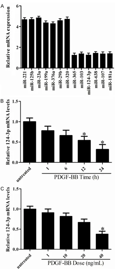
[image:3.612.92.288.68.524.2]To identify the miRNAs in the regulation of human VSMCs proliferation, we performed miRNA microarray in VSMCs after PDGF-BB stimulation (20 ng/mL). The results as shown in Figure 1A, seven miRNAs were up-regulated and six miRNAs were down-regulated in res- ponse to PDGF-BB stimulation. In agreement with previously published studies, miR-638 and
miR-365 were found to be significantly
down-regulated in response to PDGF-BB treatment. Among the six miRNAs that were down-regulat-ed by PDGF-BB stimulation, miR-124-3p was found to be highly enriched in VSMCs, and its expression was markedly down-regulated in response to PDGF-BB stimulation. Thus, these
findings suggest that miR-124-3p may be a crit -ical regulator for VSMCs proliferation.
MiR-124-3p is inhibited by PDGF-BB in VSMCs
To further confirm the expression of
miR-124-3p in proliferating VSMCs, qRT-PCR was per-formed in VSMCs stimulated by either PDGF-BB (20 ng/mL) at different time points. As shown in Figure 1B, miR-124-3p was substantially down-regulated in response to PDGF-BB treat-ment in a time dependent manner. In addition, PDGF-BB stimulation markedly reduced
the-manner (Figure 1C). These results were consis-tent with changes seen in our microarray data.
Taken together, these findings indicate an
in-verse relationship between miR-124-3p expres-sion and VSMCs proliferation.
MiR-124-3p suppressed VSMCs proliferation and migration
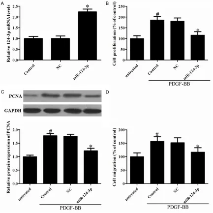
[image:4.612.92.525.73.506.2]PDGF-BB is a well-known growth factor of cell
Figure 2. The effect of miR-124-3p in VSMCs proliferation and migration. VSMCs were transfected with Negative
from the media to intima in injured vessels [11]. To further examine the functional role of miR-124-3p in VSMCs, we sought to determine whether restoration of miR-124-3p could affect PDGF-BB induced SMCCs proliferation using the CCK-8 assay. To this end, miR-124-3p was transfected into VSMCs, which caused
miR-124-3p expression levels to be significantly
increased (Figure 2A). In addition, PDGF-BB
treatment significantly increased the prolifera -tion of VSMCs compared to the non-stimulated group, and overexpression of miR-124-3p
sig-nificantly suppressed PDGF-BB-induced cell
[image:5.612.92.521.72.260.2]proliferation (Figure 2B). The proliferating cell nuclear antigen (PCNA) that plays a crucial role in the life and death decisions of cells was also
Figure 3. Identification of STAT3 as a target of miR-124-3p in human VSMCs. A. Schematic of the miR-124-3p puta -tive binding site in human STAT3 UTR and alignment of wild-type (WT) miR-124-3p and mutated (Mut) STAT3 3’-UTR binding site of miR-124-3p. B. HEK293 cells were co-transfected with the luciferase reporter carrying WT-STAT3 or Mut STAT3, together with plasmid bearing miR-124-3p or negative control. Forty-eight hours after transfection, renilla luciferase activities were measured. C. VSMCs were transfected with miR-124-3p or negative control for 24 h and then serum-deprived for 48 h, STAT3 mRNA was measured by qRT-PCR in VSMCs 3 h after stimulation with or without BB (20 ng/mL). D. Effects of miR-124-3p overexpression on STAT3 protein expression 6 h after PDGF-BB stimulation. #P<0.05 vs. untreated; *P<0.05 vs. control and NC group.
[image:5.612.91.530.372.557.2]investigated. Representative expression of
PCNA was significantly decreased in
miR-124-3p-overexpressed VSMCs compared to cells treated with PDGF-BB (Figure 2C). To
con-firm the inhibitory effect of miR-124-3p on
VSMCs migration, a Transwell assay was per-formed. As the Figure 2D shown, the number of migratory cells was suppressed in miR-124-3p-overexpressed VSMCs compared to cells treat-ed with PDGF-BB.
STAT3is a direct target of miR-124-3p
By using the TargetScan and PicTar algorithms, we found that STAT3 is a putative target of miR-124-3p (Figure 3A). To determine whether miR-124-3p directly binds to the 3’-UTR sequence of STAT3 mRNA and affects its expression, the 3’-UTR sequence of STAT3 containing the puta-tive binding site for miR-124-3p was cloned into a luciferase reporter vector. The construct vec-tor was then co-transfected with either NC or a miR-124-3p into VSMCs. The luciferase assay was inhibited in cells transfected with miR-124-3p compared to that with NC, but that does not bind to the 3’-UTR of STAT3 (Figure 3B). To fur-ther verify that STAT3 is a functional target gene of miR-124-3p in VSMCs, we determined the levels of STAT3 mRNA by RT-PCR. Inter- estingly, PDGF-BB increased mRNA levels of STAT3, but miR-124-3p effectively downregu-lated the expression of STAT3 induced with PDGF-BB (Figure 3C). As determined by west-ern blot analysis, overexpression of miR-124-3p downregulated both basal and PDGF-BB-induced STAT3 expression in VSMCs (Figure 3D).
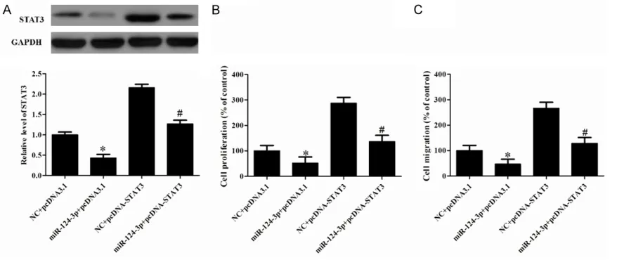
STAT3 involved in the effect of miR-124-3p in VSMCs
The data described above showed that miR-124-3p inhibited STAT3 mRNA and protein expression in VSMCs. The remaining question was whether miR-124-3p inhibits cell prolifera-tion and migraprolifera-tion by down-regulaprolifera-tion of STAT3. The results showed that the plasmid of STAT3 promoted the STAT3 protein expression detect-ed using Western blot (Figure 4A). CCK8 analy-sis demonstrated that overexpression of STAT3 promoted miR-124-3p-induced inhibition of PDGF-BB-induced VSMCs proliferation (Figure 4B). Moreover, ectopic expression of STAT3 pro-moted miR-124-3p-induced inhibition
migra-Discussion
Proliferation of VSMCs plays an essential role in the progression of atherosclerosis lesions. Inhibition of VSMCs proliferation is an impor-tant therapeutic strategy for atherosclerosis related diseases. Abnormal proliferation of PDGF-BB-stimulated VSMCs increases the risk of vascular disorders, and it is critical for the progression of restenosis and atherosclerosis
[12]. In the present study, we first detected
the miRNA expression in PDGF-BB-stimulated human VSMCs and found that seven miRNAs were up-regulated and six miRNAs were down-regulated in response to PDGF-BB stimulation. MiRNAs are small noncoding RNA molecules that post-transcriptionally or translationally reg-ulate biological processes including the modu-lation of stem cell self-renewal, differentiation, apoptosis, and proliferation by binding to the 3’-UTR of target genes [13]. However, the mech-anism by which miRNAs exert these effects remains under investigation. Recent studies have suggested important regulatory roles for miRNAs such as miR-21, miR-216, miR-217, miR-181b, miR-31b and miR-34a, which were
confirmed to be upregulated in senescing
human umbilical vascular endothelial cells (HUVECs) [14]. These miRNAs are related to cardiovascular diseases [15]. In the present study, to the best of our knowledge, we
demon-strate for the first time that miR-124-3p is sig
-nificantly down-regulated in human prolifera -tive VSMCs and restoration of miR-124-3p markedly inhibits the PDGF-BB-induced VSMCs proliferation and the expression of PCNA. In addition, miR-124-3p also suppresses the PDGF-BB-induced VSMCs migration.
endometrial cancer cells, and be involved in the miR-124-mediated suppressive effects on endometrial cancer cells [19]. In the current
study, STAT3 was identified as a direct target
of miR-124-3p and down-regulates the mRNA and protein expression of STAT3 in VSMCs. Furthermore, overexpression of STAT3 abol-ished the suppressive effects of miR-124-3p on the proliferation and migration in response to
PDGF-BB stimulation. Collectively, these find -ings demonstrated that miR-124-3p inhibits the PDGF-BB-induced proliferation and migra-tion of VSMCs through STAT3. Thus, our study provides cognate evidence implicating miR-124-3p as a novel key regulator in human VSMCs biology.
Taken together, our results for the first time
establish a functional link between miR-124-3p and STAT3 expression in VSMCs, demonstrat-ing that STAT3 is directly repressed by miR-124-3p, which subsequently inhibits its downstream signaling pathway. miR-124-3p expression was substantially down-regulated in proliferative human VSMCs and restoration of its expres-sion markedly inhibited both VSMCs prolifera-tion and migraprolifera-tion in response to PDGF stimu-lation through downregustimu-lation of STAT3. In this
regard, our study provides significant novel
insight into the molecular mechanisms associ-ated with VSMCs proliferation and migration, and suggests a potential therapeutic target for preventing and treating human vascular dis-eases, such as atherosclerosis and restenosis. Disclosure of conflict of interest
None.
Address correspondence to: Shanling Wang, De- partment of Cardiology, Henan Provincial People’s Hospital, Zhengzhou, China. Tel: +86-0371-655- 80014; Fax: +86-0371-65964376; E-mail: shanling-wzz@163.com
References
[1] Brott TG, Hobson RW, Howard G, Roubin GS, Clark WM, Brooks W, Mackey A, Hill MD, Leimgruber PP, Sheffet AJ. Stenting versus endarterectomy for treatment of carotid-artery stenosis. New Engl J Med 2010; 363: 11-23. [2] Singh RB, Mengi SA, Xu YJ, Arneja AS, Dhalla
NS. Pathogenesis of atherosclerosis: A multi-factorial process. Exp Clin Cardiol 2002; 7: 40-53.
[3] Lacolley P, Regnault V, Nicoletti A, Li Z, Michel JB. The vascular smooth muscle cell in arterial pathology: a cell that can take on multiple roles. Cardiovasc Res 2012; 95: 194-204. [4] Hata A. Functions of microRNAs in
cardiovas-cular biology and disease. Ann Rev Physiol 2013; 75: 69-93.
[5] Yu X, Li Z. The role of microRNAs expression in laryngeal cancer. Oncotarget 2015; 6: 23297-23305.
[6] Kang H, Hata A. MicroRNA regulation of smooth muscle gene expression and pheno-type. Curr Opin Hematol 2012; 19: 224-231. [7] McDonald RA, Hata A, MacLean M, Morrell
NW, Baker AH. MicroRNA and vascular remod-elling in acute vascular injury and pulmonary vascular remodelling. Cardiovasc Res 2012; 93: 594-604.
[8] Chen J, Yin H, Jiang Y, Radhakrishnan SK, Huang ZP, Li J, Shi Z, Kilsdonk EP, Gui Y, Wang DZ. Induction of microRNA-1 by myocardin in smooth muscle cells inhibits cell proliferation. Arterioscl Throm Vas 2011; 31: 368-375. [9] Liu X, Cheng Y, Zhang S, Lin Y, Yang J, Zhang C.
A necessary role of miR-221 and miR-222 in vascular smooth muscle cell proliferation and neointimal hyperplasia. Circ Res 2009; 104: 476-487.
[10] Li P, Liu Y, Yi B, Wang G, You X, Zhao X, Summer R, Qin Y, Sun J. MicroRNA-638 is highly ex-pressed in human vascular smooth muscle cells and inhibits PDGF-BB-induced cell prolif-eration and migration through targeting or-phan nuclear receptor NOR1. Cardiovasc Res 2013; 99: 185-93.
[11] Cai Y, Knight WE, Guo S, Li JD, Knight PA, Yan C. Vinpocetine suppresses pathological vascu-lar remodeling by inhibiting vascuvascu-lar smooth muscle cell proliferation and migration. J Pharmacol Exp Ther 2012; 343: 479-488. [12] Schwartz SM. Perspectives series: cell
adhe-sion in vascular biology. Smooth muscle migra-tion in atherosclerosis and restenosis. J Clin Invest 1997; 99: 2814.
[13] Adams BD, Kasinski AL, Slack FJ. Aberrant reg-ulation and function of microRNAs in cancer. Curr Biol 2014; 24: R762-R776.
[14] Menghini R, Casagrande V, Cardellini M, Martelli E, Terrinoni A, Amati F, Vasa-Nicotera M, Ippoliti A, Novelli G, Melino G. MicroRNA 217 modulates endothelial cell senescence via silent information regulator 1. Circulation 2009; 120: 1524-1532.
[15] Bronze-da-Rocha E. MicroRNAs expression
profiles in cardiovascular diseases. Bio Med
Res Int 2014; 2014: 985408-985431. [16] Cheng Y, Li Y, Nian Y, Liu D, Dai F, Zhang J.
sup-pressive effects on esophageal cancer cells. BMC Cancer 2015; 15: 306-317.
[17] Daniel JM, Dutzmann J, Bielenberg W, Widmer-Teske R, Gündüz D, Hamm CW, Sedding DG. Inhibition of STAT3 signaling prevents vascular smooth muscle cell proliferation and neointi-ma forneointi-mation. Basic Res Cardiol 2012; 107: 1-12.
[18] Cao Q, Li YY, He WF, Zhang ZZ, Zhou Q, Liu X, Shen Y, Huang TT. Interplay between microR-NAs and the STAT3 signaling pathway in hu-man cancers. Physiol Genomics 2013; 45: 1206-1214.
![N,N′ Bis[2 (methylsulfanyl)phenyl]pyridine 2,6 dicarboxamide](data:image/gif;base64,R0lGODlhAQABAIAAAP///wAAACH5BAEAAAAALAAAAAABAAEAAAICRAEAOw==)