Case Report
Mediastinal bronchial artery aneurysm treated with
aortic stent and embolization: case report
Meijun Song, Hongcheng Wu, Jingbo Jiang, Guo’an Wang, Shibo Wu, Ting Ge
Department of Respiratory Medicine, Ningbo Medical University Affiliated Li Huili Hospital, Ningbo, China
Received February 24, 2016; Accepted August 15, 2016; Epub September 15, 2016; Published September 30, 2016
Abstract: Mediastinal bronchial artery aneurysm (BAA) refers to a rare aneurysmal dilatation but can be
poten-tially life threatening. Opportune treatment is mandatory upon a confirmed diagnosis. There are several reports
of endovascular treatment of BAA with transcatheter arterial embolization (TAE) and only a few cases treated with aortic stent-graft exclusion. Here, we report on two case studies of mediastinal BAAs with a short neck treated
with a combined approach of stent-graft occlusion of the inflow and coil embolization of the outflow arteries. As a
comparison, we reviewed seven other cases that were previous reported in literature for their clinical presentation and therapeutic management. Our study demonstrated that mediastinal BAA can be successfully treated with the available endovascular techniques. Combining approaches of aortic stent-graft placement and coil embolization is a viable option in the treatment of BAA with a short neck.
Keywords: Aneurysm, angiography, embolization, covered stent-graft, thoracic aorta
Introduction
The occurrence of bronchial artery aneurysm (BAA) is rare and detected in less than 1% of all patients who undergo selective bronchial arte-riography [1]. BAA can be intrapulmonary, medi-astinal or both, and the clinical presentation depends on the size, location, and presence of concomitant disease. The main indications of mediastinal BAA is related to compression or rupture into contiguous structures [2]. Its rup-ture can cause a life-threatening hemorrhage and opportune treatment is mandatory upon a confirmed diagnosis. Open surgical procedures are preferred but recently transcatheter arteri-al embolization (TAE) has demonstrated good treatment results [3]. Currently, there are numerous reports on endovascular treatment of BAA using TAE and a few cases were treated with aortic stent-graft exclusion [3-5]. Here, we report on 2 cases of mediastinal BAAs treated with a combined approach of stent-graft occlu-sion of the inflow and coil embolization of the outflow arteries. As a comparison, we also reviewed seven other cases previous reported in literature and compared them with our cur-rent case studies.
Case report
CASE 1
A 59-year-old woman was admitted to the Emergency Room due to a sudden onset of chest pain radiating to back, with a history of constant dysphagia lasting for about 3 years. She also had a history of repeated hemoptysis for more than 10 years. Upon reaching the hos-pital, the patient was in hemodynamically sta-ble condition except for mild elevation of her respiration rate (22/min); her blood pressure was 118/76 mmHg, pulse rate was 67 beats/ min, and the heart rhythm was regular. An EKG presented a normal sinus rhythm without any ischemic signs or arrhythmia. Breath sounds were normal. Laboratory examination revealed hemoglobin of 12.5 g/dl. There was no trauma, and no sign of infection.
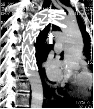
con-ducted. On the selective bronchial arteriogram, we found a bronchial artery aneurysm (18 mm) that was just in the initial part of the bronchial artery, located very close to the aorta as shown in Figures 2 and 3. The length of the neck was about 5 mm. There was no sign of active bleed-ing or extravasation. To prevent further rupture or extravasation of the aneurysm, we perfor- med a bronchial artery embolization with sev-eral microcoils. After that, the second time of selective bronchial arteriogram showed the
dis-appearance of the distal end of the bronchial artery but we still found the initial part present-ed aneurismal dilatation (Figure 4). Then an aortic stent was planted in the descending aorta to isolate the bleeding bronchial artery, and bronchial artery angiography showed the blood flow was completely blocked (Figure 5). The patient was in a stable condition during percutaneous vascular intervention. She was sent to the ICU and under close observation after the embolization procedure. During fol-low-up on the first month after operation, CT scan of the chest showed left pulmonary atel-Figure 1. Enhanced CT scan showed posterior
[image:2.612.323.521.71.243.2]medi-astinum hematoma.
Figure 2. CT scan and 3D reconstruction confirmed a
[image:2.612.89.290.72.251.2]bronchial artery aneurysm(18 mm) in the initial part of the bronchial artery, adjacent to the descending aorta.
Figure 3. CT scan and 3D reconstruction confirmed a
bronchial artery aneurysm (18 mm) in the initial part of the bronchial artery, with a neck of 5 mm in length.
[image:2.612.323.523.302.469.2] [image:2.612.88.290.303.514.2]ectasis and pleural infusion. A chest tube was inserted and 2.5 liters of unclotted blood was drained from the left pleural cavity. After that, a subsequent CT scan showed pulmonary re-expansion and no further bleeding, indicating her conditions returned to normality. In the
third month after operation, CT scan of the chest showed no stent migration or pleural effusion as shown in Figure 6.
CASE 2
[image:3.612.89.290.69.289.2]A 63-year-old man was admitted to the Em- ergency Department due to a sudden onset of sharp chest pain. The pain was located in the anterior chest wall radiating to the back. Upon reaching the hospital, the patient was in hemo-dynamically unstable condition, his respiration rate was 23/min; blood pressure was 70/40 Figure 5. An aorta stent was planted in the
[image:3.612.323.522.70.262.2]descend-ing aorta, bronchial artery angiography showed dis-appearance of the BAA but microcoils.
[image:3.612.322.523.335.509.2]Figure 6.In follow-up of the third month after opera-tion, CT scan of the chest showed no stent migraopera-tion, and disappearance of the BAA.
Figure 7. Contrast-enhanced computed tomography (CT) scan showsposterior mediastinum hematoma and bilateral pleural effusion, full of blood in the left cavity.
[image:3.612.89.290.355.587.2]mmHg, pulse rate was 200 beats/min, but the heart rhythm was regular. Left breath sounds were very low. Laboratory examination revealed hemoglobin of 8.9 g/dl. There was no trauma, no sign of infection, and no hemoptysis. A chest CT scan (Figure 7) confirmed active posterior mediastinum hematoma that was very close to the descending aorta and bilateral pleural effusion. The left chest cavity was full of blood. A chest tube was inserted into the left pleural cavity. Due to a suspicious origin of mediastinal hemorrhage on the chest CT scan, thoracic aortogram and bronchial arteriogram were conducted. On the selective bronchial ar-
teriogram, we found a bronchial artery aneu-rysm of 5 mm. The location was situated in the initial part of the bronchial artery and adjacent to the descending aorta, with a 6 mm neck length as shown in Figures 8 and 9. There was extravasation of contrast agent in the mediasti-num, which strongly suggested the presence of active bleeding. Therefore, we performed a bronchial artery embolization with 3 microcoils. After that, the second time of selective bron-chial arteriogram showed the disappearance of the distal end of the bronchial artery. However, we still found some contrast agent in the initial part of the bronchial artery near the descend-ing aorta. Then a stent (140 mm×34 mm) was planted in the descending aorta to isolate the bleeding bronchial artery, and bronchial artery angiography showed the blood flow was com-pletely blocked.
The patient was in a stable condition during percutaneous vascular intervention. He was sent to the ICU for close observation after the embolization procedure. Due to his loss of blood, the patient needed transfusion. During follow-up on the third month after operation, the patient was in good condition without fur-ther complications. His chest CT showed no pleural effusion in the left chest and no shift of the stent (Figure 10).
Discussion
Acute hemomediastinum and hemothorax are usually related to chest trauma, rupture of a thoracic aortic aneurysm, or aortic dissection [6]. The causes of spontaneous mediastinal hemorrhage can fall within four distinct catego-ries [7] that include: (1) complication of enlarg-ing mediastinal masses; (2) transient increase in intra-thoracic pressure; (3) sudden sustained hypertension; and (4) altered hemostasis. A ruptured BAA is seldom associated and rarely the causation of mediastinal hemorrhage. Mediastinal BAA is a rare condition, which has been reported only in very few cases in the lit-erature [3, 8].
The exact cause of BAA is unclear [1]. The con-dition can be congenital, as in the context of pulmonary sequestration [9] or pulmonary agenesis [10]. It can also be acquired, due largely to atherosclerosis [11, 12], inflammato-ry lung disease, bronchiectasis [1], tuberculosis or trauma (pseudoaneurysm) [13]. Other asso-Figure 9. CT scan and 3D reconstruction shows the
[image:4.612.89.290.71.259.2]short neck of the Bronchial artery aneurysm and its origin from the descending aorta.
Figure 10.CT scan showed little pleural effusion in
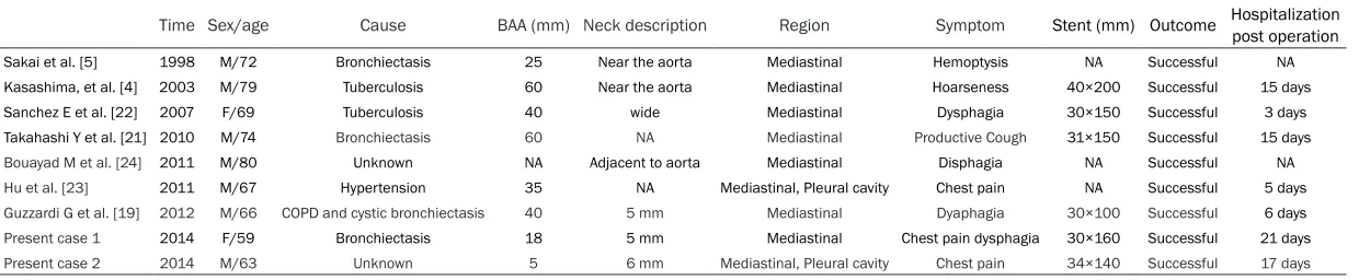
[image:4.612.89.288.321.478.2]Table 1. Comparison of published cases of ruptured mediastinal bronchial artery aneurysm treated with aortic stent
Time Sex/age Cause BAA (mm) Neck description Region Symptom Stent (mm) Outcome Hospitalization post operation
Sakai et al. [5] 1998 M/72 Bronchiectasis 25 Near the aorta Mediastinal Hemoptysis NA Successful NA Kasashima, et al. [4] 2003 M/79 Tuberculosis 60 Near the aorta Mediastinal Hoarseness 40×200 Successful 15 days Sanchez E et al. [22] 2007 F/69 Tuberculosis 40 wide Mediastinal Dysphagia 30×150 Successful 3 days Takahashi Y et al. [21] 2010 M/74 Bronchiectasis 60 NA Mediastinal Productive Cough 31×150 Successful 15 days
Bouayad M et al. [24] 2011 M/80 Unknown NA Adjacent to aorta Mediastinal Disphagia NA Successful NA
Hu et al. [23] 2011 M/67 Hypertension 35 NA Mediastinal, Pleural cavity Chest pain NA Successful 5 days
Guzzardi G et al. [19] 2012 M/66 COPD and cystic bronchiectasis 40 5 mm Mediastinal Dyaphagia 30×100 Successful 6 days
Present case 1 2014 F/59 Bronchiectasis 18 5 mm Mediastinal Chest pain dysphagia 30×160 Successful 21 days
Present case 2 2014 M/63 Unknown 5 6 mm Mediastinal, Pleural cavity Chest pain 34×140 Successful 17 days
ciations to BAA include systemic vascular ab- normalities, such as Osler-Weber-Rendu dis-ease [14] and sepsis (mycotic aneurysm) [15]. The focal weakening or injury to the vessel walls is the common link among these causes. Increased blood flow to the bronchial arteries may also play a significant role. As shown in Table 1, our review of past literature highlight-ed causes due to bronchiectasis, tuberculosis, cystic bronchiectasis and hypertension. Two of the studies were of unknown etiology. The majority of the causes were linked to chronic inflammation.
Once the bronchial artery ruptures, the clinical presentation is acute and life-threatening, with the most common symptoms being severe chest/back pain mimicking acute aortic dis-section [16] together with symptoms of shock [17]. On the other hand, the most common hemorrhagic presentation is hemoptysis, fol-lowed by hematemesis, depending on whether the aneurysm extends parenchymally or poste-riorly. Massive hemomediastinum is less com-mon and hemothorax is the least comcom-mon mode of presentation [18]. It may also be accompanied with epigastric pain, hemateme-sis, and hemoptysis resulting from the rupture into the esophagus and pulmonary parenchy-ma. Mediastinal BAA may present compressive symptoms, such as dysphagia or superior vena cava syndrome [12]. Although little is known about the process that leads to BAA rupture, the size of the aneurysm (5-40 mm) is not an incremental risk factor [3]. Prior studies have reported on radiological examination of BAAs with sizes as large as 8-10 cm in diameter, which were not as serious as cases of BAAs with sizes of 2 cm. Patients in the latter cases were manifested with life threatening bleeding conditions. As observed also in Table 1, all ca- ses with ruptured BAAs were of relatively small size ranging from 5-60 mm in diameter. This supports that the size of the aneurysm does not negatively correlate to clinical outcome. The primary diagnostic modes are computed tomographic angiography and intra-arterial an- giography. Occasionally, magnetic resonance imaging has been employed in complex cases. Here we describe mediastinal BAA with a short neck. We compared our study against other reported cases (Table 1) but most studies lack comprehensive quantitative data of the neck
description. Comparing with the study conduct-ed by Guzzardi G et al. [19], our results were consistent with their findings of measured neck length. Our measured neck length for case 1 and 2 were 5 mm and 6 mm respectively. BAA should be treated whether it is symptom-atic or not. In previous reports, TAE has been the most common approach in recent years [3]. TAE to occlude the afferent and efferent arter-ies of the BAA is considered the first line treat-ment option if the patient is stable. Surgery should be reserved for patients with contraindi-cations to embolization, such as allergy to io- dinated contrast medium or medullary artery. Open surgical treatment is a valid alternative but is associated with high morbidity and mor-tality. The advantages and disadvantages of surgery and TAE should be recognized, and the appropriate procedure should be selected based on the patient’s clinical status. Several reports commented on the failed embolization of a bronchial artery if the origin of the aneu-rysm is too close to the aorta [7, 20]. Given the clinical presentation, the optimal treatment for mediastinal BAA especially for that with short neck is combined treatment with TAE and aor-tic stent. Kasashima et al. [4] reported suc-cessful treatment of BAA by stent-graft place-ment alone. Sakai et al. [5] Takahashi et al. [21] and Giuseppe Guzzardi et al. [19] reported three cases with the treatment of BAA by stent-graft occlusion of the inflow artery and coil embolization of the outflow artery, demonstrat-ing the effectiveness of this kind of combined approach. If transcatheter coil embolization of the outflow vessels is technically difficult, it is possible to embolize these arteries using fibrin sealant, as described by Sanchez et al. [22]. Hu
et al. [23] described successful combined treatment of BAA with aortic stent-graft place-ment and embolization of the outflow arteries using sodium polymannuronate and gelatin sponge. We managed to treat these two cases of mediastinal BAA with short neck by a combi-nation of TAE and aorta stent. The average duration in the hospital after operation was 11.7 days for seven reviewed cases in Table 1 as opposed to our two cases that were hospi-talized for 19 days.
man-agement of mediastinal BAA. The endovascular method highlighted in literature is the optimal treatment for patients with suitable anatomic features. As shown in our case reports, we determined that patients with BAA and short neck length benefited most from the place-ment of aortic stent graft to exclude the aneu-rysm by closing the feeding vessels. It is also important to close the outflow arteries arising from the aneurysmal sac to prevent retrograde filling and subsequent risk of rupture of the revascularized BAA.
Conclusion
We have successfully demonstrated that medi-astinal BAA can treated with existing endovas-cular techniques. It is important that treatment is prompt even without any significant symp-toms, as mediastinal BAA is potentially life threatening. We have shown that using a com-bined approach with aortic stent-graft place-ment and coil embolization, this benefited patients suffering from BAAs with short neck length. We believe that further follow-ups on these existing cases are necessary to ensure long-term treatment effectiveness. In order to better gauge the treatment efficacy, more case studies will have to be repeated for patients with similar anatomic features.
Acknowledgements
We thank the staff of Ninbgo Medical University Affilliated Li Huili Hospital who aided in the study. We are also grateful for the two partici-pants who provided consent for this case report.
Disclosure of conflict of interest
None.
Address correspondence to: Dr. Hongcheng Wu, De- partment of Respiratory Medicine, Ningbo Medical
University Affiliated Li Huili Hospital, Ningbo
31-5040, Zhejiang, China. E-mail: smj800722@aliyun. com
References
[1] Mizuguchi S, Inoue K, Kida A, Isota M, Hige K, Aoyama T and Ishikawa T. Ruptured bronchial artery aneurysm associated with bronchiecta-sis: a case report. Ann Thorac Cardiovasc Surg 2009; 15: 115-118.
[2] Hall RJ, Miller GA and Kerr IH. Ruptured bron-chial artery aneurysm mimicking aortic dissec-tion. Br Heart J 1977; 39: 909-910.
[3] Tanaka K, Ihaya A, Horiuci T, Morioka K, Kimu-ra T, Uesaka T, Sasaki M, Uchinami M, Tsuda T and Yamada N. Giant mediastinal bronchial artery aneurysm mimicking benign esopha-geal tumor: a case report and review of 26 cases from literature. J Vasc Surg 2003; 38: 1125-1129.
[4] Kasashima F, Endo M, Kosugi I, Matsumoto Y, Abe Y, Sasaki H, Sanada J and Matsui O. Medi-astinal bronchial artery aneurysm treated with a stent-graft. J Endovasc Ther 2003; 10: 381-385.
[5] Sakai T, Razavi MK, Semba CP, Kee ST, Sze DY and Dake MD. Percutaneous treatment of bronchial artery aneurysm with use of trans-catheter coil embolization and thoracic aortic stent-graft placement. J Vasc Interv Radiol 1998; 9: 1025-1028.
[6] Vosse BAH, van Belle AF, de Vries GJ and Das M. Hemomediastinum due to spontaneous rupture of a mediastinal bronchial artery aneu-rysm-A rare cause of thoracic pain. Respir Med Case Rep 2014; 12: 27-29.
[7] Pugnale M, Portier F, Lamarre A, Halkic N, Riis H-B, Wicky S, Schnyder P and Denys A. Hemo-mediastinum caused by rupture of a bronchial artery aneurysm: successful treatment by em-bolization with N-butyl-2-cyanoacrylate. J Vasc Interv Radiol 2001; 12: 1351-1352.
[8] Wilson SR, Winger DI and Katz DS. CT visual-ization of mediastinal bronchial artery aneu-rysm. Am J Roentgenol 2006; 187: W544-W545.
[9] Abet D and Pietri J. [Ruptured bronchial artery aneurysm simulating dissection of the aorta in a patient with bronchopulmonary sequestra-tion (author’s transl)]. J Chir (Paris) 1981; 118: 743-746.
[10] Sancho C, Dominguez J, Escalante E, Hernan-dez E, Cairols M and Martinez X. Embolization of an anomalous bronchial artery aneurysm in a patient with agenesis of the left pulmonary artery. J Vasc Interv Radiol 1999; 10: 1122-1126.
[11] Shaer AH and Bashist B. Computed tomogra-phy of bronchial artery aneurysm with erosion into the esophagus. J Comput Assist Tomogr 1989; 13: 1069-1071.
[12] Hoffmann V, Ysebaert D, De Schepper A, Col-paert C and Jorens P. Acute superior vena cava obstruction after rupture of a bronchial artery aneurysm. Chest 1996; 110: 1356-1358. [13] Cearlock JR, Fontaine AB, Urbaneja A and
[14] Ishizaki N, Shimokawa S, Tanaka K, Taira A, Onohara S, Tabata M and Sakoda K. Ruptured bronchial artery aneurysm associated with pleural telangiectasis and tortuous portal ob-struction: report of a case. Surg Today 1995; 25: 852-854.
[15] Chantepie A, Robert M, Pelletier J, Gold F, Mer-cier C, Lacombe A and Laugier J. [Mycotic an-eurysm of bronchial artery. Apropos of a case in an infant]. Chir Pediatr 1979; 21: 407-410. [16] Kalangos A, Khatchatourian G, Panos A and
Faidutti B. Ruptured mediastinal bronchial ar-tery aneurysm: a dilemma of diagnosis and therapeutic approach. J Thorac Cardiovasc Surg 1997; 114: 853-856.
[17] Maisawa K and Koh E. [Ruptured bronchial ar-tery aneurysm with left hemothorax: report of a case]. Kyobu Geka 2012; 65: 419-422. [18] Karmy-Jones R, Hastreiter D and Burdick T.
He-mothorax complicating bronchial artery aneu-rysm. Can Respir J 2005; 12: 279-281. [19] Guzzardi G, Cerini P, Fossaceca R, Commodo
M, Micalizzi E and Carriero A. Endovascular treatment of bronchial artery aneurysm with aortic stent-graft placement and coil emboliza-tion. Ann Vasc Surg 2012; 26: 1013-e1015.
[20] Yanagihara K, Ueno Y, Kobayashi T, Isobe J and Itoh M. Bronchial artery aneurysm. Ann Thorac Surg 1999; 67: 854-855.
[21] Takahashi Y, Tsutsumi Y, Monta O, Kohshi K, Ohashi H, Shimamura K and Osuga K. Stent grafting for giant bronchial artery aneurysm disguised as an aneurysm with multiple feed-ing arteries. Ann Thorac Surg 2010; 89: 1652-1654.
[22] Sanchez E, Alados P, Zurera L, Canis M, Muñoz I, Casares J and Eguaras MG. Bronchial artery aneurysm treated with aortic stent graft and
fibrin sealant. Ann Thorac Surg 2007; 83: 693-695.
[23] Hu CX, Huang S, Xu ZW, Chen W, Huang JS And Fu Z. Combination of aortic stent-graft and terial embolization for ruptured bronchial ar-tery aneurysm. Ann Thorac Surg 2011; 92: e19-e21.
[24] Bouayad M, Bagan P, Brian E, Benabdesselam