Quantification of gravity-induced skin strain across the
breast surface
SANCHEZ, Amy, MILLS, Chris, HAAKE, Steve <http://orcid.org/0000-0002-4449-6680>, NORRIS, Michelle and SCURR, Joanna
Available from Sheffield Hallam University Research Archive (SHURA) at:
http://shura.shu.ac.uk/17113/
This document is the author deposited version. You are advised to consult the publisher's version if you wish to cite from it.
Published version
SANCHEZ, Amy, MILLS, Chris, HAAKE, Steve, NORRIS, Michelle and SCURR, Joanna (2017). Quantification of gravity-induced skin strain across the breast surface. Clinical Biomechanics, 50, 47-55.
Copyright and re-use policy
See http://shura.shu.ac.uk/information.html
1
Quantification of gravity-induced skin strain across the breast surface. 1
Amy Sanchez1, Chris Mills1, Steve Haake2, Michelle Norris1 and Joanna Scurr1 2
3
1
Department of Sport and Exercise Science, Spinnaker Building, University of Portsmouth, 4
PO1 2ER, UK. 5
2
Centre for Sports Engineering Research, Sheffield Hallam University, Sheffield, S10 2BP, 6
UK. 7
8
Corresponding Author: 9
Dr Chris Mills 10
Department of Sport and Exercise Sciences 11
University of Portsmouth 12
Spinnaker Building 13
Portsmouth 14
PO1 2ER 15
United Kingdom 16
Email: [email protected]
17
18
Word Count (abstract): 250 words 19
2 Abstract
21
Background 22
Quantification of the magnitude of skin strain in different regions of the breast may help to 23
estimate possible gravity-induced damage whilst also being able to inform the selection of 24
incision locations during breast surgery. The aim of this study was to quantify static skin 25
strain over the breast surface and to estimate the risk of skin damage caused by gravitational 26
loading. 27
Methods 28
Fourteen participants had 21 markers applied to their torso and left breast. The non-gravity 29
breast position was estimated as the mid-point of the breast positions in water and soybean 30
oil (higher and lower density than breast respectively). The static gravity-loaded breast 31
position was also measured. Skin strain was calculated as the percentage extension between 32
adjacent breast markers in the gravity and non-gravity loaded conditions. 33
Findings 34
Gravity induced breast deformation caused peak strains ranging from 14 to 75% across 35
participants, with potentially damaging skin strain (>60%) in one participant and skin 36
strains above 30% (skin resistance zone) in a further four participants. These peak strain 37
values all occurred in the longitudinal direction in the upper region of the breast skin. In the 38
latitudinal direction, smaller-breasted participants experienced greater strain on the outer 39
(lateral) breast regions and less strain on the inner (medial) breast regions, a trend which 40
was reversed in the larger breasted participants (above size 34D). 41
Interpretation 42
To reduce tension on surgical incisions it is suggested that preference should be given to 43
medial latitudinal locations for smaller breasted women and lateral latitudinal locations for 44
3 46
Keywords 47
Breast; surgery; strain; skin; damage; density 48
49
50
51
Highlights 52
Quantification of breast skin strain to inform incision locations during surgery 53
Up to 75% skin strain in the longitudinal direction in upper region of breast 54
Smaller-breasted participants experienced greater strain on lateral breast regions 55
Larger-breasted participants experienced greater strain on medial breast regions 56
57
58
59
60
4 1.0 Introduction
62
The female breast is a highly malleable structure that is easily deformed by external forces 63
(Rajagopal et al., 2008). Deformation of the breast has been hypothesised to damage the 64
breast structure, which may lead to breast sag (ptosis) (Page & Steele 1999). Measurements 65
of strain can be used to evaluate the magnitude and reversibility of a biological tissue’s 66
response to external loading (Gao & Desai 2010; Hull et al., 1996; Lim et al., 2008; Miller 67
2001; Toms et al., 2002). One of the breast’s primary support systems is the skin (Hindle 68
1991) and during breast surgery an incision must be made in this supporting tissue. 69
70
Previous research has investigated numerous methods of identifying the correct placement 71
and direction of surgical incisions, to minimise tissue damage and long term scarring (Seo, 72
Kim, Cordier, Choi, & Hong, 2013). These have included the identification of Langer’s Lines 73
(where surgical incisions are performed in the direction of maximum skin tension) (Gibson, 74
1978), Kraissl’s Lines (where surgical incisions coincide with wrinkle lines) (Kraissl, 1951), 75
and relaxed tissue lines (similar to Kraissl’s lines, however performed when the skin is 76
relaxed) (Borges & Alexander, 1962). The aforementioned are a select few of many 77
guidelines currently available to surgeons, when performing surgical incisions (Seo et al., 78
2013). However, with further information as to skin strain properties surgeons may be better 79
informed when selecting incision location and direction. This is of particular interest across 80
the breast surface as recent studies have reported an increase in breast augmentation surgery 81
(Mahmood et al., 2013), and an increase in mastectomy rates in those with breast cancer or 82
benign breast lump removal (Albornoz et al., 2013). Surgical incisions performed in areas of 83
high skin strain, when gravity loaded, may cause stretching of scars and increased healing 84
times as well as increased incidence of scar repair / removal. 85
5
The biomechanical properties of the skin vary directionally, regionally, and between 87
individuals (Clark et al., 1996, Finlay 1970). At low strains the collagen fibres are loosely 88
interwoven and there is little resistance to deformation. At increasing strains the collagen 89
fibres align in the direction of loading and begin to resist extension, until eventually failure 90
occurs (Daly 1982). Skin failure studies are typically conducted on porcine or cadaver skin 91
samples rather than in vivo (Winter 2006; Gallagher et al., 2012), and results have shown that 92
skin resistance and skin failure can occur at a range of different strain values. The onset of 93
skin resistance has been reported to occur at strains between 16% and 48% (Stark 1977), with 94
skin failure occurring at strains between 16% (Lim et al., 2011) and 126% (Gallagher et al., 95
2012; Ní Annaidh et al., 2012). The wide-ranging results presented for the different stages of 96
skin extension may be due to differences in skin sampling techniques, sample preservation 97
procedures, and strain measurement systems. For the purpose of this study strain limits were 98
defined as 30% for skin resistance and 60% for skin failure based on the representative strain 99
values for human skin reported by Silver et al., (2001). 100
101
When evaluating the risk of strain-induced damage to the breast skin it is imperative that 102
measurements of strain are taken from the unloaded (neutral) position of the breast. 103
However, the continuous deforming effect of gravity on the breast makes it difficult to 104
identify the neutral breast position from which to take measurements of strain (Gao & Desai 105
2010). Previously reported strain measurements taken from the gravity-loaded breast 106
position have produced the counter-intuitive result that larger-breasted women experienced 107
less breast strain than their smaller-breasted counterparts (Scurr et al., 2009). Subsequent 108
studies have considered the effect of gravity, but have only included two markers to measure 109
breast strain (one on the nipple and one on the torso) (Haake & Scurr 2011, Haake et al., 110
6
values may not represent the strain on any particular breast structure, making it difficult to 112
apply the appropriate strain failure limits to assess damage. Despite the limitations associated 113
with the two-marker method, Haake et al., (2012) reported static gravity-induced breast 114
strains up to 80%, which indicate that gravity may induce considerable static strains on the 115
breast skin. 116
117
This study uses a novel approach for assessing breast skin strain from the neutral (unloaded) 118
position using a marker array over the breast surface. The method used the buoyant force of 119
the fluid to counteract the effect of gravity on the breast. As breast mass-density can vary 120
between women, a single fluid may not completely counteract the effect of gravity across 121
different participants. Instead, the boundaries of the neutral breast position may be identified 122
by immersing the breast and body in two fluids with densities above (water) and below 123
(soybean oil) the range of reported breast mass-densities (Sanchez et al., 2016). The mid-124
point between these two immersion conditions may then be used to identify a more accurate 125
neutral breast position than could be achieved using either fluid in isolation (Mills et al., 126
2016). 127
128
The second novel aspect of this study was the implementation of a marker array on the breast 129
skin. Although an array has been implemented in previous research assessing the effect of 130
gravity on the breast (Rajagopal 2007), there have been no attempts to calculate skin strain. 131
Application of a marker array over the breast skin provides a better representation of the 132
breast’s curved surface, which enables measurements of strain to better replicate the strain 133
experienced by the breast skin. This is important for evaluating the risk of skin damage 134
7
evaluation of skin strain in different regions of the breast, which may enable identification of 136
breast regions that are most susceptible to excessive levels of skin strain. 137
138
Measurements of strain on the breast skin could be used to assess the risk of damage 139
associated gravitational loading and also act as a starting point from which to subsequently 140
help inform the selection of incision locations during breast surgery. The aim of this study 141
was to quantify static skin strain over the breast surface and to estimate the risk of skin 142
damage caused by gravitational loading. 143
144
2.0 Methods 145
Following institutional ethical approval (SFEC 2013-001), a convenience sample of 14 146
females gave written informed consent to take part in this study. All participants were aged 147
between 20 and 27 years, were nulliparous, had not exposed their breasts to UV radiation 148
within the last three months, and had not undergone surgical procedures on their breasts. 149
These criteria were imposed in an attempt to ensure the participants’ breast skin was elastic 150
and would return to its neutral position when supported by the buoyant forces from water and 151
soybean oil (Gambichler et al., 2006, Fujimura et al., 2007, Smalls et al., 2006, Fisher et al., 152
1997). Participants had their bra size assessed by a trained bra fitter using best-fit criteria 153
(McGhee & Steele 2010), and were assigned a participant number in ascending bra cross-154
grading size. 155
156
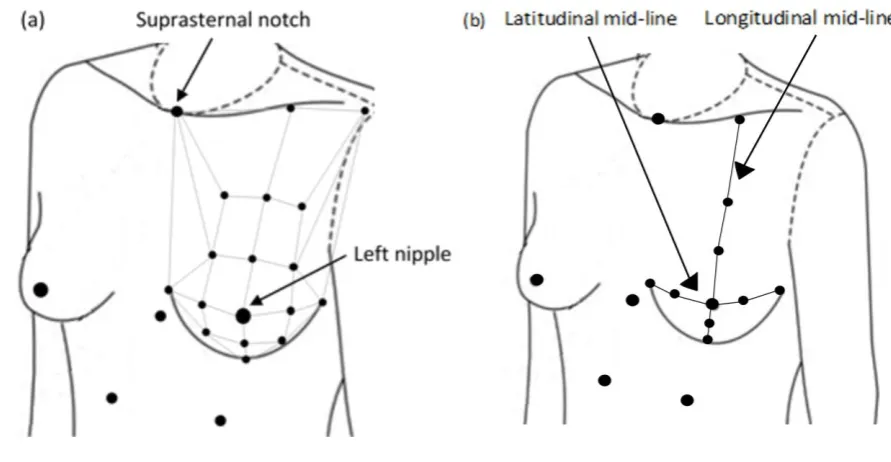
Retro-reflective markers (12 mm diameter flat markers) were applied to the participants’ 157
suprasternal notch, xiphoid process, right and left anterior-inferior aspect of the 10th ribs, and 158
left nipple using hypoallergenic tape, based on the torso marker set described by Scurr et al. 159
8
breast (6 mm diameter flat markers) (Figure 1), which was based on the rectangular 161
segmentation of the breast described by Rajagopal et al., (2008). The total mass of the 162
markers on the breast was 0.17 g, and was assessed using a Mettler PC400 balance (Mettler 163
Toledo, Switzerland). 164
165
166
167
Figure 1: (a) Torso marker set, breast marker array, and inter-marker pairings (grey lines) 168
used to calculate skin strain; and (b) longitudinal and latitudinal breast mid-lines. 169
170
The neutral position of the breast was obtained using immersion in both water and soybean 171
oil. Three synchronised underwater cameras (25 Hz, VB5C6 Submersible Colour Camera, 172
Videcon PLC) were attached to the inside of a D-shaped tank. The tank was first filled with 173
water, and all participants were tested, then the tank was emptied, cleaned and filled with 174
soybean oil. The cameras were calibrated before testing each participant using a custom-175
made 36-point calibration frame. A 16 order DLT was used to correct for image distortion 176
caused by the fluids. In each fluid, participants sat on an adjustable stool so that their 177
[image:9.595.66.512.211.442.2]9
position with their arms by their sides while the static positions of the breast markers were 179
recorded for three 1 s trials in each fluid. Participants also had their static gravity-loaded 180
breast positions recorded in six 1 s trials (three before each fluid immersion) using a 181
calibrated optoelectronic camera system (200 Hz, Oqus, Qualisys, Sweden). 182
183
The 3D co-ordinates of the torso and breast markers in the two immersion conditions were 184
identified and reconstructed using SIMI software (version 8.5.5, Tracksys Ltd), and the 185
gravity-loaded marker co-ordinates were identified using Qualisys Track Manager (QTM) 186
(Qualisys, Sweden). The mean reconstruction errors for the SIMI and QTM software were 187
0.7 mm and 0.4 mm, respectively. All co-ordinate data were then exported to Visual 3D 188
(v4.96.4, C-motion) for further analysis. Within Visual 3D, a torso segment was created for 189
each participant using the suprasternal notch marker and the two rib markers to define the 190
proximal and distal segment ends respectively (Mills et al., 2014). The torso segment origin 191
was defined at the proximal end of the segment and the xiphoid process marker was added to 192
aid segment tracking. The 3D marker co-ordinate data were filtered using a generalised 193
cross-validatory quintic spline and the position of each breast marker was calculated relative 194
to the torso segment in each condition (water, soybean oil, and gravity-loaded). A total of 35 195
inter-marker distances were calculated for each participant, in each condition, using the 196
resultant separation between the breast marker pairings shown in Figure 1. 197
198
The neutral (unloaded) inter-marker separation (L0) was defined as the mean of the water and
199
soybean oil conditions. Strain was calculated using, 200
Equation 1: Strain = 100 . ((𝐿− 𝐿𝐿 𝑂)
𝑂 ) = 100 . ( (∆𝐿)
𝐿𝑂) 201
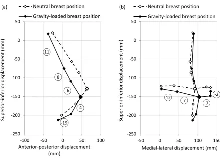
where L was defined as the mean inter-marker separation calculated from the six gravity-202
10
was estimated by comparing the static skin strain values for each participant to the strain 204
limits reported by Silver (2001) (30% representing skin resistance and 60% representing the 205
onset of skin failure). 206
207
To evaluate the potential improvement in skin strain estimation using a breast marker array, 208
and for comparison to previously published data, strain was also calculated using the two-209
marker method described by Haake and Scurr (2011). For this analysis, strain was calculated 210
using Equation 1 where the neutral and loaded breast lengths were defined as the superior-211
inferior displacement of the left nipple from the suprasternal notch in the neutral (L0) and
212
gravity-loaded (L) conditions respectively (Figure 1) (Haake & Scurr 2011). 213
214
215
3.0 Results 216
In the neutral position the breast shape was conical or hemispherical, with the breast bulk 217
distributed symmetrically behind the nipple (Figure 2). Gravitational loading caused the 218
breast bulk to fall inferiorly, leading to flattening of the upper breast and distortion of the 219
lower breast to form the typically observed tear-drop breast shape (Figure 2). This breast 220
deformation led to a posterior and inferior displacement of the nipple (Figure 2), with most 221
participants also experiencing a small lateral shift of the breast bulk in the gravity-loaded 222
condition, particularly below the nipple (Figure 3). Example gravity-induced skin strains 223
resulting from deformation of the breast mid-lines are shown for Participant 11 (breast size 224
32DD) in Figure 4. These strain data reflect the changes in breast shape, with the inferior and 225
lateral displacement of the breast causing positive strain (tension) to occur on the upper and 226
medial skin segments, and negative strain (compression) to occur on the lower and lateral 227
12
Anterior-posterior displacement (mm)
Anterior-posterior displacement (mm) -250 -210 -170 -130 -90 -50 -10 30
-20 20 60
Su p eri o r-in fer io r d is p lac me en t (mm) Participant 1 (32B)
-30 10 50 Participant 2
(32B)
-30 10 50 Participant 3
(32B)
-40 0 40 Participant 4
(34B)
-20 20 60 Participant 5
(32C)
-20 20 60 Participant 6
(32C)
-30 10 50 Participant 7
(32D)
-50 -10 30 70 Participant 8 (32D) -250 -210 -170 -130 -90 -50 -10 30
-50 -10 30
Su p eri o r-in fer io r d is p lac me en t (mm) Participant 9 (32D)
-40 0 40 Participant 10
(34D)
-50 -10 30 70 Participant 11
(32DD)
-50 -10 30 70 Participant 12
(30E)
-20 20 60 100 Participant 13
(34DD)
-50 -10 30 70 Participant 14
13
Figure 2: Position of the markers along the longitudinal breast mid-line in the neutral (dashed) and gravity-loaded (grey) conditions, in the
14
Medial-lateral displacement (mm)
Medial-lateral displacement (mm) -250 -210 -170 -130 -90 -50 -10 30
0 40 80 120
Su p eri o r-in fer io r d is p lac me en t (mm)
Participant 1 (32B)
-10 30 70 110 150 Participant 2 (32B)
0 40 80 120 160 Participant 3 (32B)
0 40 80 120 160 Participant 4 (34B)
0 40 80 120 160 Participant 5 (32C)
0 40 80 120 Participant 6 (32C)
0 40 80 120 Participant 7 (32D)
-250 -210 -170 -130 -90 -50 -10 30
0 40 80 120 160
Su p eri o r-in fer io r d is p lac me en t (mm)
Participant 8 (32D)
0 40 80 120 Participant 9 (32D)
0 40 80 120 160 Participant 10 (34D)
0 40 80 120 Participant 11 (32DD)
-10 30 70 110 150 Participant 12 (30E)
0 40 80 120 160 Participant 13 (34DD)
15
Figure 3: Position of the markers along the longitudinal and latitudinal breast mid-lines in the neutral (dashed) and gravity-loaded (grey)
16 230
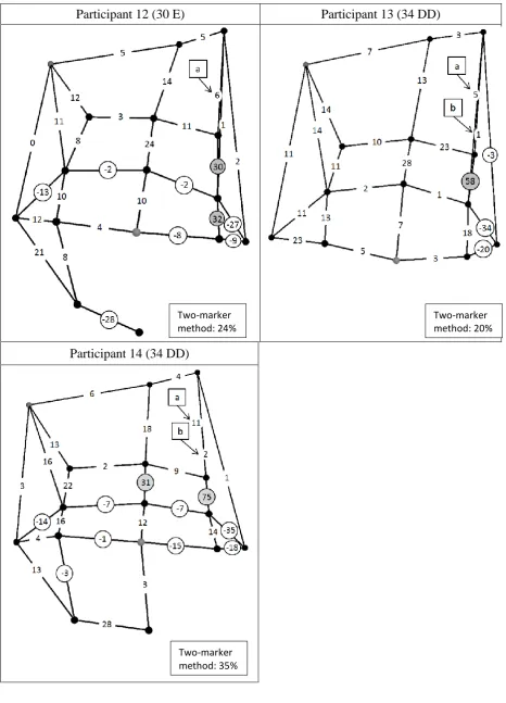
Skin strains across the surface of the breast are shown for each participant in Figure 5 and 231
peak skin strain ranged from 14 to 75% across participants. Errors in the calculated strain 232
values were estimated using the quotient rule (Taylor 1982), and the mean maximum error in 233
the static strain data was 3%. One participant (Participant 14) experienced potentially 234
damaging gravity-induced skin strain (75%), and four participants (Participants 1, 4, 12 and 235
13) experienced skin strains above 30% (skin resistance zone) (Figure 5). Participant-236
specific strain data demonstrate that the highest longitudinal breast strains generally occurred 237
in the second row of skin segments on the upper region of the breast (Figure 5). In the 238
[image:17.595.76.519.82.399.2]latitudinal direction contrasting results were observed for smaller- and larger-breasted 239
Figure 4: Static deformation of the breast mid-lines in the (b) sagittal plane and (a) frontal plane
(Participant 11, 32DD). The numbers indicate the strain on the segments shown. -250 -200 -150 -100 -50 0 50
-100 -50 0 50 100
Su p erio r-in ferio r d isp lacem ent (m m ) Anterior-posterior displacement (mm)
Neutral breast position
Gravity-loaded breast position
11 8 6 4 -19 (a) -250 -200 -150 -100 -50 0 50
-50 0 50 100 150
Su p erio r-in ferio r d isp lacem ent (m m )
Medial-lateral displacement (mm) Neutral breast position
Gravity-loaded breast position
12 7
7 -2
(b)
17
participants. With the exception of two participants (Participants 2 and 8), peak latitudinal 240
skin strains occurred on the medial side of the breast for participants with a breast size of 34D 241
or smaller, but on the lateral side of the breast for the larger-breasted (34DD or greater) 242
participants (Figure 5). 243
244
Comparison of individual static strain data revealed high between-participant variation in 245
strain values across the breast skin, with differences of up to 74% in strain for the same 246
marker pairing between individuals (Participants 1 and 6, and participant 14 in the upper 247
outer breast, Figure 5). Furthermore, differences of up to 110% strain were observed across 248
the breast skin of a single participant (Participant 14, Figure 5), highlighting the importance 249
of implementing a marker array when calculating breast skin strain. 250
251
A comparison of the results obtained using the two-marker and breast array method (Figure 252
5) demonstrates that the two-marker method produced static strain values of the same order 253
of magnitude as those presented previously (Haake et al., 2012, Haake and Scurr 2011), and 254
that these values could be used to approximate the longitudinal strain on the upper breast 255
mid-line (Figure 5). However, the two-marker method consistently underestimated the peak 256
static strain on the breast skin (by up to 59%) assessed using a marker array, as these peak 257
strains typically occurred on the upper-outer breast regions. 258
259
260
261
262
263
18 265
Generic array Participant 1 (32 B)
Participant 2 (32 B) Participant 3 (32 B)
266
Two-marker method: 15%
Two-marker method: 8%
Two-marker method: 17% Upper-Inner Upper-Outer
Lower-Inner
19
Participant 4 (34 B) Participant 5 (32 C)
Participant 6 (32 C) Participant 7 (32 D)
267
Two-marker method: 13%
Two-marker method: 21%
20
Participant 8 (32 D) Participant 9 (32 D)
Participant 10 (34 D) Participant 11 (32 DD)
268
Two-marker
method: 22% Two-marker method: 17%
Two-marker method: 9%
21
Participant 12 (30 E) Participant 13 (34 DD)
Participant 14 (34 DD)
[image:22.595.66.533.69.717.2]269
Figure 5: Static left breast skin strain for 14 participants with breast sizes ranging from 32 to 270
34 under band and B to E cup size. The grey marker represents the nipple. Strains above the 271
Two-marker method: 24%
Two-marker method: 20%
22
skin resistance limit (30%) are in grey circles, and negative strains (compression) are in white 272
circles. Strains calculated using the two-marker method are also shown for each participant. 273
Breast regions are identified on the generic array, and strain lines ‘a’ and ‘b’ are marked on 274
the generic array, and subsequent participant arrays, to aid clarification of the strain line as 275
these can superimpose over each other. 276
277
4.0 Discussion 278
Marker array data obtained within this study provided an opportunity to investigate the 279
deforming and strain-inducing effects of gravity over the breast surface for the first time in 280
breast research. The results demonstrate that gravity-induced breast deformation caused 281
potentially damaging breast skin strain (up to 75%) for one participant (Participant 14), and 282
that four further participants (Participants 1, 4, 12 and 13) experienced gravity-induced skin 283
strains above 30% (skin resistance zone) (Figure 5). These peak strain values all occurred in 284
the longitudinal direction in upper-outer region of the breast skin for the three largest-285
breasted participants, suggesting that this region of the breast skin may be particularly prone 286
to damage in larger-breasted women. Excessive gravity-induced skin strain in the upper-287
outer region of the breast may lead to failure of the collagen fibres and a permanent extension 288
of the skin in this breast region. This skin extension may allow the breast bulk to move 289
inferiorly and laterally on the torso; a position change which has previously been associated 290
with breast ptosis (Brown et al.,, 1999). 291
292
It was initially anticipated that the highest static strains would occur along the longitudinal 293
breast lines for all participants as gravity was assumed to act predominantly in this direction 294
in the static standing position. However, aside from the three largest breasted participants, 295
23
line or in the lower regions of the breast. Interestingly, individual static strain data (Figure 5) 297
demonstrated that the smaller-breasted participants experienced greater strain on the outer 298
(lateral) breast regions and less strain on the inner (medial) breast regions, a trend which was 299
reversed in their larger breasted counterparts (above size 34D). This new information could 300
be combined with existing knowledge on the lines of natural tension in the skin (Jatoi et al., 301
2006) to inform the selection of incision locations during breast surgery. There are multiple 302
factors taken into consideration when selecting the incision location, such as surgeon 303
visibility and control, and patient choice (Tebbetts & Adams, 2005). Interestingly, possible 304
injury to neighbouring soft tissue is also a factor taken into consideration (Tebbetts & Adams, 305
2005), and results in the current study indicate that for smaller breasted women it may be 306
preferential to select more medially positioned incision locations, whilst for larger breasted 307
women it may be preferential to select more laterally positioned incision sites. Surgeons 308
would thereby be selecting incision locations with reduced skin tension or strain. 309
310
In the longitudinal direction, strain data demonstrate that the greatest breast strain generally 311
occurred in the second row of skin segments on the upper region of the breast (Figure 5). 312
This may be explained by considering the hemispherical shape of the breast (Figure 2) and 313
the underlying breast anatomy. Breast tissue typically extends from the second to the sixth or 314
seventh rib in the superior-inferior direction (Macéa & Fregnani 2006). The breast is 315
broadest at its contact point on the torso and is generally narrowest at the nipple (the apex of 316
the breast). The most superior row of longitudinal skin segments may have predominantly 317
overlaid the soft tissue of the torso rather than the breast, meaning that the second row of skin 318
segments may have overlaid the broadest cross-section of the breast and experienced larger 319
strains during gravitational breast loading. 320
24
The results of this study demonstrate diverse strain values across the breast skin, which could 322
not be measured using the previously published two-marker method for estimating breast 323
strain (Haake & Scurr 2011). Although the two-marker method could approximate the 324
longitudinal strain on the upper breast mid-line, it was not appropriate for identifying peak 325
skin strain or for estimating the risk of skin damage. For example, if the two-marker method 326
alone had been implemented in this study then the potentially damaging skin strain (75%) 327
experienced by Participant 14 would not have been identified (Figure 5). Consequently, the 328
two-marker method is not recommended for assessing breast skin strain in future research. 329
Furthermore, the magnitude of static skin strains observed within this study (up to 75% for 330
Participant 14, Figure 3) demonstrate the importance of identifying the neutral breast position 331
before calculating breast strain, particularly if assessing the risk of skin damage. Measuring 332
skin strain from the gravity loaded position, as performed by Scurr in 2009, may lead to the 333
omission of potentially damaging skin strain caused by static gravitational loading of the 334
breast (Scurr et al., 2009). 335
336
Peak skin strain values observed in this study were higher than anticipated. The implication 337
that gravity alone could be causing permanent damage to the breast skin is surprising, and the 338
lack of existing static breast strain data makes it is difficult to assess the credibility of these 339
results. On one hand the prevalence of ptosis among mature women (Rinker et al., 2010) , 340
and the reports of markedly elongated breasts among tribal women who do not wear breast 341
support (Morgan 1997, Gunkel & Handler 1969), suggest that the breast can experience 342
damaging skin strains. However, it was acknowledged that the straight-line approximation 343
method used to calculate strain within this study may have led to an over-estimation of breast 344
skin strain. Although the marker array used to represent the breast surface was more detailed 345
25
large to negate the possibility of skin curvature between markers in the neutral position (L0).
347
Consequently, some degree of inter-marker extension (∆L) may have been caused by 348
flattening of the breast surface. 349
350
5.0 Conclusion 351
This exploratory study provides a novel contribution to breast research by quantifying 352
regional skin strain caused by external gravitational loading on the breast. The key outcome 353
of this work was the observation of potentially damaging static skin strains (up to 75% peak 354
strain) caused by gravitational loading. Particularly high skin strains were observed 355
longitudinally in the upper-outer breast region for larger-breasted women. In the latitudinal 356
direction, smaller-breasted participants experienced more strain on the outer (lateral) breast 357
regions and less strain on the inner (medial) breast regions, a trend which was reversed in 358
their larger breasted counterparts (above size 34D). These initial results suggest that to 359
reduce tension on latitudinal surgical incisions the preference should be given to medial 360
locations for smaller breasted women and lateral locations for larger breasted women. 361
Finally, this study also demonstrated the importance of considering the deforming effect of 362
gravity in breast research, and that a marker array is required to assess strain on the breast 363
skin. 364
365
366
367
Acknowledgments 368
The authors would like to thank Hanes Brands Inc. for funding this research; however the 369
26 References
371
Albornoz C R, Bach P B, Mehrara B J, Disa J J, Pusic A L, McCarthy C M, … Matros E 372
2013 A paradigm shift in U.S. Breast reconstruction: increasing implant rates. Plas 373
Recon Surg.131, 15–23 374
Borges A F, Alexander J E 1962 Relaxed skin tension lines, z-plasties on scars, and fusiform 375
excision of lesions. Brit J Plas Surg.15, 242–254. 376
Brown T, Ringrose C, Hyland R, Cole A and Brotherston T 1999 A method of assessing 377
female breast morphometry and its clinical application. Br. J. Plast. Surg.52 355–9 378
Clark J, Cheng J and Leung K 1996 Mechanical properties of normal skin and hypertrophic 379
scars. Burns22 443–6 380
Daly C 1982 Biomechanical properties of dermis. J. Invest. Dermatol.79 Suppl 1 17s – 20s 381
Finlay B 1970 Dynamic mechanical testing of human skin “in vivo”. J. Biomech.3 557–68 382
Fisher G, Wang Z, Datta S, Varani J, Kang S and Voorhees J 1997 Pathophysiology of 383
premature skin aging induced by ultraviolet light. N. Engl. J. Med.337 1419–28 384
Fujimura T, Haketa K, Hotta M and Kitahara T 2007 Loss of skin elasticity precedes to rapid 385
increase of wrinkle levels. J. Dermatol. Sci.47 233–9 386
Gallagher A, Ní Anniadh A, Bruyere K, Otténio M, Xie H and Gilchrist M 2012 Dynamic 387
tensile properties of human skin. 2012 IRCOBI Conference Proceedings. International 388
Research Council on the Biomechanics of Injury 389
Gambichler T, Matip R, Moussa G, Altmeyer P and Hoffmann K 2006 In vivo data of 390
epidermal thickness evaluated by optical coherence tomography: effects of age, gender, 391
skin type, and anatomic site. J. Dermatol. Sci.44 145–52 392
Gao Z and Desai J 2010 Estimating zero-strain states of very soft tissue under gravity loading 393
using digital image correlation. Med. Image Anal.14 126–37 394
27
Gunkel A and Handler J 1969 A Swiss medical doctor’s description of Barbados in 1661. J. 396
Barb. Museun Hist. Soc. 3–13 397
Haake S, Milligan A and Scurr J 2012 Can measures of strain and acceleration be used to 398
predict breast discomfort during running? J. Sport. Eng. Technol.227 209–16 399
Haake S and Scurr J 2011 A method to estimate strain in the breast during exercise. Sport. 400
Eng.14 49–56 401
Hindle W 1991 The breast and exercise. Caring for the exercising woman. (New York: 402
Elsevier Science Publishing.) pp 83–92 403
Hull M, Berns G, Varma H and Patterson H 1996 Strain in the medial collateral ligament of 404
the human knee under single and combined loads. J. Biomech.29 199–206 405
Jatoi I, Kaufmann M and Petit J 2006 Atlas of breast surgery. (Heidelberg: Springer.) 406
Kraissl C J 1951 The selection of appropriate lines for elective surgical incisions. Plas Recon 407
Surg (1946).8, 1–28. 408
Lim J, Hong J, Chen W and Weerasooriya T 2011 Mechanical response of pig skin under 409
dynamic tensile loading. Int. J. Impact Eng.38 130–5 Online: 410
Lim K, Chew C, Chen P, Jeyapalina S, Ho H, Rappel J and Lim B 2008 New extensometer to 411
measure in vivo uniaxial mechanical properties of human skin. J. Biomech.41 931–6 O 412
Macéa J and Fregnani J 2006 Anatomy of the thoracic wall, axilla and breast. Int. J. Morphol. 413
24 691–704 414
Mahmood U, Hanlon A L, Koshy M, Buras R, Chumsri S, Tkaczuk K H, … Feigenberg S J 415
2013 Increasing national mastectomy rates for the treatment of early stage breast cancer. 416
Annals of Surgical Oncology. 20, 1436–43. 417
McGhee D and Steele J 2010 Optimising breast support in female patients through correct bra 418
fit. A cross-sectional study. J. Sci. Med. Sport13 568–72 419
28
Mills C, Loveridge A, Milligan A, Risius D and Scurr J 2014 Can axes conventions of the 421
trunk reference frame influence breast displacement calculation during running? J. 422
Biomech.47 575–8 423
Mills C, Sanchez A and Scurr J 2016 Estimating the gravity induced three dimensinal 424
deformation of the breast. J. Biomech. 49 4134-4137. 425
Morgan J 1997 “Some Could Suckle over Their Shoulder”: Male Travelers, Female Bodies, 426
and the Gendering of Racial Ideology, 1500-1770. William Mary Q.54 167–92 427
Ní Annaidh A, Bruyère K, Destrade M, Gilchrist M and Otténio M 2012 Characterization of 428
the anisotropic mechanical properties of excised human skin. J. Mech. Behav. Biomed. 429
Mater.5 139–48 430
Page K-A and Steele J 1999 Breast motion and sports brassiere design implications for future 431
research. Sport. Med.27 205–11 432
Rajagopal V 2007 Modelling breast tissue mechanics under gravity loading. (Unpublished 433
doctoral thesis) (The University of Auckland) 434
Rajagopal V, Lee A, Chung J-H, Warren R, Highnam R, Nash M and Nielsen P 2008 435
Creating individual-specific biomechanical models of the breast for medical image 436
analysis. Acad. Radiol.15 1425–36 437
Rinker B, Veneracion M and Walsh C 2010 Breast ptosis: Causes and cure. Ann. Plast. Surg. 438
64 579–84 439
Sanchez A, Mills C and Scurr J 2016 Estimating breast mass-density: A retrospecitive 440
analysis of readiological data. The Breast J. in press. 441
Scurr J, Bridgman C, Hedger W and White J 2009 Multi-planar breast strain during 442
incremental treadmill activity. J. Sports Sci.27 S29 443
Scurr J, White J and Hedger W 2011 Supported and unsupported breast displacement in three 444
29
Seo H, Kim S, Cordier F, Choi J, Hong K. 2013 Estimating dynamic skin tension lines in 446
vivo using 3D scans. Computer-Aided Design.45, 551–555 447
Silver F, Freeman J and DeVore D 2001 Viscoelastic properties of human skin and processed 448
dermis. Ski. Res. Technol.7 18–23 449
Smalls L, Wickett R and Visscher M 2006 Effect of dermal thickness, tissue composition, 450
and body site on skin biomechanical properties. Ski. Res. Technol.12 43–9 451
Stark H 1977 Directional variations in the extensibility of human skin. Br. J. Pastic Surg.30 452
105–14 453
Taylor J R 1982 An introduction to error analysis: The study of uncertainties in physical 454
measurements. (University Science Books) 455
Tebbetts J B, Adams W P 2005 Five critical decisions in breast augmentation using five 456
measurements in 5 minutes: the high five decision support process. Plas Recon Surg. 457
116, 2005–16 458
Toms S, Lemons J, Bartolucci A and Eberhardt A 2002 Nonlinear stress-strain behavior of 459
periodontal ligament under orthodontic loading. Am. J. Orthod. Dentofac. Orthop.122 460
174–9 461
Winter G 2006 Some factors affecting skin and wound healing. J. Tissue Viability16 20–3 462
463