organic papers
Acta Cryst.(2005). E61, o1001–o1003 doi:10.1107/S1600536805007865 Vratislav Langeret al. C
20H26O8
o1001
Acta Crystallographica Section EStructure Reports Online
ISSN 1600-5368
erythro
-2-(2,6-Dimethoxy-4-methylphenoxy)-
1-(4-hydroxy-3,5-dimethoxyphenyl)propane-1,3-diol
Vratislav Langer,a* Knut Lundquistband Gerhard E. Mikschec
aDepartment of Chemical and Biological
Engi-neering, Chalmers University of Technology, SE-41296 Go¨teborg, Sweden,bDepartment of
Forest Products and Chemical Engineering, Chalmers University of Technology, SE-41296 Go¨teborg, Sweden, andcConimar AB, Box 2086, SE-141 02 Huddinge, Sweden
Correspondence e-mail: [email protected]
Key indicators
Single-crystal X-ray study
T= 173 K
Mean(C–C) = 0.004 A˚ Disorder in main residue
Rfactor = 0.049
wRfactor = 0.120
Data-to-parameter ratio = 13.8
For details of how these key indicators were automatically derived from the article, see http://journals.iucr.org/e.
#2005 International Union of Crystallography Printed in Great Britain – all rights reserved
In the title compound, C20H26O8, the molecules adopt a
conformation in which the torsion angle C(aryl)—C—C—
O(aryloxy) is70.8 (2) and the torsion angle C(aryl)—O—
C—C(benzylic) is 152.71 (18), leading to a
C(aryl) C(aryl) distance of 4.426 (3) A˚ . This distance is
identical, within experimental error, to the calculated maximum value obtained when the torsion angle C(aryl)— O—C—C(benzylic) is varied and the torsion angle C(aryl)— C—C—O(aryloxy) is kept constant. The angle between the
aromatic ring planes is 64.13 (7). The conformation is
compared with those of related lignin model compounds. The hydrogen-bonding pattern reveals intramolecular inter-actions, and the formation of centrosymmetric dimers and chains of molecules by intermolecular interactions.
Comment
This paper describes the crystal structure of the lignin model
compound (I), which is representative of erythro forms of
structural elements in lignins of the syringylglycerol-syringyl
ether type. The crystal structures of three compounds of this
type,viz. (II)–(IV), have been reported previously (Stomberg
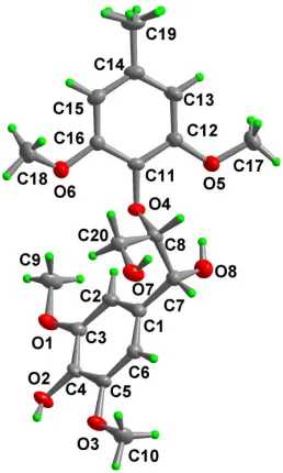
& Lundquist, 1989; Langer & Lundquist, 2001; Langeret al., 2002). A perspective drawing of (I) and the atomic numbering are shown in Fig. 1. The methyl H atoms at C19 are disordered
with two orientations rotated from each other by 60.
Interestingly, the conformations of (I) and (II) are very
similar. The torsion angle C1—C7—C8—O4 is 70.8 (2) in
(I) and the corresponding angle in (II) is 71.0 (4).
Furthermore, the magnitude of the torsion angle C11—O4—
C8—C7 [152.71 (18)] is very similar to the corresponding
angle in (II) [150.5 (4)]. The angle between the aromatic
ring planes is 64.13 (7) in (I) and 63.1 (1) in (II). The
conformations of (III) and (IV) are similar but differ drama-tically from those of (I) and (II). The torsion angles
corre-sponding to C1—C7—C8—O4 are, for instance, close to 180.
The distance C1 C11 [4.426 (3) A˚ ] can be taken as a
measure of the separation of the bulky 2,6-dimethoxy-4-methylphenyl and 4-hydroxy-3,5-dimethoxyphenyl groups.
This distance is not close to the maximum possible value (ca
5 A˚ ), but agrees with the calculated maximum value of 4.43 A˚
obtained when the torsion angle C11—O4—C8—C7 is varied and the torsion angle C1—C7—C8—O4 is kept constant at 70.8 (2).
In the crystal structure of (I), there are three intramolecular
and three intermolecular hydrogen bonds of the O—H O
type and one intramolecular and two intermolecular hydrogen
bonds of the weak C—H O type (Table 1). Intermolecular
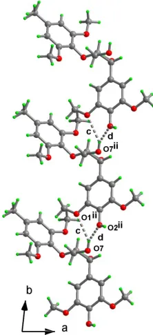
hydrogen bond b (see Table 1 and Fig. 2) gives rise to
centrosymmetric dimers. On the first-level graph-set (Bern-steinet al., 1995; Grellet al., 1999), the hydrogen bonds are classified asS(5) for bondsaande, S(8) for fandS(6) forg.
The centrosymmetric dimer, formed by hydrogen bondb, is
classified as ringR22(20), while hydrogen bondscand d form
C(9) andC(10) chains, respectively (Fig. 3). The weak
intra-molecular hydrogen bonds h and i form rings R2
2(20) and R2
2(16), respectively. On the second-level graph-set, many
chains and rings could be identified, the most important being ringsR4
4(8), formed by bondsbandd, andR
1
2(5), formed by
hydrogen bonds c and d. The assignment of graph-set
descriptors was performed using PLUTO, as described by
Motherwellet al.(1999).
It is notable that the conformations adopted by (I) and (II) in the crystal structures are those that are most favored, as
judged from computational studies (Besombes et al., 2003).
The conformations adopted by (III) and (IV) are conceivable
but not favored according to such studies (Besombeset al.,
2003). A plausible assumption is that the conformations of
-ether models are largely governed by hydrogen bonding. Attempts to obtain unequivocal proof of this have so far failed. In a previous paper (Langer & Lundquist, 2001), it was proposed that the different hydrogen-bonding patterns are the main reason for the conformational differences between (II) and (III). However, compounds (I) and (II) adopt very similar conformations in spite of a difference in hydrogen-bonding patterns (see above, also Stomberg & Lundquist, 1989), due to the fact that a hydroxymethyl group in (II) has been exchanged for a methyl group in (I). In a recent paper, it is concluded, based on NMR studies, that intramolecular hydrogen bonding is of minor importance for the
conforma-tions adopted by-ethers in solution (Besombeset al., 2004).
Experimental
The synthesis of (I) has been described by Miksche (1973). Crystals suitable for X-ray analysis were obtained from ethyl acetate (m.p. 420–422 K).
Crystal data
C20H26O8
Mr= 394.41
Triclinic,P1
a= 8.6930 (2) A˚
b= 8.8976 (2) A˚
c= 13.1057 (3) A˚
= 73.852 (1)
= 86.150 (1)
= 83.062 (1)
V= 965.96 (4) A˚3
Z= 2
Dx= 1.356 Mg m
3
MoKradiation Cell parameters from 4159
reflections
= 1.6–25.5
= 0.11 mm1
T= 173 (2) K Prism, colorless 0.240.120.08 mm
Data collection
Siemens SMART CCD area-detector diffractometer
!scans
Absorption correction: multi-scan (SADABS; Sheldrick, 2002)
Tmin= 0.715,Tmax= 0.992
10 905 measured reflections
3607 independent reflections 2409 reflections withI> 2(I)
Rint= 0.047
max= 25.5
h=10!10
k=10!10
l=15!15
organic papers
o1002
Vratislav Langeret al. C [image:2.610.109.238.71.286.2]20H26O8 Acta Cryst.(2005). E61, o1001–o1003
Figure 2
The hydrogen-bonding (dashed lines) pattern of (I). Centrosymmetric dimers are formed [symmetry code: (i) x+ 2, y+ 1, z+2]. The hydrogen-bond labels match those given in Table 1.
Figure 1
[image:2.610.121.219.474.716.2]Refinement
Refinement onF2 R[F2> 2(F2)] = 0.049
wR(F2) = 0.121
S= 1.02 3607 reflections 261 parameters
H-atom parameters constrained
w= 1/[2
(Fo2) + (0.0497P)2
+ 0.3785P]
whereP= (Fo2+ 2Fc2)/3
(/)max< 0.001
max= 0.24 e A˚ 3
[image:3.610.385.496.73.316.2]min=0.27 e A˚ 3
Table 1
Hydrogen-bonding geometry (A˚ ,).
Label D—H A D—H H A D A D—H A a O2—H2A O3 0.84 2.29 2.666 (2) 108
b O2—H2A O7i
0.84 1.92 2.676 (2) 148
c O7—H7A O1ii
0.84 2.61 3.316 (2) 142
d O7—H7A O2ii
0.84 1.91 2.662 (2) 149
e O8—H8A O4 0.84 2.35 2.766 (2) 112
f O8—H8A O5 0.84 2.43 3.201 (3) 154
g C2—H2 O4 0.95 2.50 3.022 (3) 114
h C19—H19B O8iii
0.98 2.59 3.428 (3) 144
i C20—H20B O3i
0.99 2.57 3.323 (3) 132
Symmetry codes: (i) 2x, 1y, 2z; (ii)x, 1 +y, z; (iii) 1x, 2y, 1z.
H atoms were constrained to ideal geometry using an appropriate riding model. The C—H distance was kept fixed at 0.95 A˚ for aromatic H atoms, at 0.99 A˚ for secondary H atoms and at 1.00 A˚ for tertiary H atoms. For the hydroxyl groups, the O—H distance (0.84 A˚ ) and C—O—H angle (109.5) were kept fixed, while the torsion angle was allowed to refine with the starting position based on a circular Fourier synthesis. For methyl groups, the C—H distances (0.98 A˚ ) and C—C—H angles (109.5) were kept fixed, while the torsion angles were allowed to refine with the starting position based on threefold-averaged circular Fourier syntheses. The methyl H atoms on C19 were treated as disordered with two orientations rotated from each other by 60with half occupancy.U
isovalues were fixed at 1.2Ueqof the pivot atom for aromatic, secondary and tertiary H atoms, while for hydroxyl and methyl H atoms, theUisovalues were fixed at 1.5Ueqof the pivot atom.
Data collection:SMART(Siemens, 1995); cell refinement:SAINT (Siemens, 1995); data reduction: SAINT (Siemens, 1995) and SADABS (Sheldrick, 2002; program(s) used to solve structure: SHELXTL (Bruker, 2001); program(s) used to refine structure: SHELXTL; molecular graphics:DIAMOND (Brandenburg, 2004); software used to prepare material for publication:SHELXTL.
References
Bernstein, J., Davis, R. E., Shimoni, L. & Chang, N.-L. (1995).Angew. Chem. Int. Ed. Engl.34, 1555–1573.
Besombes, S., Robert, D., Utille, J.-P., Taravel, F. R. & Mazeau, K. (2003).J. Agric. Food Chem.51, 34–42.
Besombes, S., Utille, J.-P., Mazeau, K., Robert, D. & Taravel, F. R. (2004).
Magn. Reson. Chem.42, 337–347.
Brandenburg, K. (2004).DIAMOND. Version 3.0a. Crystal Impact GbR, Bonn, Germany.
Bruker (2001). SHELXTL. Version 6.10. Bruker AXS Inc., Madison, Wisconsin, USA.
Grell, J., Bernstein, J. & Tinhofer, G. (1999).Acta Cryst.B55, 1030–1043. Langer, V., Li, S. & Lundquist, K. (2002).Acta Cryst.E58, o42–o44. Langer, V. & Lundquist, K. (2001).Acta Cryst.E57, o1219–o1221. Miksche, G. E. (1973).Acta Chem. Scand.27, 1355–1368.
Motherwell, W. D. S., Shields, G. P. & Allen, F. H. (1999).Acta Cryst.B55, 1044–1056.
Sheldrick, G. M. (2002).SADABS.Version 2.03. University of Go¨ttingen, Germany.
Siemens (1995).SMARTandSAINT.Siemens Analytical X-ray Instruments Inc., Madison, Wisconsin, USA.
Stomberg, R. & Lundquist, K. (1989).J. Crystallogr. Spectrosc. Res.19, 331– 339.
organic papers
Acta Cryst.(2005). E61, o1001–o1003 Vratislav Langeret al. C
[image:3.610.44.297.205.303.2]20H26O8
o1003
Figure 3supporting information
sup-1
Acta Cryst. (2005). E61, o1001–o1003
supporting information
Acta Cryst. (2005). E61, o1001–o1003 [https://doi.org/10.1107/S1600536805007865]
erythro
-2-(2,6-Dimethoxy-4-methylphenoxy)-1-(4-hydroxy-3,5-dimethoxy-phenyl)propane-1,3-diol
Vratislav Langer, Knut Lundquist and Gerhard E. Miksche
erythro-2-(2,6-Dimethoxy-4-methylphenoxy)-1-(4-hydroxy-3,5- dimethoxyphenyl)propane-1,3-diol
Crystal data
C20H26O8
Mr = 394.41 Triclinic, P1 Hall symbol: -P 1 a = 8.6930 (2) Å b = 8.8976 (2) Å c = 13.1057 (3) Å α = 73.852 (1)° β = 86.150 (1)° γ = 83.062 (1)° V = 965.96 (4) Å3
Z = 2 F(000) = 420 Dx = 1.356 Mg m−3
Melting point = 420–422 K Mo Kα radiation, λ = 0.71073 Å Cell parameters from 4159 reflections θ = 1.6–25.5°
µ = 0.11 mm−1
T = 173 K Prism, colourless 0.24 × 0.12 × 0.08 mm
Data collection
Bruker SMART CCD area-detector diffractometer
Radiation source: fine-focus sealed tube Graphite monochromator
ω scans
Absorption correction: multi-scan (SADABS; Sheldrick, 2002) Tmin = 0.975, Tmax = 0.992
10905 measured reflections 3607 independent reflections 2409 reflections with I > 2σ(I) Rint = 0.047
θmax = 25.5°, θmin = 1.6°
h = −10→10 k = −10→10 l = −15→15
Refinement
Refinement on F2
Least-squares matrix: full R[F2 > 2σ(F2)] = 0.049
wR(F2) = 0.121
S = 1.02 3607 reflections 261 parameters 0 restraints
Primary atom site location: structure-invariant direct methods
Secondary atom site location: difference Fourier map
Hydrogen site location: inferred from neighbouring sites
H-atom parameters constrained w = 1/[σ2(F
o2) + (0.0497P)2 + 0.3785P]
where P = (Fo2 + 2Fc2)/3
(Δ/σ)max < 0.001
Δρmax = 0.24 e Å−3
supporting information
sup-2
Acta Cryst. (2005). E61, o1001–o1003
Special details
Experimental. Data were collected at low temperature using a Siemens SMART CCD diffractometer equiped with a LT-2 device. A full sphere of reciprocal space was scanned by 0.3° steps in ω with a crystal–to–detector distance of 3.97 cm, 20 s per frame. Preliminary orientation matrix was obtained from the first 100 frames using SMART (Siemens, 1995). The collected frames were integrated using the preliminary orientation matrix which was updated every 100 frames. Final cell parameters were obtained by refinement on the position of 4159 reflections with I>10σ(I) after integration of all the frames data using SAINT (Siemens, 1995).
Geometry. All e.s.d.'s (except the e.s.d. in the dihedral angle between two l.s. planes) are estimated using the full covariance matrix. The cell e.s.d.'s are taken into account individually in the estimation of e.s.d.'s in distances, angles and torsion angles; correlations between e.s.d.'s in cell parameters are only used when they are defined by crystal symmetry. An approximate (isotropic) treatment of cell e.s.d.'s is used for estimating e.s.d.'s involving l.s. planes.
Refinement. Refinement of F2 against ALL reflections. The weighted R-factor wR and goodness of fit S are based on F2,
conventional R-factors R are based on F, with F set to zero for negative F2. The threshold expression of F2 > σ(F2) is used
only for calculating R-factors(gt) etc. and is not relevant to the choice of reflections for refinement. R-factors based on F2
are statistically about twice as large as those based on F, and R- factors based on ALL data will be even larger.
Fractional atomic coordinates and isotropic or equivalent isotropic displacement parameters (Å2)
x y z Uiso*/Ueq Occ. (<1)
O1 0.74929 (19) 0.31626 (18) 0.79907 (13) 0.0336 (4) O2 0.9884 (2) 0.20449 (17) 0.91973 (13) 0.0327 (4)
H2A 1.0410 0.1890 0.9739 0.049*
O3 1.20416 (18) 0.38230 (18) 0.94010 (13) 0.0345 (4) O4 0.65180 (16) 0.90028 (17) 0.68637 (12) 0.0237 (4) O5 0.65548 (18) 1.13536 (19) 0.50803 (12) 0.0332 (4) O6 0.41150 (18) 0.88773 (19) 0.82912 (13) 0.0347 (4) O7 0.89908 (19) 0.94500 (19) 0.88914 (14) 0.0338 (4)
H7A 0.8993 1.0414 0.8833 0.051*
O8 0.9347 (2) 0.8642 (2) 0.58124 (13) 0.0386 (5)
H8A 0.8515 0.9129 0.5548 0.058*
C1 0.9447 (3) 0.6773 (3) 0.75319 (18) 0.0248 (5) C2 0.8366 (3) 0.5799 (3) 0.74325 (18) 0.0269 (5)
H2 0.7529 0.6209 0.6969 0.032*
C3 0.8505 (3) 0.4231 (3) 0.80077 (18) 0.0252 (5) C4 0.9752 (3) 0.3612 (2) 0.86679 (17) 0.0231 (5) C5 1.0841 (3) 0.4580 (3) 0.87512 (18) 0.0245 (5) C6 1.0681 (3) 0.6159 (3) 0.81996 (18) 0.0254 (5)
H6 1.1416 0.6824 0.8278 0.031*
C7 0.9296 (3) 0.8506 (3) 0.69182 (18) 0.0273 (5)
H7 1.0224 0.8962 0.7069 0.033*
C8 0.7862 (2) 0.9463 (3) 0.72375 (18) 0.0237 (5)
H8 0.7937 1.0606 0.6891 0.028*
C9 0.6210 (3) 0.3727 (3) 0.7312 (2) 0.0406 (7)
H9A 0.5623 0.4632 0.7496 0.061*
H9B 0.5535 0.2890 0.7403 0.061*
H9C 0.6592 0.4046 0.6571 0.061*
C10 1.3161 (3) 0.4762 (3) 0.9572 (2) 0.0386 (6)
H10A 1.3663 0.5265 0.8891 0.058*
supporting information
sup-3
Acta Cryst. (2005). E61, o1001–o1003
H10C 1.2647 0.5573 0.9891 0.058*
C11 0.5262 (2) 1.0153 (3) 0.66575 (18) 0.0224 (5) C12 0.5235 (3) 1.1349 (3) 0.57048 (18) 0.0258 (5) C13 0.3936 (3) 1.2437 (3) 0.54675 (19) 0.0304 (6)
H13 0.3918 1.3244 0.4816 0.036*
C14 0.2664 (3) 1.2357 (3) 0.61720 (19) 0.0314 (6) C15 0.2704 (3) 1.1183 (3) 0.71258 (19) 0.0311 (6)
H15 0.1843 1.1130 0.7616 0.037*
C16 0.3995 (3) 1.0087 (3) 0.73664 (18) 0.0259 (5) C17 0.6645 (3) 1.2631 (3) 0.4143 (2) 0.0410 (7)
H17A 0.5868 1.2585 0.3647 0.062*
H17B 0.7682 1.2555 0.3806 0.062*
H17C 0.6447 1.3629 0.4332 0.062*
C18 0.3190 (3) 0.9136 (3) 0.9180 (2) 0.0435 (7)
H18A 0.3320 1.0177 0.9259 0.065*
H18B 0.3517 0.8326 0.9826 0.065*
H18C 0.2097 0.9084 0.9066 0.065*
C19 0.1238 (3) 1.3521 (3) 0.5894 (2) 0.0452 (7)
H19A 0.0545 1.3435 0.6524 0.068* 0.50
H19B 0.0698 1.3293 0.5330 0.068* 0.50
H19C 0.1546 1.4591 0.5646 0.068* 0.50
H19D 0.1314 1.4111 0.5143 0.068* 0.50
H19E 0.1162 1.4253 0.6337 0.068* 0.50
H19F 0.0314 1.2955 0.6021 0.068* 0.50
C20 0.7640 (3) 0.9206 (3) 0.84382 (18) 0.0277 (5)
H20A 0.6760 0.9940 0.8587 0.033*
H20B 0.7383 0.8119 0.8772 0.033*
Atomic displacement parameters (Å2)
U11 U22 U33 U12 U13 U23
supporting information
sup-4
Acta Cryst. (2005). E61, o1001–o1003
C10 0.0292 (14) 0.0394 (15) 0.0446 (16) −0.0063 (12) −0.0142 (12) −0.0031 (13) C11 0.0205 (12) 0.0219 (12) 0.0257 (12) 0.0016 (9) −0.0073 (10) −0.0079 (10) C12 0.0250 (12) 0.0298 (13) 0.0228 (12) 0.0011 (10) −0.0022 (10) −0.0086 (10) C13 0.0352 (14) 0.0295 (13) 0.0231 (13) 0.0056 (11) −0.0079 (11) −0.0038 (11) C14 0.0277 (13) 0.0360 (14) 0.0309 (14) 0.0070 (11) −0.0087 (11) −0.0122 (12) C15 0.0237 (13) 0.0384 (14) 0.0323 (14) 0.0009 (11) 0.0005 (11) −0.0136 (12) C16 0.0239 (12) 0.0270 (13) 0.0255 (13) −0.0034 (10) −0.0025 (10) −0.0043 (10) C17 0.0470 (16) 0.0377 (15) 0.0298 (15) −0.0012 (13) 0.0064 (12) 0.0017 (12) C18 0.0465 (16) 0.0470 (17) 0.0304 (15) −0.0037 (14) 0.0091 (13) −0.0026 (13) C19 0.0355 (15) 0.0516 (17) 0.0447 (17) 0.0170 (13) −0.0099 (13) −0.0143 (14) C20 0.0295 (13) 0.0256 (13) 0.0285 (13) −0.0025 (10) −0.0044 (11) −0.0073 (10)
Geometric parameters (Å, º)
O1—C3 1.377 (3) C9—H9A 0.9800
O1—C9 1.427 (3) C9—H9B 0.9800
O2—C4 1.368 (2) C9—H9C 0.9800
O2—H2A 0.8400 C10—H10A 0.9800
O3—C5 1.378 (3) C10—H10B 0.9800
O3—C10 1.423 (3) C10—H10C 0.9800
O4—C11 1.388 (2) C11—C16 1.389 (3)
O4—C8 1.441 (3) C11—C12 1.397 (3)
O5—C12 1.364 (3) C12—C13 1.386 (3)
O5—C17 1.428 (3) C13—C14 1.388 (3)
O6—C16 1.380 (3) C13—H13 0.9500
O6—C18 1.427 (3) C14—C15 1.388 (3)
O7—C20 1.416 (3) C14—C19 1.511 (3)
O7—H7A 0.8400 C15—C16 1.386 (3)
O8—C7 1.418 (3) C15—H15 0.9500
O8—H8A 0.8400 C17—H17A 0.9800
C1—C2 1.389 (3) C17—H17B 0.9800
C1—C6 1.390 (3) C17—H17C 0.9800
C1—C7 1.522 (3) C18—H18A 0.9800
C2—C3 1.384 (3) C18—H18B 0.9800
C2—H2 0.9500 C18—H18C 0.9800
C3—C4 1.394 (3) C19—H19A 0.9800
C4—C5 1.383 (3) C19—H19B 0.9800
C5—C6 1.385 (3) C19—H19C 0.9800
C6—H6 0.9500 C19—H19D 0.9800
C7—C8 1.526 (3) C19—H19E 0.9800
C7—H7 1.0000 C19—H19F 0.9800
C8—C20 1.528 (3) C20—H20A 0.9900
C8—H8 1.0000 C20—H20B 0.9900
C3—O1—C9 116.76 (17) H10A—C10—H10C 109.5
C4—O2—H2A 109.5 H10B—C10—H10C 109.5
supporting information
sup-5
Acta Cryst. (2005). E61, o1001–o1003
C12—O5—C17 117.47 (18) O4—C11—C12 119.9 (2) C16—O6—C18 115.87 (18) O5—C12—C13 125.0 (2)
C20—O7—H7A 109.5 O5—C12—C11 115.07 (19)
C7—O8—H8A 109.5 C13—C12—C11 119.9 (2)
C2—C1—C6 119.6 (2) C14—C13—C12 120.7 (2)
C2—C1—C7 120.7 (2) C14—C13—H13 119.7
C6—C1—C7 119.7 (2) C12—C13—H13 119.7
C3—C2—C1 120.1 (2) C13—C14—C15 119.4 (2)
C3—C2—H2 120.0 C13—C14—C19 120.3 (2)
C1—C2—H2 120.0 C15—C14—C19 120.4 (2)
O1—C3—C2 125.3 (2) C16—C15—C14 120.3 (2)
O1—C3—C4 114.40 (19) C16—C15—H15 119.8
C2—C3—C4 120.3 (2) C14—C15—H15 119.8
O2—C4—C5 121.8 (2) O6—C16—C11 115.85 (19)
O2—C4—C3 118.8 (2) O6—C16—C15 123.7 (2)
C5—C4—C3 119.46 (19) C11—C16—C15 120.4 (2)
O3—C5—C6 125.5 (2) O5—C17—H17A 109.5
O3—C5—C4 114.09 (19) O5—C17—H17B 109.5
C6—C5—C4 120.4 (2) H17A—C17—H17B 109.5
C5—C6—C1 120.1 (2) O5—C17—H17C 109.5
C5—C6—H6 119.9 H17A—C17—H17C 109.5
C1—C6—H6 119.9 H17B—C17—H17C 109.5
O8—C7—C1 109.34 (19) O6—C18—H18A 109.5
O8—C7—C8 111.09 (18) O6—C18—H18B 109.5
C1—C7—C8 114.07 (19) H18A—C18—H18B 109.5
O8—C7—H7 107.3 O6—C18—H18C 109.5
C1—C7—H7 107.3 H18A—C18—H18C 109.5
C8—C7—H7 107.3 H18B—C18—H18C 109.5
O4—C8—C20 107.87 (17) C14—C19—H19A 109.5
O4—C8—C7 108.24 (17) C14—C19—H19B 109.5
C20—C8—C7 113.42 (18) H19A—C19—H19B 109.5
O4—C8—H8 109.1 C14—C19—H19C 109.5
C20—C8—H8 109.1 H19A—C19—H19C 109.5
C7—C8—H8 109.1 H19B—C19—H19C 109.5
O1—C9—H9A 109.5 C14—C19—H19D 109.5
O1—C9—H9B 109.5 C14—C19—H19E 109.5
H9A—C9—H9B 109.5 H19D—C19—H19E 109.5
O1—C9—H9C 109.5 C14—C19—H19F 109.5
H9A—C9—H9C 109.5 O7—C20—C8 111.56 (19)
H9B—C9—H9C 109.5 O7—C20—H20A 109.3
O3—C10—H10A 109.5 C8—C20—H20A 109.3
O3—C10—H10B 109.5 O7—C20—H20B 109.3
H10A—C10—H10B 109.5 C8—C20—H20B 109.3
O3—C10—H10C 109.5 H20A—C20—H20B 108.0
C6—C1—C2—C3 0.9 (3) C1—C7—C8—O4 −70.8 (2)
supporting information
sup-6
Acta Cryst. (2005). E61, o1001–o1003
C9—O1—C3—C4 −178.8 (2) C8—O4—C11—C16 −103.0 (2) C1—C2—C3—O1 178.7 (2) C8—O4—C11—C12 80.0 (2) C1—C2—C3—C4 −1.7 (4) C17—O5—C12—C13 4.1 (3) O1—C3—C4—O2 1.9 (3) C17—O5—C12—C11 −174.5 (2) C2—C3—C4—O2 −177.8 (2) C16—C11—C12—O5 177.5 (2) O1—C3—C4—C5 −179.7 (2) O4—C11—C12—O5 −5.5 (3) C2—C3—C4—C5 0.6 (3) C16—C11—C12—C13 −1.2 (3) C10—O3—C5—C6 4.1 (3) O4—C11—C12—C13 175.7 (2) C10—O3—C5—C4 −176.6 (2) O5—C12—C13—C14 −178.1 (2) O2—C4—C5—O3 0.2 (3) C11—C12—C13—C14 0.5 (4) C3—C4—C5—O3 −178.1 (2) C12—C13—C14—C15 0.6 (4) O2—C4—C5—C6 179.6 (2) C12—C13—C14—C19 −178.4 (2) C3—C4—C5—C6 1.2 (3) C13—C14—C15—C16 −0.9 (4) O3—C5—C6—C1 177.3 (2) C19—C14—C15—C16 178.1 (2) C4—C5—C6—C1 −1.9 (3) C18—O6—C16—C11 155.3 (2) C2—C1—C6—C5 0.8 (3) C18—O6—C16—C15 −25.3 (3) C7—C1—C6—C5 −179.2 (2) O4—C11—C16—O6 3.4 (3) C2—C1—C7—O8 −60.2 (3) C12—C11—C16—O6 −179.65 (19) C6—C1—C7—O8 119.8 (2) O4—C11—C16—C15 −176.0 (2) C2—C1—C7—C8 64.8 (3) C12—C11—C16—C15 0.9 (3) C6—C1—C7—C8 −115.2 (2) C14—C15—C16—O6 −179.2 (2) C11—O4—C8—C20 84.2 (2) C14—C15—C16—C11 0.1 (4) C11—O4—C8—C7 −152.71 (18) O4—C8—C20—O7 172.91 (16) O8—C7—C8—O4 53.4 (2) C7—C8—C20—O7 53.0 (2)
Hydrogen-bond geometry (Å, º)
D—H···A D—H H···A D···A D—H···A
O2—H2A···O3 0.84 2.29 2.666 (2) 108
O2—H2A···O7i 0.84 1.92 2.676 (2) 148
O7—H7A···O1ii 0.84 2.61 3.316 (2) 142
O7—H7A···O2ii 0.84 1.91 2.662 (2) 149
O8—H8A···O4 0.84 2.35 2.766 (2) 112
O8—H8A···O5 0.84 2.43 3.201 (2) 154
C2—H2···O4 0.95 2.50 3.022 (3) 114
C19—H19B···O8iii 0.98 2.59 3.428 (3) 144
C20—H20B···O3i 0.99 2.57 3.323 (3) 132