inorganic papers
Acta Cryst.(2005). E61, i253–i255 doi:10.1107/S1600536805034811 Jianrong Chenet al. Al
2SiO4(OD)2
i253
Acta Crystallographica Section EStructure Reports Online
ISSN 1600-5368
A Rietveld refinement using neutron powder
diffraction data of a fully deuterated topaz,
Al
2SiO
4(OD)
2Jianrong Chen,a* George A. Lager,aMartin Kunz,bThomas C. Hansencand Peter Ulmerd
aDepartment of Geography and Geosciences,
University of Louisville, Kentucky 40292, USA,
bLawrence-Berkeley Laboratory, Berkeley, CA
94720, USA,cInstitut Laue–Langevin, BP 156,
F-38042 Grenoble Cedex 9, France, and
d
Laboratorium fu¨r Kristallographie, ETH Zentrum, Sonneggstrasse 5, CH-8092 Zurich, Switzerland
Correspondence e-mail: [email protected]
Key indicators
Powder neutron study T= 295 K
Mean(Al–O) = 0.003 A˚ Disorder in main residue Rfactor = 0.037 wRfactor = 0.046
Data-to-parameter ratio = 106.9
For details of how these key indicators were automatically derived from the article, see http://journals.iucr.org/e.
#2005 International Union of Crystallography
Printed in Great Britain – all rights reserved
The structure of topaz-OD, dialuminium orthosilicate di-hydroxide, Al2SiO4(OD)2, was refined in the space group
Pbnm by Rietveld analysis of constant wavelength neutron powder diffraction data. Two non-equivalent half-occupied deuterium positions were located. Each D atom is character-ized by an irregular trifurcated hydrogen-bond geometry. The refined hydrogen-bond distances are in the ranges 2.038 (5)– 2.281 (6) and 2.280 (5)–2.524 (6) A˚ for the two D atoms. Hydrogen-bond angles range from 83.6 (4) to 151.9 (4).
Results indicate that it is feasible to characterize the hydrogen bonding in small-volume samples (25 mg) synthesized at high pressure and temperature.
Comment
This study was undertaken in order to provide more reliable information on the hydrogen-bonding geometry in a fully deuterated topaz (topaz-OD) and determine the feasibility of characterizing the hydrogen bonding in small-volume samples (Chenet al., 2004). A previous X-ray single-crystal refinement showed that H atoms are located in two non-equivalent half-occupied sites, leading to the reduction of symmetry from
Pbnm to Pbn21, at least locally (Northrup et al., 1994).
However, owing to the weak X-ray scattering power of hydrogen, the H-atom positions could not be determined with a high degree of accuracy. Neutron diffraction, on the other hand, is well suited to this investigation because of the large neutron scattering length of deuterium.
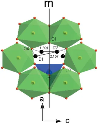
Topaz-OD is an orthosilicate consisting of chains of AlO6
octahedra linked by silicate tetrahedra. D atoms reside in a cavity, lying in the (010) plane, and are disordered over four sites, which occur in pairs related by mirror symmetry (Fig. 2). The trifurcated hydrogen-bond geometry differs slightly from the previously reported arrangement (Northrupet al., 1994.). D1 is hydrogen bonded to atoms O3, O2 and O4, whereas D2 is bonded to atoms O4, O1 and O2(x + 1
2, y 1 2, z+
1 2]
(Fig. 3), in contrast to the X-ray data with hydrogen bonds to atoms O4, O1 and O2(x+1
2,y 1 2,z+
1
2] [see Fig. 2 of the
original paper by Northrup et al. (1994); note also that the H1 O3 hydrogen bond is incorrectly represented in the original Fig. 2]. The weaker hydrogen bonds are associated with D2, resulting in shorter O—D2 bond length. The refined hydrogen-bond distances are in excellent agreement with the theoretical calculation and our experimental IR spectra (Churakov & Wunder, 2004; Lageret al., 2005).
Experimental
A polycrystalline sample (25 mg) of topaz-OD was prepared from SiO2,-Al2O3and D2O at 1023 K, 7.5 GPa, for 21 h using a rocking multi-anvil press.
Crystal data
Al2SiO4(OD)2
Mr= 182.07 Orthorhombic,Pbnm a= 4.7282 (1) A˚ b= 8.9320 (2) A˚ c= 8.4309 (2) A˚ V= 356.06 (1) A˚3
Z= 4
Dx= 3.396 Mg m
3
Neutron radiation
Wavelength of incident radiation: 1.37404 A˚
T= 295 K White
Specimen shape: cylinder 444 mm
Particle morphology: equi-dimen-sional 10mm crystals
Data collection
PSD powder diffractometer D20, at ILL
Specimen mounting: loose powder in can
Specimen mounted in reflection mode
2min= 5.0231, 2max= 154.0119
Increment in 2= 0.025
Refinement
Rp= 0.0368
Rwp= 0.0464
Rexp= 0.0353
S= 1.32
2min= 15.0175, 2max= 154.0119
Excluded region(s): <15.0/2
CW profile function number 2 with 18 terms; profile coefficients for
Simpson’s rule integration of pseudo-Voigt function (Howard, 1982; Thompsonet al., 1987). 5560 reflections
52 parameters (/)max= 0.01
Preferred orientation correction: none
Table 1
Selected geometric parameters (A˚ ,).
Al—O1 1.974 (3)
Al—O2i
1.951 (3)
Al—O3ii 1.918 (3)
Al—O3iii
1.917 (3)
Al—O4 1.829 (3)
Al—O4iv
1.825 (3)
Si—O1v
1.631 (4)
Si—O2 1.666 (4)
Si—O3 1.656 (3)
Si—O3vi
1.656 (3)
D1—O4 0.971 (5)
D2—O4 0.941 (5)
Al—O4—D1 104.0 (3)
Al—O4—D2 111.3 (4)
Alvii
—O4—D1 101.7 (4)
Alvii
—O4—D2 107.1 (4)
Symmetry codes: (i) 3 2x;y
1
2;z; (ii) 1þx;y1;z; (iii) 1x;1y;z; (iv) 1
2þx; 1
2y;z; (v)x;1þy;z; (vi)x;y; 1
2z; (vii)x 1 2;
[image:2.610.47.298.68.229.2]1 2y;z.
Table 2
Hydrogen-bonding geometry (A˚ ,).
D—H A D—H H A D A D—H A
O4—D1 O4vii 0.971 (5) 2.253 (6) 2.614 (2) 100.7 (4) O4—D1 O2viii
0.971 (5) 2.281 (6) 2.966 (2) 126.8 (4) O4—D1 O3ix
0.971 (5) 2.038 (5) 2.930 (2) 151.9 (4) O4—D2 O1 0.941 (5) 2.524 (6) 2.593 (2) 83.6 (4) O4—D2 O2viii
0.941 (5) 2.363 (6) 2.966 (2) 121.6 (4) O4—D2 O4vi
0.941 (5) 2.280 (5) 3.101 (2) 145.3 (4)
Symmetry codes: (vi) x;y;1
2z; (vii) x 1 2;
1
2y;z; (viii) 1 2x;y
1 2;
1 2z; (ix) x;y1;z.
Constant wavelength (CW) neutron diffraction data were collected for about 2 h on the D20 powder diffractometer at Institut Laue—Langevin, Grenoble, France. The 25 mg sample was contained in a vanadium can. Data were analysed by the Rietveld method using the programGSAS(Larson & Von Dreele, 2000). Neutron scattering lengths of 0.3349, 0.4149, 0.5803 and 0.66711012cm for Al, Si, O and D, respectively, were used (Rauch & Waschowski, 2003). The structure of topaz–OH (Northrup et al., 1994) was used as a trial model. Refinements were carried out in space groups Pbnm and
Pbn21. However, the refinement in Pbn21 did not converge,
inorganic papers
i254
Jianrong Chenet al. Al [image:2.610.89.250.287.492.2]2SiO4(OD)2 Acta Cryst.(2005). E61, i253–i255
Figure 3
(010) projection of the topaz-OD structure, showing the trifurcated hydrogen-bond arrangement.
Figure 1
Observed, calculated and difference neutron powder diffraction profiles for topaz-OD. Tick marks below the profile indicate the allowed Bragg reflections.
Figure 2
[image:2.610.316.565.534.607.2] [image:2.610.92.245.547.718.2]suggesting that the powder data cannot be used to resolve the possible symmetry reduction. Refined occupancies of D1 and D2 were within one s.u. of 0.5 and were fixed to this value in subsequent refinements. The 52 parameters in the refinement comprised a scale factor, six profile coefficients (Thompson–Cox–Hastings pseudo-Voigt function), 12 background coefficients (shifted Chebyshev function), the zero error, the unit-cell parameters, the atomic posi-tional parameters and isotropic displacement parameters.
Data collection:MAD(local program in D20 diffractometer); cell refinement: GSAS (Larson & Von Dreele, 2000); data reduction:
LAMP (Richardet al., 2004); program(s) used to refine structure:
GSAS; molecular graphics: DIAMOND(Brandenburg, 2005); soft-ware used to prepare material for publication: GSAS; program(s) used to solve structure: used coordinates from X-ray work.
This research was supported by the National Science Foundation through grant No. EAR-0337534.
References
Brandenburg, K. (2005).DIAMOND.Demo Version 3.0. Crystal Impact GbR, Bonn, Germany.
Chen, J. R., Lager, G. A., Kunz, M., Ulmer, P. & Hansen, T. (2004).Eos Trans. AGU, Vol. 85, No. 47.
Churakov, S. V. & Wunder, B. (2004).Phys Chem Miner.31, 131–141. Howard, C. J. (1982).J. Appl. Cryst.15, 615–620.
Lager, G. A., Chen, J. R., Liu, Z. X., Hu, J. Z. & Ulmer, P. (2005). GSAAbstr. Prog.Vol. 37, No. 7.
Larson, A. C. & Von Dreele, R. B. (2000).GSAS.Report No. LAUR 86–748. Los Alamos National Laboratory, New Mexico, USA.
Northrup, P. A., Leinenweber, K. & Parize, J. B. (1994).Am. Mineral.79, 401– 404.
Rauch, H. & Waschowski, W. (2003).Neutron Data Booklet, edited by A.-J. Dianoux & G. Lander. Philadelphia: Old City Publishing.
Richard, D., Ferrand, M. & Kearley, G. J. (2004).LAMP. Institut Laue-Langevin, Grenoble, France.
Thompson, P., Cox, D. E. & Hastings, J. B. (1987).J. Appl. Cryst.20, 79– 83.
inorganic papers
Acta Cryst.(2005). E61, i253–i255 Jianrong Chenet al. Al
supporting information
sup-1
Acta Cryst. (2005). E61, i253–i255
supporting information
Acta Cryst. (2005). E61, i253–i255 [https://doi.org/10.1107/S1600536805034811]
A Rietveld refinement using neutron powder diffraction data of a fully
deuterated topaz, Al2SiO4(OD)2
Jianrong Chen, George A. Lager, Martin Kunz, Thomas C. Hansen and Peter Ulmer
Dialuminium orthosilicate dihydroxide
Crystal data
Al2SiO4(OD)2 Mr = 182.07
Orthorhombic, Pbnm a = 4.7282 (1) Å b = 8.9320 (2) Å c = 8.4309 (2) Å V = 356.06 (1) Å3 Z = 4
Dx = 3.396 Mg m−3
Neutron radiation, λ = 1.37404 Å T = 295 K
Particle morphology: equii-dimensional 10µm crystals
white
cylinder, 4 × 4 mm
Data collection
The PSD powder
diffractometer D20, at ILL Germanium monochromator
Specimen mounting: loose powder in can
Data collection mode: reflection Scan method: step
2θmin = 15.018°, 2θmax = 154.012°, 2θstep = 0.025°
Refinement
Least-squares matrix: full Rp = 0.037
Rwp = 0.046 Rexp = 0.035 R(F2) = 0.07382 5960 data points
Excluded region(s): <15.0 °/2θ
Profile function: CW Profile function number 2 with 18 terms Profile coefficients for Simpson's rule integration of pseudovoigt function (Howard, 1982; Thompson et al., 1987). #1(GU) = 217.007 #2(GV) = -511.054 #3(GW) = 407.749 #4(LX) = 2.951 #5(LY) = 0.000 #6(trns) = 7.080 #7(asym) = 18.0696 #8(shft) = 0.0000 #9(GP) = 0.000 #10(stec)= 0.00 #11(ptec)= 0.00 #12(sfec)= 0.00 #13(L11) = 0.000 #14(L22) = 0.000 #15(L33) = 0.000 #16(L12) = 0.000 #17(L13) = 0.000 #18(L23) = 0.000 Peak tails are ignored where the intensity is below 0.0010 times the peak Aniso.
broadening axis 0.0 0.0 1.0 52 parameters
0 restraints 0 constraints
(Δ/σ)max = 0.01
Background function: GSAS Background function number 1 with 12 terms. Shifted Chebyshev function of 1st kind 1: 15.5910 2: -1.30168 3: 1.59919 4: 1.754620E-02 5: -0.429047 6: -0.549426 7: 0.155696 8: 2.576300E-02 9: 0.372468 10: -0.107973 11: -0.172470 12: -0.149335
supporting information
sup-2
Acta Cryst. (2005). E61, i253–i255
Fractional atomic coordinates and isotropic or equivalent isotropic displacement parameters (Å2)
x y z Uiso*/Ueq Occ. (<1)
Al 0.9071 (6) 0.1325 (3) 0.0779 (3) 0.0030 (4)
Si 0.4032 (6) 0.9404 (3) 0.25 0.0002 (5)
O1 0.7080 (5) 0.0260 (2) 0.25 0.0015 (4)
O2 0.4431 (4) 0.7551 (3) 0.25 0.0017 (5)
O3 0.2129 (3) 0.9922 (2) 0.0946 (2) 0.0031 (3)
O4 0.5917 (3) 0.2504 (2) 0.0661 (2) 0.0048 (3)
D1 0.440 (1) 0.1858 (5) 0.1031 (8) 0.045 (1) 0.5
D2 0.532 (1) 0.2811 (7) 0.1675 (6) 0.048 (1) 0.5
Geometric parameters (Å, º)
Al—O1 1.974 (3) Si—O1v 1.631 (4)
Al—O2i 1.951 (3) Si—O2 1.666 (4)
Al—O3ii 1.918 (3) Si—O3 1.656 (3)
Al—O3iii 1.917 (3) Si—O3vi 1.656 (3)
Al—O4 1.829 (3) D1—O4 0.971 (5)
Al—O4iv 1.825 (3) D2—O4 0.941 (5)
Al—O4—D1 104.0 (3) Alvii—O4—D1 101.7 (4)
Al—O4—D2 111.3 (4) Alvii—O4—D2 107.1 (4)
Symmetry codes: (i) −x+3/2, y−1/2, z; (ii) x+1, y−1, z; (iii) −x+1, −y+1, −z; (iv) x+1/2, −y+1/2, −z; (v) x, y+1, z; (vi) x, y, −z+1/2; (vii) x−1/2, −y+1/2, −z.
Hydrogen-bond geometry (Å, º)
D—H···A D—H H···A D···A D—H···A
O4—D1···O4vii 0.97 (1) 2.25 (1) 2.614 (2) 101 (1)
O4—D1···O2viii 0.97 (1) 2.28 (1) 2.966 (2) 127 (1)
O4—D1···O3ix 0.97 (1) 2.04 (1) 2.930 (2) 152 (1)
O4—D2···O1 0.94 (1) 2.52 (1) 2.593 (2) 84 (1)
O4—D2···O2viii 0.94 (1) 2.36 (1) 2.966 (2) 122 (1)
O4—D2···O4vi 0.94 (1) 2.28 (1) 3.101 (2) 145 (1)