Original Article
Double
CEBPA
mutations are prognostically favorable in
non-M3 acute myeloid leukemia patients with wild-type
NPM1
and
FLT3-ITD
Xiang-Mei Wen
1*, Jiang Lin
2*, Jing Yang
1, Dong-Ming Yao
2, Zhao-Qun Deng
2, Chun-Yan Tang
1, Gao-Fei Xiao
2,
Lei Yang
1, Ji-Chun Ma
2, Jia-Bo Hu
3, Wei Qian
2, Jun Qian
11Department of Hematology, Affiliated People’s Hospital of Jiangsu University, Jiangsu 212002, Zhenjiang, People’s Republic of China; 2Laboratory Center, Affiliated People’s Hospital of Jiangsu University, Jiangsu 212002,
Zhenjiang, People’s Republic of China; 3School of Medical Science and Laboratory Medicine, Jiangsu University, Jiangsu 212013, Zhenjiang, People’s Republic of China. *Equal contributors.
Received August 1, 2014; Accepted September 18, 2014; Epub September 15, 2014; Published October 1, 2014
Abstract: This study is aimed to investigate the pattern of CEBPA mutations and its clinical significance in Chinese
non-M3 acute myeloid leukemia (AML) patients. The entire coding region of CEBPA gene was amplified by PCR and then sequenced in samples from 233 non-M3 AML patients. Fifty mutations were identified in 37 (15.8%) patients with eleven (4.7%) double mutated CEBPA (dmCEBPA) and twenty-six (11.1%) single mutated CEBPA (smCEBPA). dmCEBPA was exclusively observed in M1 and M2 subtypes of FAB classification (P = 0.008), whereas smCEBPA occurred in almost all subtypes (P = 0.401). Patients with dmCEBPA had significantly younger age and higher WBC
counts than those with wtCEBPA (P = 0.016 and 0.043, respectively). Both dmCEBPA and smCEBPA were mainly present in cytogenetically normal patients. Patients with dmCEBPA achieved higher rate of complete (CR) than wtCEBPA patients (88% vs. 51%, P = 0.037), whereas smCEBPA and wtCEBPA groups are similar (47% vs. 51%, P = 0.810). Patients with dmCEBPA had a superior overall survival (OS) compared with patients with wtCEBPA (P = 0.033), whereas patients with smCEBPA had a similar OS as patients with wtCEBPA (P = 0.976). dmCEBPA but not smCEBPA was also associated with favorable outcome in patients with wild-type NPM1 and FLT3-ITD (NPM1wt FLT3-ITDwt). Our data confirm that dmCEBPA but not smCEBPA is prognostically favorable in NPM1wtFLT3-ITDwt AML, and suggest that the entity AML with mutated CEBPA should be definitely designated as AML with dmCEBPA in WHO
classification and smCEBPA should be excluded from the favorable risk of molecular abnormalities. Keywords: CEBPA, mutation, prognosis, acute myeloid leukemia
Introduction
The hallmark of acute myeloid leukemia (AML)
is the differentiation arrest and neoplastic
accumulation of myeloid precursor cells in the
bone marrow. Researches on the pathogenesis
of AML have identified the molecular changes
caused by acquired cytogenetic abnormalities
in leukemic patients [1, 2]. Inappropriately
acti-vated transcription factors involved in these
structural rearrangements disturb normal
pro-grams of myeloid cell proliferation,
differentia-tion, and survival [1-3]. Other genetic and
epi-genetic lesions accumulate and act in concert
with aberrant transcription factors in multiple
pathways of leukemogenesis [4-6]. Recurrent
cytogenetic abnormalities have been
demon-strated as powerful predictors of the outcome
in AML patients [7-9]. Aberrant molecular
events have further refined the prognosis in
AML [10].
CCAAT/enhancer binding protein alpha (
CEBPA
)
gene, located on chromosome 19q13.1,
encodes a protein of 358 amino acids which
consists of a N-terminal transcriptional
activa-tion domain (TAD) and a C-terminal basic
leu-cine zipper (bZIP) domain. The
CEBPA
transcrip-tion factor is widely expressed during the
dif-ferentiation of myelopoiesis and is involved in
cell cycle block, proliferation inhibition, and
repression of self-renewal [11-13].
CEBPA
majority are cytogenetically normal (CN)
[14-17]. Although
CEBPA
mutations can occur
across the whole coding region, two types of
mutations are predominant: (1) N-terminal
frame-shift mutations that lead to loss of
trans-lation of the full-length 42-kDa CEBPA protein
(p42 CEBPA) and to the overexpression of a
truncated, dominant negative 30-kDa CEBPA
isoform (p30 CEBPA); (2) C-terminal in-frame
insertions/deletions that prevent
homodimer-ization or hetero-dimerhomodimer-ization of CEBPA
[15-18]. These
CEBPA
mutation types can occur as
mono- or bi-allelic mutations. More than half of
[image:2.612.94.524.84.589.2]CEBPA
-mutated patients harbor bi-allelic
muta-tions with a N-terminal frame-shift mutation on
one allele and a C-terminal in-frame mutation
on the other allele [19]. The clinical significance
of
CEBPA
mutations has been extensively
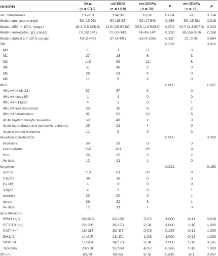
Table 1.
Patient characteristics according to
CEBPA
mutation status in non-M3 AML
Variables (n = 233)Total (n = 196)wtCEBPA smCEBPA(n = 26) P dmCEBPA(n = 11) P
Sex, male/female 135/98 114/82 16/10 0.834 5/6 0.534
Median age, years (range) 53 (15-93) 54 (15-93) 52 (17-85) 0.489 35 (15-61) 0.016
Median WBC, × 109/L (range) 16.1 (0.6-528.0) 14.5 (0.8-528.0) 35.5 (1.3-528.0) 0.072 44.7 (2.4-507.0) 0.043
Median hemoglobin, g/L (range) 73 (32-147) 72 (32-142) 74 (45-147) 0.220 85 (56-108) 0.168 Median platelets, × 109/L (range) 40 (3-447) 37 (3-447) 53 (4-203) 0.137 21 (3-76) 0.084
FAB 0.158 0.022
M0 1 1 0 0
M1 27 18 6 3
M2 115 95 12 8
M4 51 49 2 0
M5 28 24 4 0
M6 11 9 2 0
WHO 0.005 0.007
AML with t (8; 21) 37 37 0 0
AML with inv (16) 1 1 0 0
AML with 11q23 4 3 0 1
AML without maturation 23 15 6 2
AML with maturation 82 62 12 8
Acute myelomonocytic leukemia 50 48 2 0
Acute monoblastic and monocytic leukemia 25 21 4 0
Acute erythroid leukemia 11 9 2 0
Karyotype classification 0.005 0.099
Favorable 39 39 0 0
Intermediate 153 121 23 9
Poor 29 25 2 2
No data 12 11 1 0
Karyotype 0.021 0.180
normal 119 91 20 8
t (8;21) 38 38 0 0
inv (16) 1 1 0 0
11q23 4 3 0 1
complex 23 20 2 1
others 36 32 3 1
No data 12 11 1 0
Gene Mutation
NPM1 (+/-) 26/203 23/169 3/23 1.000 0/11 0.618
FLT3 ITD (+/-) 22/207 19/173 2/24 1.000 1/10 1.000
C-KIT (+/-) 15/214 15/177 0/26 0.226 0/11 1.000
IDH1/2 14/207 13/171 1/25 1.000 0/11 1.000
DNMT3A 17/204 14/170 2/24 1.000 1/10 0.595
N/K-RAS 20/201 15/169 4/22 0.266 1/10 1.000
explored. AML with mutated
CEBPA
has
recent-ly been included in the current WHO classifica
-tion as a provisional entity due to its
prognosti-cally favorable influence in normal karyotype
[20]. However, more recent studies have
identi-fied that the favorable impact on AML outcome
is predicted by double mutated
CEBPA
(
dmCEB
-PA
) rather than single mutated
CEBPA
(
smCEB
-PA
) [21-24]. In the present study, we evaluated
the frequency, the main associated features,
and the prognostic significance of
CEBPA
muta-tions in a cohort of Chinese de novo non-M3
AML patients.
Materials and methods
Patients and samples
This study was approved by the Ethics
Committee Board of the Affiliated People’s
Hospital of Jiangsu University. Bone marrow
aspirates of 233 non-M3 AML patients were
collected after informed consent written by
patients or their guardians.
The diagnosis and
classification were conducted according to the
French-American-British Cooperative Group
Criteria and the 2008 World Health Organization
(WHO) proposal [20, 25].
Treatment protocol
was described as reported previously [26]. The
main clinical and laboratory features of the
patient cohort were summarized in
Table 1
.
Cytogenetic analysis
Conventional cytogenetic analysis was
per-formed in the cytogenetics laboratory of our
hospital. Chromosomes were prepared
routine-ly by the direct method or 24h short-term
cul-ture of bone marrow cells. Karyotypes were
analyzed on R-banded metaphases.
The defini
-tion of a cytogenetic clone and descrip-tions of
karyotypes followed the International System
for Human Cytogenetic Nomenclature [27].
Karyotypes were classified according to the
revised MRC prognostic classification [9].
Cell separation and DNA isolation
The mononuclear cells were separated by
den-sity-gradient centrifugation using Ficoll. Subseq-
uently, genomic DNA was extracted using the
Genomic DNA Purification Kit (Gentra,
Minn-eapolis, MN, USA) according to the
manufac-turer’s instructions.
Gene mutation detection
Mutation of the
CEBPA
gene was detected in
genomic DNA by PCR and direct sequencing.
Two overlapping primer pairs were used to
amplify the entire coding region of
CEBPA
[image:3.612.93.524.73.249.2]seconds, and 68°C for 3 minutes; then 72°C
for 7 minutes. PCR products were
electropho-resed on 2% agarose gels, purified using
Axygen AP-GS500 kit (Axygen, CA, USA) and
then sequenced in both directions with PCR
primers on an ABI 3730 Prism Sequencer
(Applied Biosystems, CA, USA).
In samples with
a
CEBPA
sequence variation, the entire coding
region was amplified with primers P1F and P2R
under the previously described PCR conditions
except for annealing at 65°C and was cloned
into the pMD19-T vector (TaKaRa, Japan). 5 to
10 clones were sequenced in each patient with
the primers used to amplify the entire coding
region
of
CEBPA
.
NPM1,
FLT3
internal tandem duplication (ITD),
C-KIT
,
DNMT3A
,
IDH1
/
IDH2
, and
N/K
-
RAS
mutations were detected as described
previ-ously [28-30]. Briefly, genomic DNA was ampli
-fied using gene-specific primers. Mutation
scanning was performed for PCR products of all
genes except for
FLT3-ITD
using HRMA with the
LightScanner
TMplatform (Idaho, Utah, USA). All
positive samples were directly DNA sequenced
to confirm the results of HRMA.
FLT3
internal
tandem duplication (ITD) was detected using
direct DNA sequencing.
Statistical analysis
All statistical analyses were performed using
the SPSS 17.0 software package (SPSS,
Chicago, IL). Statistical significance of the dif
-ference between groups for continuous
vari-ables was determined by the Mann-Whitney
test. Statistical significance of the difference
between groups for categoric variables was
determined by Fisher exact test or Chi-square
analysis. Overall survival (OS) was estimated
using the Kaplan-Meier method and were
com-pared using the log-rank test. Multivariate
anal-yses were performed using Cox proportional
hazards regression. The significance of results
was defined as a level of
P
<0.05 at both tails.
Results
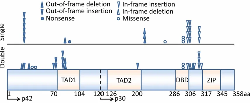
Frequency and types of CEBPA mutations in
Chinese AML patients
Fifty mutations were identified in 37 (15.8%)
patient samples (
Figure 1
; Table S1). Eleven
patients (4.7%) had
dmCEBPA
, whereas
twenty-six (11.1%) had
smCEBPA
. We observed four
[image:4.612.92.524.72.283.2]leading to premature truncation, the majority of
mutations (15/26, 58%) were located in the
C-terminal resulting in in-frame duplication/
insertion/substitution, while three mutations
were located in between TAD2 and DNA binding
domain (DBD) regions resulting in frame-shift
premature truncation.
Patient characteristics related to CEBPA muta
-tion status
Because double
CEBAP
mutations were
consid-ered as a prognostic factor according to
previ-ous studies, we divided our patient cohort into
the following three groups: patients with
dmCEBPA
(n = 11), patients with
smCEBPA
(n =
26), and patients with wild-type
CEBPA
(
wtCEB
-PA
, n = 196). The comparison of the clinical and
laboratory features between three groups is
summarized in
Table 1
.
smCEBPA
was distributed in almost all
sub-types of FAB classification (Table 1
). Although
the majority of patients with
smCEBPA
had M1
or M2 subtypes (18/26, 69%), there was no dif
-ference in the distribution of
smCEBPA
in the
whole cohort [18/142 (13%) patients with M1
or M2 versus 8/91 (9%) patients with other
subtypes,
P
= 0.401]. There was a trend that
the patients with
smCEBPA
had higher WBCs
than those with
wtCEBPA
(
P
= 0.072).
smCEB
-PA
was predominantly present in
cytogeneti-cally normal patients (
P
= 0.005). Concurrent
other molecular mutations including
NPM1
,
FLT3
,
IDH1
,
N/K-RAS
, and
DNMT3A
occurred in
patients with
wtCEBPA
, but no correlation was
observed (
P
> 0.05).
dmCEBPA
was exclusively observed in M1 and
M2 subtypes (
Table 1). Overall, 11 (8%) out of
142 patients with M1 or M2 subtypes harbored
dmCEBPA
, while none of 91 patients with other
subtypes who did so (
P
= 0.008). Patients with
dmCEBPA
had significantly younger age and
higher WBC counts at diagnosis than those
with
wtCEBPA
(
P = 0.016 and 0.043,
respec-tively). There was a trend that
dmCEBPA
group
had lower platelet counts than
wtCEBPA
group
(
P
= 0.084).
dmCEBPA
was mainly present in
cytogenetically normal patients except for two
patients harboring poor-risk karyotypes. Con-
current
FLT3-ITD
,
N-RAS
, and
DNMT3A
muta-tions occurred in two patients with
dmCEBPA
.
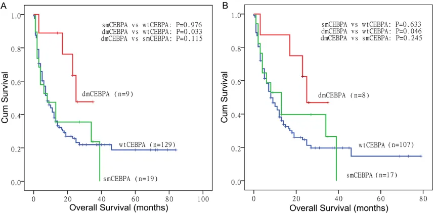
Prognostic impact of CEBPA mutation status
Survival data were obtained for 157 AML
patients with mean follow-up time of 14 months
(range, 1-84 months). There was no significant
difference in the rate of complete remission
(CR) after induction chemotherapy between
smCEBPA
and
wtCEBPA
groups (47% vs 51%,
P
= 0.810), however, more patients with
dmCEB
-PA
achieved CR than
wtCEBPA
patients (88%
vs 51%,
P
= 0.037,
Table 1
). Patients with
dmCEBPA
had a superior overall survival (OS)
compared with patients with
wtCEBPA
(esti-mated median 25
vs
8 months, respectively,
P
= 0.033;
Figure 2A
), whereas
smCEBPA
patients had a similar OS as
wtCEBPA
patients
(estimated median 8
vs
8 months, respectively,
[image:5.612.91.530.86.272.2]P
= 0.976;
Figure 2A
). Furthermore, in
multi-variate analysis that included sex,
age (≤60 yrs
vs >60 yrs), WBC count
(≤30 vs >30 × 10
9/L),
karyotype risk group, and mutational status of
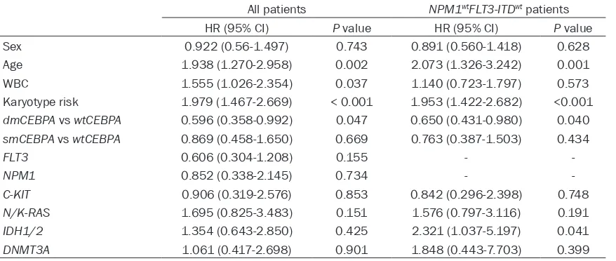
Table 2.
Multivariate analyses of prognostic factors for OS in non-M3 AML
All patients NPM1wtFLT3-ITDwt patients
HR (95% CI) P value HR (95% CI) P value
Sex 0.922 (0.56-1.497) 0.743 0.891 (0.560-1.418) 0.628
Age 1.938 (1.270-2.958) 0.002 2.073 (1.326-3.242) 0.001
WBC 1.555 (1.026-2.354) 0.037 1.140 (0.723-1.797) 0.573
Karyotype risk 1.979 (1.467-2.669) < 0.001 1.953 (1.422-2.682) <0.001 dmCEBPA vs wtCEBPA 0.596 (0.358-0.992) 0.047 0.650 (0.431-0.980) 0.040 smCEBPA vs wtCEBPA 0.869 (0.458-1.650) 0.669 0.763 (0.387-1.503) 0.434
FLT3 0.606 (0.304-1.208) 0.155 -
-NPM1 0.852 (0.338-2.145) 0.734 -
-C-KIT 0.906 (0.319-2.576) 0.853 0.842 (0.296-2.398) 0.748
N/K-RAS 1.695 (0.825-3.483) 0.151 1.576 (0.797-3.116) 0.191
IDH1/2 1.354 (0.643-2.850) 0.425 2.321 (1.037-5.197) 0.041
DNMT3A 1.061 (0.417-2.698) 0.901 1.848 (0.443-7.703) 0.399
seven genes as covariates, the presence of a
dmCEBPA
mutation remained an independent
favorable prognostic factor for OS (
Table 2
).
Because of the small size of
CEBPA
mutations,
patients with
dmCEBPA
, compared with
patients with
smCEBPA
, showed the trend
towards longer OS (
P
= 0.115,
Figure 2A
).
The impact of
CEBPA
mutations was also
evalu-ated in patients with wild-type
NPM1
and
FLT3-ITD
(
NPM1
wtFLT3-ITD
wt),
dmCEBPA
but not
smCEBPA
was associated with favorable
out-come (
Figure 2B
). Estimated median OS in
dmCEBPA
,
smCEBPA, and wtCEBPA
groups
was 25, 13 and 8 months, respectively.
Multivariate analysis also confirmed
dmCEBPA
only as an independent favorable factor in
NPM1
wtFLT3-ITD
wtpatients (
Table 2
).
Discussion
A total of 37 (15.8%) out of 233 Chinese non-3
AML patients were identified to carry 50
CEBPA
mutations after excluding all known
CEBPA
polymorphisms (data not shown). The
frequen-cy of
CEBPA
mutations detected in our study is
in good accordance with previous studies [24,
31]. Three mutational hot spots were identified,
one 5’ of the TAD1 region, the other in bZIP and
the third in the DBD of the
CEBPA
gene, which
was comparable with previous studies [22-24,
32]. The proportion of
smCEBPA
and
dmCEBPA
was different from previous studies in which
the percentage of
dmCEBPA
was more than
that of
smCEBPA
[22-24]. However, a recent
large-scale study has identified
dmCEBPA
in
104 (42.6%) out 2296 AML patient [33].
The observations are highly consistent across
all studies: double
CEBPA
mutations occur
mainly in M1/M2 subtypes and are highly
asso-ciated with normal karyotypes. However, there
is a discrepancy about clinical characteristics
of AML with
CEBPA
mutations. Although the
majority of studies did not find the correlation
of
CEBPA
mutations with peripheral leukocytes
in AML [22, 23, 32], two groups observed that
CEBPA
mutated patients had higher peripheral
leukocytes than wild-type patients [34, 35],
which was consistent with our results.
Moreover, similar to two previous reports [24,
36], the present study found the association of
double
CEBPA
mutation with younger age,
whereas other studies did no find this correla
-tion. However, a recent study on
CEBPA
muta-tions, the largest size of AML patients (a total of
2296 cases) till now, confirmed the association
of
dmCEBPA
with younger age [33].
The impact of
CEBPA
mutations on outcome
has been extensively evaluated. Earlier studies
have led to the introduction of ‘AML with
mutat-ed
CEBPA’
, which includes both
smCEBPA
and
smCEBPA
, into the current WHO classification
as a provisional entity among ‘AML with
recur-rent genetic abnormalities’ [20]. However, later
studies have shown only
dmCEBPA
but not
smCEBPA
is a favorable prognostic factor. A
recent study has confirmed that survival of AML
with
dmCEBPA
is similar with those with
PML-RARA
[37]. Although in the present study
patients with
dmCEPA
harbored higher
periph-eral leukocytes which also affected patients’
outcome, multivariate COX analysis verified
dmCEBPA
as an independent favorable
predic-tor after adjusting for other covariates including
peripheral leukocytes. Because of limited case
numbers the difference in outcome was not
sig-nificant between
dmCEBPA
and
smCEBPA
groups in this study. Initial study revealed that
the presence of additional
FLT3-ITD significant
-ly worsen overall survival in the
CEBPA
-mutated
group [38]. However, a subsequent study did
not find the influence of FLT3-ITD
on survival in
the
CEBPA
-mutated group [34]. The
aforemen-tioned studies did not differentiate
dmCEBPA
from
smCEBPA
. Concurrent
FLT3-ITD
or
NPM1
mutations are significantly less frequent in
patients with
dmCEBPA
compared with those
with
smCEBPA
, therefore, the impact of
FLT3-ITD
in AML with
dmCEBP
still needs to be
deter-mined. Further endeavor has been made to
investigate the clinically relevant aspect of
whether the favorable prognosis of
dmCEBPA
is
influenced by other additional molecular mark
-ers. Concurrent
TET2
mutations were adversely
prognostic for OS [33, 39], whereas
GATA-2
mutations improved OS [39, 40].
Although it has been shown that AML patients
carrying single
CEBPA
mutation have similar
outcome as those with wild-type
CEBPA
,
fur-ther risk stratification has been tried in AML
with single
CEBPA
mutation. Two studies
revealed that
FLT3-ITD significantly impaired
the survival of AML patients with
smCEBPA
[24,
33]. Furthermore, Fasan et al revealed that
NPM1
mt/CEBPA
smgenotype showed a trend
toward favorable outcome compared with
NPM1
wt/CEBPA
sm[24, 33]. Although
smCEBPA
was considered beneficial for outcome of AML
with
NPM1
mutation by Dufour et al [41], the
later results from the same group did not
con-firm this observation [33]. As for the role of
smCEBPA
in
NPM1
wtFLT-ITD
wtAML, it is also
controversial. Park et al found
smCEBPA
sub-group had longer survival than
wtCEBPA
sub-group in
NPM1
wtFLT-ITD
wtAML [42], however,
similar with the results of the large-scale study
of Fasan et al [33], we did not identify the
difference.
In conclusion, the results of our present study
confirm that AML with
dmCEBPA
but not
smCEBPA
is associated with a favorable
out-come in
NPM1
wtFLT3-ITD
wtnon-M3 AML. Our
data suggest that the entity AML with mutated
CEBPA
should be definitely designated as AML
with
dmCEBPA
in WHO classification and
smCEBPA
should be excluded from the
favor-able risk of molecular abnormalities [10].
Acknowledgements
This study was supported by National Natural
Science foundation of China (81270630,
81172592), Science and Technology Special
Project in Clinical Medicine of Jiangsu Province
(BL2012056), 333 Project of Jiangsu Province
(BRA2011085, BRA2013136), Science and
Technology Infrastructure Program of Zhenjiang
(SS2012003), Social Development Foundation
of Zhenjiang (SH2013042, SH2013082,
SH2014044), Research and Development
Foundation of Clinical Medicine of Jiangsu
University (JLY20120013), Key Medical Talent
Program of Zhenjiang City, and Jiangsu
Government Scholarship for Overseas Studies.
Disclosure of conflict of interest
None.
Address correspondence to: Dr. Jun Qian, Depar-
tment of Hematology, Affiliated People’s Hospital of
Jiangsu University, 8 Dianli Road, Jiangsu 212002,
Zhenjiang, People’s Republic of China. Tel: +86-511-88915303; Fax: +86-511-85234387; E-mail: qian-jun0007@hotmail.com; Dr. Jia-Bo Hu, School of Medical Science and Laboratory Medicine, Jiangsu University, 301 Xuefu Rd., Jiangsu 212013,
Zhenjiang, People’s Republic of China. Tel:
+86-511-85038449; Fax: +86-511-85038483; E-mail: hu@ ujs.edu.cn
References
[1] Look AT. Oncogenic transcription factors in the human acute leukemias. Science 1997; 278: 1059-64.
[2] Rowley JD. Chromosomal translocations: revis-ited yet again. Blood 2008; 112: 2183-9. [3] Tenen DG, Hromas R, Licht JD, Zhang DE.
Tran-scription factors, normal myeloid develop-ment, and leukemia. Blood 1997; 90: 489-519.
[4] Renneville A, Roumier C, Biggio V, Nibourel O, Boissel N, Fenaux P, Preudhomme C. Cooper-ating gene mutations in acute myeloid leuke-mia: a review of the literature. Leukemia 2008; 22: 915-31.
[5] Shih AH, Abdel-Wahab O, Patel JP, Levine RL. The role of mutations in epigenetic regulators in myeloid malignancies. Nat Rev Cancer 2012; 12: 599-612.
[6] Chen J, Odenike O, Rowley JD. Leukaemogen-esis: more than mutant genes. Nat Rev Cancer 2010; 10: 23-36.
[7] Slovak ML, Kopecky KJ, Cassileth PA, Har-rington DH, Theil KS, Mohamed A, Paietta E, Willman CL, Head DR, Rowe JM, Forman SJ, Appelbaum FR. Karyotypic analysis predicts outcome of preremission and postremission therapy in adult acute myeloid leukemia: a Southwest Oncology Group/Eastern Coopera-tive Oncology Group Study. Blood 2000; 96: 4075-83.
[8] Byrd JC, Mrózek K, Dodge RK, Carroll AJ, Ed-wards CG, Arthur DC, Pettenati MJ, Patil SR, Rao KW, Watson MS, Koduru PR, Moore JO, Stone RM, Mayer RJ, Feldman EJ, Davey FR,
Schiffer CA, Larson RA, Bloomfield CD; Cancer
and Leukemia Group B (CALGB 8461). Pre-treatment cytogenetic abnormalities are pre-dictive of induction success, cumulative inci-dence of relapse, and overall survival in adult patients with de novo acute myeloid leukemia: results from Cancer and Leukemia Group B (CALGB 8461). Blood 2002; 100: 4325-36. [9] Grimwade D, Hills RK, Moorman AV, Walker H,
Chatters S, Goldstone AH, Wheatley K, Harri-son CJ, Burnett AK; National Cancer Research Institute Adult Leukaemia Working Group.
Re-finement of cytogenetic classification in acute
myeloid leukemia: determination of prognostic
[10] Döhner H, Estey EH, Amadori S, Appelbaum FR, Büchner T, Burnett AK, Dombret H, Fenaux P, Grimwade D, Larson RA, Lo-Coco F, Naoe T, Niederwieser D, Ossenkoppele GJ, Sanz MA,
Sierra J, Tallman MS, Löwenberg B, Bloomfield
CD; European LeukemiaNet. Diagnosis and management of acute myeloid leukemia in adults: recommendations from an internation-al expert panel, on behinternation-alf of the European Leu-kemiaNet. Blood 2010; 115: 453-74.
[11] Scott LM, Civin CI, Rorth P, Friedman AD. A novel temporal expression pattern of three C/ EBP family members in differentiating myelo-monocytic cells. Blood 1992; 80: 1725-35. [12] Wang X, Scott E, Sawyers CL, Friedman AD. C/
EBPα bypasses granulocyte colony-stimulating
factor signals to rapidly induce PU.1 gene ex-pression, stimulate granulocytic differentia-tion, and limit proliferation in 32D cl3 myelo-blasts. Blood 1999; 94: 560-71.
[13] Zhang P, Iwasaki-Arai J, Iwasaki H, Fenyus ML, Dayaram T, Owens BM, Shigematsu H, Levan-tini E, Huettner CS, Lekstrom-Himes JA, Akashi K, Tenen DG. Enhancement of hematopoietic stem cell repopulating capacity and self-re-newal in the absence of the transcription
fac-tor C/EBPα. Immunity 2004; 21: 853-63.
[14] Pabst T, Mueller BU, Zhang P, Radomska HS, Narravula S, Schnittger S, Behre G, Hiddemann W, Tenen DG. Dominant-negative mutations of CEBPA, encoding CCAAT/enhancer binding protein-alpha (C/EBPalpha), in acute myeloid leukemia. Nat Genet 2001; 27: 263-70. [15] Snaddon J, Smith ML, Neat M,
Cambal-Par-rales M, Dixon-McIver A, Arch R, Amess JA, Ro-hatiner AZ, Lister TA, Fitzgibbon J. Mutations of CEBPA in acute myeloid leukemia FAB types M1 and M2. Genes Chromosomes Cancer 2003; 37: 72-8.
[16] Gombart AF, Hofmann WK, Kawano S, Takeu-chi S, Krug U, Kwok SH, Larsen RJ, Asou H,
Miller CW, Hoelzer D, Koeffler HP. Mutations in
the gene encoding the transcription factor CCAAT/enhancer binding protein alpha in my-elodysplastic syndromes and acute myeloid leukemias. Blood 2002; 99: 1332-40.
[17] Barjesteh van Waalwijk van Doorn-Khosrovani S, Erpelinck C, Meijer J, van Oosterhoud S, van Putten WL, Valk PJ, Berna Beverloo H, Tenen DG, Löwenberg B, Delwel R. Biallelic mutations in the CEBPA gene and low CEBPA expression levels as prognostic markers in intermediate-risk AML. Hematol J 2003; 4: 31-40.
[18] Schwieger M, Löhler J, Fischer M, Herwig U, Te-nen DG, Stocking C. A dominant-negative mu-tant of C/EBPalpha, associated with acute my-eloid leukemias, inhibits differentiation of myeloid and erythroid progenitors of man but not mouse. Blood 2004; 103: 2744-52.
[19] Mueller BU, Pabst T. C/EBPalpha and the pathophysiology of acute myeloid leukemia. Curr Opin Hematol 2006; 13: 7-14.
[20] In: Swerdlow SH, Campo E, Harris NL, Jaffe ES, Pileri SA, Stein H, Thiele J, Vardiman JW,
edi-tors. WHO classification of tumours of haema -topoietic and lymphoid tissues. Lyon, France: IARC Press; 2008.
[21] Wouters BJ, Lowenberg B, Erpelinck-Ver-schueren CA, van Putten WL, Valk PJ, Delwel R. Double CEBPA mutations but not single CEBPA
mutations, define a subgroup of acute myeloid
leukemia with a distinctive gene expression
profile that is uniquely associated with a favor -able outcome. Blood 2009; 113: 3088-91. [22] Pabst T, Eyholzer M, Fos J, Mueller BU.
Hetero-geneity within AML with CEBPA mutations: Only CEBPA double mutations, but not single CEBPA mutations are associated with favourable prognosis. Br J Cancer 2009; 100: 1343-6. [23] Dufour A, Schneider F, Metzeler KH, Hoster E,
Schneider S, Zellmeier E, Benthaus T, Sauer-land MC, Berdel WE, Büchner T, Wörmann B, Braess J, Hiddemann W, Bohlander SK, Spiekermann K. Acute myeloid leukemia with biallelic CEBPA gene mutations and normal karyotype represents a distinct genetic entity associated with a favorable clinical outcome. J Clin Oncol 2010; 28: 570-7.
[24] Taskesen E, Bullinger L, Corbacioglu A, Sand-ers MA, Erpelinck CA, WoutSand-ers BJ, van der Poel-van de Luytgaarde SC, Damm F, Krauter J, Ganser A, Schlenk RF, Löwenberg B, Delwel R, Döhner H, Valk PJ, Döhner K. Prognostic im-pact, concurrent genetic mutations, and gene expression features of AML with CEBPA muta-tions in a cohort of 1182 cytogenetically nor-mal AML patients: further evidence for CEBPA double mutant AML as a distinctive disease entity. Blood 2011; 117: 2469-75.
[25] Bennett JM, Catovsky D, Daniel MT, Flandrin G, Galton DA, Gralnick HR, Sultan C. Proposed
re-vised criteria for the classification of acute my -eloid leukaemia. A report of the French-Ameri-can-British Cooperative Group. Ann Intern Med 1985; 103: 620-5.
[26] Li Y, Lin J, Yang J, Qian J, Qian W, Yao DM, Deng ZQ, Liu Q, Chen XX, Xie D, An C, Tang CY. Over-expressed let-7a-3 is associated with poor out-come in acute myeloid leukemia. Leuk Res 2013; 37: 1642-7.
[27] Mitelman F. An International system for human cytogenetic nomenclature. In: ISCN, editor. Ba-sel: Karger; 1995.
[29] Lin J, Yao DM, Qian J, Chen Q, Qian W, Li Y, Yang J, Wang CZ, Chai HY, Qian Z, Xiao GF, Xu WR. IDH1 and IDH2 mutation analysis in Chi-nese patients with acute myeloid leukemia and myelodysplastic syndrome. Ann Hematol 2012; 91: 519-25.
[30] Yang X, Qian J, Sun A, Lin J, Xiao G, Yin J, Chen S, Wu D. RAS mutation analysis in a large co-hort of Chinese patients with acute myeloid leukemia. Clin Biochem 2013; 46: 579-83. [31] Kihara R, Nagata Y, Kiyoi H, Kato T, Yamamoto
E, Suzuki K, Chen F, Asou N, Ohtake S, Miyawa-ki S, MiyazaMiyawa-ki Y, Sakura T, Ozawa Y, Usui N, Kanamori H, Kiguchi T, Imai K, Uike N, Kimura F, Kitamura K, Nakaseko C, Onizuka M, Takeshita A, Ishida F, Suzushima H, Kato Y, Miwa H, Shiraishi Y, Chiba K, Tanaka H, Miyano S, Ogawa S, Naoe T. Comprehensive analysis of genetic alterations and their prognostic im-pacts in adult acute myeloid leukemia pa-tients. Leukemia 2014: 28; 1586-95.
[32] Lin LI, Chen CY, Lin DT, Tsay W, Tang JL, Yeh YC, Shen HL, Su FH, Yao M, Huang SY, Tien HF. Characterization of CEBPA mutations in acute myeloid leukemia: most patients with CEBPA mutations have biallelic mutations and show a distinct immunophenotype of the leukemic cells. Clin Cancer Res 2005; 11: 1372-9. [33] Fasan A, Haferlach C, Alpermann T, Jeromin S,
Grossmann V, Eder C, Weissmann S, Dicker F, Kohlmann A, Schindela S, Kern W, Haferlach T,Schnittger S. The role of different genetic subtypes of CEBPA mutated AML. Leukemia 2014; 28: 794-803.
[34] Fröhling S, Schlenk RF, Stolze I, Bihlmayr J, Benner A, Kreitmeier S, Tobis K, Döhner H, Döhner K. CEBPA mutations in younger adults with acute myeloid leukemia and normal cyto-genetics: prognostic relevance and analysis of cooperating mutations. J Clin Oncol 2004; 22: 624-33.
[35] Shen Y, Zhu YM, Fan X, Shi JY, Wang QR, Yan XJ, Gu ZH, Wang YY, Chen B, Jiang CL, Yan H, Chen FF, Chen HM, Chen Z, Jin J, Chen SJ. Gene mutation patterns and their prognostic impact in a cohort of 1185 patients with acute myeloid leukemia. Blood 2011; 118: 5593-603.
[36] Green CL, Koo KK, Hills RK, Burnett AK, Linch
DC, Gale RE. Prognostic significance of CEBPA
mutations in a large cohort of younger adult patients with acute myeloid leukemia: impact of double CEBPA mutations and the interaction with FLT3 and NPM1 mutations. J Clin Orthod 2010; 28: 2739-47.
[37] Grossmann V, Schnittger S, Kohlmann A, Eder C, Roller A, Dicker F, Schmid C, Wendtner CM, Staib P, Serve H, Kreuzer KA, Kern W, Hafer-lach T, HaferHafer-lach C. A novel hierarchical prog-nostic model of AML solely based on molecular mutations. Blood 2012; 120: 2963-72. [38] Preudhomme C, Sagot C, Boissel N, Cayuela
JM, Tigaud I, de Botton S, Thomas X, Raffoux E, Lamandin C, Castaigne S, Fenaux P, Dombret
H; ALFA Group. Favorable prognostic signifi -cance of CEBPA mutations in patients with de novo acute myeloid leukemia: a study from the Acute Leukemia French Association (ALFA). Blood 2002; 100: 2717-23.
[39] Grossmann V, Haferlach C, Nadarajah N, Fasan A, Weissmann S, Roller A, Eder C, Stopp E, Kern W, Haferlach T, Kohlmann A, Schnittger S. CEBPA double-mutated acute myeloid leu-kaemia harbours concomitant molecular
mu-tations in 76·8% of cases with TET2 and GATA2
alterations impacting prognosis. Br J Haematol 2013; 161: 649-58.
[40] Fasan A, Eder C, Haferlach C, Grossmann V, Kohlmann A, Dicker F, Kern W, Haferlach T, Schnittger S. GATA2 mutations are frequent in intermediate-risk karyotype AML with biallelic CEBPA mutations and are associated with fa-vorable prognosis. Leukemia 2013; 27: 482-5. [41] Dufour A, Schneider F, Hoster E, Benthaus T,
Ksienzyk B, Schneider S, Kakadia PM, Sauer-land MC, Berdel WE, Büchner T, Wörmann B, Braess J, Subklewe M, Hiddemann W, Boh-lander SK, Spiekermann K; AML CG study group. Monoallelic CEBPA mutations in normal karyotype acute myeloid leukemia: indepen-dent favorable prognostic factor within NPM1 mutated patients. Ann Hematol 2012; 91: 1051-63.
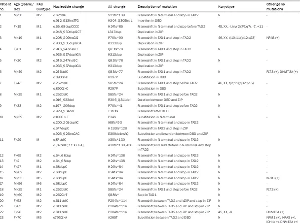
Table S1.
Description of
CEBPA
mutations in 37 AML patients
Patient
no. Age (years)/Sex FAB Subtype Nucleotide change AA change Description of mutation Karyotype Other gene mutations
1 M/50 M2 c.62delG S21fs*139 Frameshift in N-terminal and stop in TAD2 N
-c.912_913insTTG K304_Q305insL Insertion in DBD
2 F/15 M1 c.65_68dupCCCC H24fs*85 Frameshift in N-terminal and stop before TAD2 45, XX, l, inv(2)(P?,q?), 7, +11
-c.948_950dupGCT L317dup Duplication in ZIP
3 M/19 M1 c.208_209insGG P70fs*90 Frameshift in TAD1 and stop in TAD2 46, XY, t(10;11)(p12;q23) NRAS (+)
c.933_935dupGCA K313dup Duplication in ZIP
4 F/61 M2 c.246_247insGC Q83fs*78 Frameshift in TAD1 and stop in TAD2 N
-c.935_937dupAGA K313dup Duplication in ZIP
5 F/30 M2 c.246_247insGC Q83fs*78 Frameshift in TAD1 and stop in TAD2 N
-c.935_937dupAGA K313dup Duplication in ZIP
6 M/49 M2 c.246delC Q83fs*77 Frameshift in TAD1 and stop in TAD2 N FLT3 (+), DNMT3A (+)
c.890G>C R297P Substitution in DBD
7 F/47 M2 c.252delC S85fs*24 Frameshift in TAD1 and stop before TAD2 46, XX, t(2;11)(q32;p15)
-c.890G>C R297P Substitution in DBD
8 M/35 M1 c.252delC S85fs*24 Frameshift in TAD1 and stop before TAD2 N
-c.916_933del R306_Q311del Deletion between DBD and ZIP
9 F/33 M2 c.197_206dup P70fs*41 Frameshift in TAD1 and stop before TAD2 N
-c.929_934del T310fs Frameshift after DBD
10 M/39 M2 c.100C > T P34S Substitution in N-terminal N
-c.200_201dupAC I68fs*93 Frameshift in N-terminal and stop in TAD2 c.577dupC H193fs*128 Frameshift in TAD2 and stop in ZIP
c.925_926insCAC E309delinsAQ Substitution and insertion between DBD and ZIP
11 F/29 M c.87delC A30fs*130 Frameshift in N-terminal and stop in TAD2 N
-c.[87delC; 113G > A] A30fs*130, A38T Frameshift and substitution in N-terminal and stop in TAD2
12 F/65 M2 c.64_68dup H24fs*138 Frameshift in N-terminal and stop in TAD2 N
-13 F/2 M2 c.64_68dup H24fs*138 Frameshift in N-terminal and stop in TAD2 N
-14 F/27 M1 c.68dupC H24fs*84 Frameshift in N-terminal and stop in TAD2 N
-15 M/62 M2 c.68dupC H24fs*84 Frameshift in N-terminal and stop in TAD2 N
-16 M/53 M5 c.68dupC H24fs*84 Frameshift in N-terminal and stop in TAD2 N KRAS (+)
17 M/56 M6 c.68dupC H24fs*84 Frameshift in N-terminal and stop in TAD2 N
-18 M/35 M1 c.252delC S85fs*24 Frameshift in TAD1 and stop before TAD2 N FLT3 (+)
19 M/60 M2 c.262C>T Q88fs* Stop in TAD1 N
-20 F/53 M2 c.611delC P204fs*114 Frameshift between TAD2 and bZIP and stop in ZIP N
-21 F/85 M2 c.611delC P204fs*114 Frameshift between TAD2 and ZIP and stop in ZIP N
-22 F/28 M2 c.611delC P204fs*114 Frameshift between TAD2 and ZIP and stop in ZIP 45, XX, -8 DNMT3A (+)
23 F/70 M5 c793G>A A265T Substitution between TAD2 and DBD N NPM1 (+), NRAS (+),
24 F/39 M1 c.890G>C R297P Substitution in DBD N FLT3 (+)
25 M/57 M2 c.890G>C R297P Substitution in DBD N
-26 M/42 M6 c.890G>C R297P Substitution in DBD N NPM1 (+)
27 M/78 M2 c.912_913insTTG K304_Q305insL Insertion in DBD N
-28 M/41 M2 c.912_913insTTG K304_Q305insL Insertion in DBD 47, XY, +17
-9 M/55 M1 c.912_913insTTG K304_Q305insL Insertion in DBD N NRAS (+)
30 M/69 M2 c.912_913insTTG K304_Q305insL Insertion in DBD 49, XY, +3, +8, +11
-31 F/41 M5 c.918_919dupAAGGCCAAGCAGCGC K302_R306dup Duplication in DBD ND KRAS (+)
32 M/51 M2 c.918_919dupAAGGCCAAGCAGCGC K302_R306dup Duplication in DBD N
-33 M/22 M1 c.918_919dupAAGGCCAAGCAGCGC K302_R306dup Duplication in DBD N
-34 M/31 M4 c.918_919dupAAGGCCAAGCAGCGC K302_R306dup Duplication in DBD N
-35 F/66 M1 c.935_937dupAGA K313dup Duplication in ZIP N NPM1 (+)
36 M/28 M5 c.935_937dupAGA K313dup Duplication in ZIP 47, XY, del(1)(p22), +1