Original Article
Analysis of the factors affecting lymph node metastasis
and the prognosis of rectal neuroendocrine tumors
Peng Li, Fan Wu, Hong Zhao, Lizhou Dou, Yang Wang, Chunguang Guo, Guiqi Wang, Dongbing Zhao Department of Abdominal Surgery, Cancer Hospital, Chinese Academy of Medical Sciences & Peking Union Medical College, Beijing 100021, China
Received August 13, 2015; Accepted September 22, 2015; Epub October 1, 2015; Published October 15, 2015
Abstract: Objective: To analyze the factors affecting lymph node metastasis and the prognosis of rectal neuroendo-crine tumors after surgical treatment. Methods: A retrospective analysis was conducted using the clinical data from 156 cases of rectal neuroendocrine tumors during the period of January 1999 to December 2013. The Kaplan-Meier method was used to calculate the survival time, Cox regression analysis was performed for statistical analysis of clinicopathological factors that may be associated with lymph node metastasis and prognosis, and correlation analysis was carried out using binary logistic regression. Results: The overall 5-year survival rate of the entire group was 95.7%. Multivariate analysis showed that the depth of invasion was an independent prognostic factor
(P < 0.001). The incidence of lymph node metastasis was 7.7% (12/156), and logistic regression analysis showed that lymph node metastasis was related to the depth of invasion (P = 0.003) and tumor diameter (P = 0.006). Conclusion: The surgical approach of rectal neuroendocrine tumors should be selected based on a comprehensive consideration of factors such as tumor size, depth of invasion and lymph node metastasis.
Keywords: Neuroendocrine tumors, rectal neuroendocrine, lymph node metastasis
Introduction
Originating from chromaffin-like cells, neuroen-docrine tumors (NENs) are a type of tumor with neuroendocrine functions and malignant potential. The clinical manifestations of NENs are significantly heterogeneous depending on the disease site and endocrine function [1, 2]. According to the US Surveillance, Epidemiology, and End Results (SEER) database, NEN inci-dence rose from 1.09/100,000 population per year in 1973 to 5.25/100,000 population per year in 2004 [3], and the incidence rate has increased annually [2]. Several large-scale epi-demiological surveys have confirmed that NENs primarily occur in the digestive tract [4-6]. Differing from the European and American populations, studies based on populations in Taiwan and Japan revealed that the rectum is the most likely site of the digestive tract affect-ed by NENs in Asian populations [7, 8]. Similar to colorectal cancer, surgical resection is the standard treatment of NENs, but lymph node metastasis is an important factor affecting the
choice between radical resection and local ex- cision. Therefore, the investigation of factors related to lymph node metastasis and progno-sis has a great clinical significance for rectal NENs. The purposes of this study were to ana-lyze the factors affecting lymph node metasta-sis and the prognometasta-sis of rectal neuroendocrine tumors after surgical treatment.
Patients and methods
colonoscopy, and pathological confirmation was also obtained. The clinical and pathologi-cal data were obtained through medipathologi-cal records and databases. Pathological sections under-went immunohistochemical staining, and the Ki-67 index and mitotic images under high-power fields were also recorded. According to the seventh edition of the American Joint Committee on Cancer (AJCC) staging manual and pathological grading system of the World Health Organization 2010 criteria of tumors of the digestive system [9, 10], we re-staged and
Results
Clinical and pathological characteristics
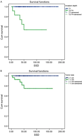
[image:2.612.92.370.72.533.2]This study collected 181 rectal NEN cases that received treatment at the Chinese Academy of Medical Sciences Cancer Hospital during the period of January 1999 to December 2013. Of these cases, 25 were not included in the study, including 15 cases associated with a second primary cancer, 5 cases that received palliative surgery and 5 cases that did not receive sur-gery. The final analysis included 156 cases. Figure 1. Survival curves stratified by tumor invasion depth (A. P < 0.001)
and tumor size (B. P < 0.001).
graded the lesions. The study was conducted with the app- roval of the institutional ethics board of our institute.
Surgical approaches were divided into two categories, local excision and radical sur-gery. Local tumor excision in- cluded transanal excision (TAE) and endoscopic resection [en- doscopic mucosal resection (EMR) or endoscopic submu- cosal dissection (ESD)]. Radi- cal surgery included low ante-rior resection (LAR), abdomi- noperineal resection (APR), and transsacral resection (TSR). Follow-up studies were conducted based on outpa-tient re-examination and tele-phone follow-up.
There were a total of 104 males and 52 fe- males, with a median age of 50.82 ± 11.566 years. Approximately 34% (n = 54) of the pati- ents had no clinical symptoms, and the disease was identified during routine physical examina-tion. Changes in bowel movement habits (n = 43) and hematochezia (n = 44) were the most common clinical manifestations. None of the patients showed symptoms associated with carcinoid syndrome. Tumor location was deter-mined by preoperative colonoscopy, and the pathological diagnosis was also obtained. The
median distance between the lower edge of the tumor and anus was 5.798 ± 4.044 cm. The median lesion diameter was 9.79 ± 11.34 mm. Among the 156 cases, 126 had a lesion diam-eter ≤ 1 cm, 19 cases had a lesion diamdiam-eter between 1-2 cm, and 11 cases had a lesion diameter > 2 cm.
[image:3.612.93.527.86.560.2]Lesions in 140 cases were confined to the submucosa, of which lesions in 4 cases were accompanied by lymph node metastasis. Lesions in five cases invaded the muscular
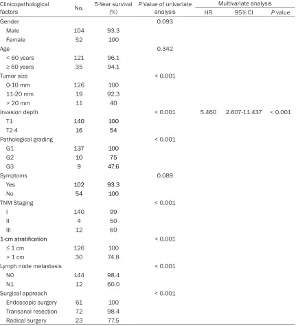
Table 1. Analysis of prognostic factors of overall survival
Clinicopathological
factors No. 5-Year survival (%) P Value of univariate analysis
Multivariate analysis HR 95% CI P value
Gender 0.093
Male 104 93.3
Female 52 100
Age 0.342
< 60 years 121 96.1
≥ 60 years 35 94.1
Tumor size < 0.001
0-10 mm 126 100
11-20 mm 19 92.3
> 20 mm 11 40
Invasion depth < 0.001 5.460 2.607-11.437 < 0.001
T1 140 100
T2-4 16 54
Pathological grading < 0.001
G1 137 100
G2 10 75
G3 9 47.6
Symptoms 0.089
Yes 102 93.3
No 54 100
TNM Staging < 0.001
I 140 99
II 4 50
III 12 60
1-cmstratification < 0.001
≤ 1 cm 126 100
> 1 cm 30 74.8
Lymph node metastasis < 0.001
N0 144 98.4
N1 12 60.0
Surgical approach < 0.001
layer, including one case that also had lymph node metastasis; lesions in seven cases in- volved the subserosa, of which four cases had intestinal lymph node or distant metastases; lesions in four cases invaded the outer serosa, of which three cases had lymph node metasta-sis. According to the seventh edition of the AJCC staging manual, the cases were catego-rized as 140 stage I cases (89.7%), 4 stage II cases (2.56%), and 12 stage III cases (7.69%). According to pathological grading, there were 87.8% of grade 1 (G1) lesions (n = 137), 6.4% of G2 lesions (n = 10) and 5.8% of G3 lesions (n = 9).
Surgical approaches included local excision and radical surgery. A total of 85.3% of patients underwent local excision, including TAE (n = 72) and endoscopic resection (n = 61). Radical resection included LAR (n = 15), TSR (n = 2), APR (n = 5), and Hartmann’s operation (n = 1). There were no postoperative complications and postoperative mortality (≤ 30 days).
Overall survival
As of December 2014, the average follow-up time was 65 ± 43.31 months (range, 3-185 months). The overall 5-year survival rate was 95.7%. The 5-year survival rates for tumors at different stages were 99.0% for stage I tumors, 50.0% for stage II tumors, and 60.0% for stage III tumors. The 5-year survival rates for tumors of different pathological grades were 100% for G1 tumors, 75% for G2 tumors, and 47.6% for G3 tumors. After the tumors were grouped according to the tumor diameter of 0-10 mm, 11-20 mm, or > 20 mm, there were significant differences (P < 0.001) in the prognosis bet- ween the groups. There were also significant differences in prognosis between different surgical procedures (P < 0.001). Lymph node metastasis was a prognostic factor (P < 0.001). Univariate analysis showed that factors asso- ciated with prognosis of overall survival includ-ed TNM stage (P < 0.001), grade (P < 0.001), depth of invasion (P<0.001), tumor size (P < 0.001) (Figure 1), surgical approach (P < 0.001), and lymph node metastasis (P < 0.001). Multivariate analysis via Cox regression showed that the depth of invasion was an independent prognostic factor (P < 0.001; Table 1). The 133 cases with local excision were all G1-G2 grade lesions, and the lesion diameter was less than
2 cm. The 5-year survival rates of patients in the TRG and ECG groups were 98.4% and 100%, and there was no significant difference between the two groups (P = 0.452).
Lymph node metastasis and influencing fac-tors
There were 12 cases in this group with lymph node metastasis, and the metastasis rate was 7.69%. Logistic binary regression analysis showed that a tumor size larger than 1 cm in diameter (P = 0.006) and depth of invasion (P = 0.003) were independent factors associated with lymph node metastasis (Table 2).
Discussion
Rectal NENs account for 60-89% of all NENs of the entire digestive tract [11], and the NEN inci-dence shows a gradual upward trend due to the wide application of endoscopic examina-tion. NENs of the rectum show no specific clini-cal symptoms. Additionally, 89% of the newly diagnosed patients in this group were acciden-tally discovered in colonoscopy. Weinstock et al. showed that the prognosis of asymptomatic patients was significantly better than that of symptomatic patients [12]. In this study, asymp-tomatic patients accounted for 33.9% of the patients, and there were no significant differ-ence in the prognosis between the asymptom-atic and symptomasymptom-atic groups (P = 0.119). We found that 81.7% of the symptomatic patients had stage I disease, and the early-disease stage is the main cause of the lack of differ-ence in the survival between the symptomatic and asymptomatic groups. Therefore, improv-ing the early detection of rectal NENs by colo-noscopy is an important factor to improve prognosis.
impor-tant reference for the selection of clinical treat-ment methods.
Due to the low incidence of rectal NENs, statis-tical analyses with large sample sizes are lack-ing. There remain many inconsistent reports on the prognostic factors of rectal NENs. For example, multiple retrospective studies have suggested that factors such as tumor stage, tumor grade, tumor size, lymph node metasta-sis, depth of invasion, and surgical methods are associated with prognosis. Garcia-Car- bonero et al. [14] found that only pathological stage (P = 0.0001, hazard ratio (HR) = 3.96, 95% CI: 1.97-7.96) and Ki-67 index (P = 0.008, HR = 6.69, 95% CI: 1.96-22.88) were indepen-dent prognostic factors. Chi et al. [15] found that tumor grade was an independent factor associated with prognosis (HR = 2.797, 95% CI:
[image:5.612.92.527.82.479.2]1.676-4.668, P = 0.004). Chagpar et al. [16] found that the depth of invasion, tumor size, lymph node metastasis, and distant meta- stasis were all independent prognostic factors (P < 0.001). The 5-year survival rate of this study group was 92.6%, a value that is similar to that reported in the literature. Univariate prognostic factor analysis focusing on this group of 156 NEN cases revealed that the prognostic factors included tumor stage, grade, depth of invasion, tumor size, surgical approach, and lymph node metastasis. However, multi-variate analysis showed that only the depth of invasion was an independent prognostic factor. Wang et al. [17] conducted a retrospec-tive analysis of 106 cases of rectal NEN and obtained conclusions similar to this study. Therefore, based on the above studies, preop-erative staging and lymph node status have
Table 2. Analysis of factors associated with lymph node metastasis
Clinicopathological factors N0 metastasis (%)Lymph node univariate analysisP Value of Multivariate analysis HR 95% CI P Value
Gender 1.000
Male 104 8 (7.69)
Female 52 4 (7.69)
Age 0.352
< 60 years 121 8 (6.61)
≥ 60 years 35 4 (11.43)
Tumor size < 0.001
0-10 mm 126 2 (1.59) 11-20 mm 19 2 (10.53) > 20 mm 11 8 (72.73)
Invasion depth < 0.001 3.295 1.508-7.200 0.003
T1 140 4 (2.86)
T2-4 16 8 (50.00)
Pathological grading < 0.001
G1 137 4 (2.92)
G2 10 2 (20.00)
G3 9 6 (66.67)
Symptoms 0.997
Yes 102 12 (11.76)
No 54 0 (0)
Staging 0.991
I 140 0 (0)
II 4 0 (0)
III 12 12 (100)
1-cmstratification < 0.001 13.124 2.068-71.073 0.006
≤ 1 cm 126 2 (1.59)
decisive significance on the selection of rectum NEN treatment methods.
Previous literature has suggested that the metastasis rate of T1 lesions with a diameter < 1 cm was less than 3%. When the tumor diam-eter was greater than 1 cm, the metastasis rate may increase to 10-15% [18]. Gleeson et al. [13] found that the metastasis rates of tumors with a diameter ≤ 10 mm, between 11 and 19 mm, and ≥ 20 mm were 3%, 66%, and 73%, respectively. In this study, 12 patients exhibited lymph node metastasis. Among them, two had a diameter < 1 cm, of which 1 was a case of G1 mucosal lesion and 1 was a case of a G2 lesion invading the muscular layer. There were two cases with a diameter of 1-2 cm; both were con-fined to the mucosa: one case was a G3 lesion, and the other was a G1 lesion. Logistic bivari-ate analysis showed that a tumor diameter greater than 1 cm and depth of invasion are independent factors associated with lymph node metastasis. Currently, endoscopic ultra-sonography (EUS) has a high accuracy for eval-uating per-intestinal lymph node status. It has been reported that EUS has an accuracy of 74.6% in the preoperative assessment of rectal adenocarcinoma lymph node metastasis, a value that dropped to 43.3% after postopera-tive pathological correction [19]. Chen et al. reported that the accuracy rate of ultrasonog-raphy in rectal NENs was approximately 94.4% [20]. Therefore, paying attention to the preop-erative evaluation of lymph node metastasis in rectal NEN patients and improving EUS are of important guiding significance, particularly for tumors greater than 1 cm in diameter and those suspected of deep invasion.
Surgical resection is the primary treatment approach for rectal NENs, and the surgical methods primarily include endoscopic surgical resection, local excision and radical resection. The determination of the surgical approach is based on the evaluation of lymph node metas-tasis, while tumor size and depth of invasion are also important determinants of the surgical approach. The present study separated the cases into different groups based on the invad-ed muscular layers and then conductinvad-ed bivari-ate logistic regression. We found that histo- logical grade (P = 0.002, HR = 9.429, 95% CI: 2.243-39.632) and a tumor diameter greater than 2 cm (P = 0.004, HR = 49.999, 95% CI: 3.609-692.687) are independent factors
asso-ciated with muscular layer invasion. For rectal NENs less than 1 cm in diameter, because the probability of metastasis is less than 5% [21-24], local excision is sufficient [25]. There has been no international consensus on the treat-ment of lesions with a diameter of 11-20 mm. The European Neuroendocrine Tumor Society (ENETS) guidelines recommend endoscopic ultrasound assessment of lymph nodes and depth of invasion, and G1 lesions of stage T1N0 can be treated by local excision [21]. The National Comprehensive Cancer Network (NCCN) guidelines recommend local excision for tumors with a diameter less than 2 cm, while tumors 1-2 cm in diameter require the removal of external muscular layer invasion and lymph node metastasis. Tsukamoto et al. [26] suggested that tumors with a diameter greater than 10 mm had a significantly in- creased rate of lymph node metastasis and should be recommended to receive radical treatment in accordance with the principle of radical treatment for colorectal cancer. In this study, 19 cases corresponded to a tumor diameter in the range of 11-20 mm, 16 cases received local excision, and 1 case received salvage radiation due to a positive surgical margin after TAE, but the patient died due to cancer 3 years after the surgery. There were no significant differences in long-term survival between radical resection and local excision (P = 0.670), and between endoscopic resection (n = 6) and TAE (n = 10) (P = 0.752). These results suggest that, for tumors less than 2 cm in diameter, local excision can be conducted after the removal of lymph node metastasis and muscular invasion, and these tumors can also be treated with the less invasive endoscopic treatment. However, this study had limited data, and further research is required to con-firm and clarify the above observations.
In conclusion, a rectal NEN is a relatively rare type of tumor, with a good prognosis after sur- gical treatment. Fully assessing and compre-hensively considering the tumor size, depth of invasion, lymph node status and other factors can help select the appropriate treatment approach, and therefore improve the prognosis of patients.
Disclosure of conflict of interest
Address correspondence to: Dr. Dongbing Zhao, Department of Abdominal Surgery, Cancer Hospital,
Chinese Academy of Medical Sciences & Peking
Union Medical College, 17, South Panjiayuan Rd, Beijing 100021, China. E-mail: drzhaodb@yeah.net
References
[1] Landry CS, Cavaness K, Celinski S, Preskitt J.
Biochemical prognostic indicators for pancre-atic neuroendocrine tumors and small bowel neuroendocrine tumors. Gland Surg 2014; 3: 215-218.
[2] Wang YZ, Chauhan A, Hall MA. Adjuvant intra-operative post-dissectional tumor bed chemo-therapy-A novel approach in treating midgut neuroendocrine tumors. J Gastrointest Oncol 2015; 6: 254-258.
[3] Modlin IM, Lye KD, Kidd M. A 5-decade analy-sis of 13,715 carcinoid tumors. Cancer 2003; 97: 934-959.
[4] Clawson GA. From devils to jobs: tracking neu -roendocrine tumors. Transl Cancer Res 2013; 2: 3-5.
[5] Helland SK, Prøsch AM, Viste A. Carcinoid tu-mours in the gastrointestinal tract--a popula-tion-based study from Western Norway. Scand J Surg 2006; 95: 158-161.
[6] Eftekhari A, Worsley D, Klass D, Liu DM.
Technical note: simultaneous 90Y and 99mTc-MAA injection for two-stage selective internal radiation therapy (SIRT) of liver metastases. Transl Cancer Res 2014; 3: 138-145.
[7] Li AF, Hsu CY, Li A, Tai LC, Liang WY, Li WY, Tsay SH, Chen JY. A 35-year retrospective study of carcinoid tumors in Taiwan: differences in dis-tribution with a high probability of associated second primary malignancies. Cancer 2008; 112: 274-283.
[8] Soga J. Carcinoids of the rectum: an evaluation of 1271 reported cases. Surg Today 1997; 27: 112-119.
[9] Rindi G, Klöppel G, Couvelard A, Komminoth P, Körner M, Lopes JM, McNicol AM, Nilsson O, Perren A, Scarpa A, Scoazec JY, Wiedenmann B. TNM staging of midgut and hindgut (neuro) endocrine tumors: a consensus proposal in-cluding a grading system. Virchows Arch 2007; 451: 757-762.
[10] Bosman FT, World Health Organization. Inter- national Agency for Research on Cancer. WHO
classification of tumours of the digestive sys -tem. 4th edition. Lyon: International Agency for Research on Cancer; 2010. pp. 417.
[11] Ito T, Sasano H, Tanaka M, Osamura RY, Sasaki I, Kimura W, Takano K, Obara T, Ishibashi M, Nakao K, Doi R, Shimatsu A, Nishida T, Komoto I, Hirata Y,Nakamura K, Igarashi H, Jensen RT,
Wiedenmann B, Imamura M. Epidemiological study of gastroenteropancreatic neuroendo-crine tumors in Japan. J Gastroenterol 2010; 45: 234-243.
[12] Weinstock B, Ward SC, Harpaz N, Warner RR, Itzkowitz S, Kim MK. Clinical and prognostic
features of rectal neuroendocrine tumors. Neuroendocrinology 2013; 98: 180-187. [13] Gleeson FC, Levy MJ, Dozois EJ, Larson DW,
Wong Kee Song LM, Boardman LA. Endo-
scopically identified well-differentiated rectal
carcinoid tumors: impact of tumor size on the natural history and outcomes. Gastrointest Endosc 2014; 80: 144-151.
[14] Garcia-Carbonero R, Capdevila J, Crespo-Herrero G, Díaz-Pérez JA, Martínez Del Prado MP, Alonso Orduña V, Sevilla-García I, Villa- bona-Artero C, Beguiristain-Gómez A, Llanos-Muñoz M, Marazuela M, Alvarez-Escola C, Castellano D, Vilar E, Jiménez-Fonseca P, Teulé A, Sastre-Valera J, Benavent-Viñuelas M, Monleon A, Salazar R. Incidence, patterns of care and prognostic factors for outcome of gastroenteropancreatic neuroendocrine tu-mors (GEP-NETs): results from the National Cancer Registry of Spain (RGETNE). Ann Oncol 2010; 21: 1794-1803.
[15] Chi Y, Du F, Zhao H, Wang JW, Cai JQ. Characteristics and long-term prognosis of patients with rectal neuroendocrine tumors. World J Gastroenterol 2014; 20: 16252-16257.
[16] Chagpar R, Chiang YJ, Xing Y, Cormier JN, Feig BW, Rashid A, Chang GJ, You YN. Neuro- endocrine tumors of the colon and rectum: prognostic relevance and comparative perfor-mance of current staging systems. Ann Surg Oncol 2013; 20: 1170-1178.
[17] Wang M, Peng J, Yang W, Chen W, Mo S, Cai S. Prognostic analysis for carcinoid tumours of the rectum: a single institutional analysis of 106 patients. Colorectal Dis 2011; 13: 150-153.
[18] Shields CJ, Tiret E, Winter DC; International Rectal Carcinoid Study Group. Carcinoid tu-mors of the rectum: a multi-institutional inter-national collaboration. Ann Surg 2010; 252: 750-755.
[19] Spinelli P, Schiavo M, Meroni E, Di Felice G, Andreola S, Gallino G, Belli F, Leo E. Results of EUS in detecting perirectal lymph node
metas-tases of rectal cancer: the pathologist makes
the difference. Gastrointest Endosc 1999; 49: 754-758.
[21] Caplin M, Sundin A, Nillson O, Baum RP, Klose
KJ, Kelestimur F, Plöckinger U, Papotti M,
Salazar R, Pascher A; Barcelona Consensus Conference participants. ENETS Consensus Guidelines for the management of patients with digestive neuroendocrine neoplasms: colorectal neuroendocrine neoplasms. Neuro- endocrinology 2012; 95: 88-97.
[22] Kim DH, Lee JH, Cha YJ, Park SJ, Cheon JH,
Kim TI, Kim H, Kim WH, Hong SP. Surveillance strategy for rectal neuroendocrine tumors
ac-cording to recurrence risk stratification. Dig Dis
Sci 2014; 59: 850-856.
[23] Fahy BN, Tang LH, Klimstra D, Wong WD, Guillem JG, Paty PB, Temple LK, Shia J, Weiser
MR. Carcinoid of the rectum risk stratification
(CaRRs): a strategy for preoperative outcome assessment. Ann Surg Oncol 2007; 14: 1735-1743.
[24] Holinga J, Khalid A, Fasanella K, Sanders
M, Davison J, McGrath K. Metastatic risk of
diminutive rectal carcinoid tumors: a need for surveillance rectal ultrasound? Gastrointest Endosc 2012; 75: 913-916.
[25] Kwaan MR, Goldberg JE, Bleday R. Rectal car-cinoid tumors: review of results after endo-scopic and surgical therapy. Arch Surg 2008; 143: 471-475.
[26] Tsukamoto S, Fujita S, Yamaguchi T, Yamamoto S, Akasu T, Moriya Y, Taniguchi H, Shimoda T.