Original Article
Development of a new real-time PCR system for
simultaneous detection of bacteria and
fungi in pathological samples
Hitomi Fukumoto
1,2, Yuko Sato
1, Hideki Hasegawa
1, Hidehisa Saeki
2, Harutaka Katano
11
Department of Pathology, National Institute of Infectious Diseases, 1-23-1 Toyama, Shinjuku-ku, Tokyo
162-8640, Japan;
2Department of Dermatology, Nippon Medical School, 1-1-5 Sendagi, Bunkyo-ku, Tokyo 113-8603,
Japan
Received July 17, 2015; Accepted October 20, 2015; Epub November 1, 2015; Published November 15, 2015
Abstract:
A novel system for simultaneous detection of pathogenic bacteria and fungi in pathological samples was
developed using a real-time polymerase chain reaction (PCR) system. This system, designated the “multi-microbial
real-time PCR”, has the potential to simultaneously detect 68 bacterial and 9 fungal species in a 96-well plate
format. All probe-primer sets were designed to produce amplicons smaller than 210 bp using formalin-fixed
paraf-fin-embedded samples as input. The specificity and sensitivity of each probe-primer set were tested against DNA
extracted from pure cultures of specific pathogens. The multi-microbial real-time PCR system revealed profiles of
microorganism infection in lung samples collected at autopsy from 10 patients with acquired immunodeficiency
syndrome.
Staphylococcus aureus
was the most common microbe detected (n=8), but with low copy numbers. High
copy numbers of
Pseudomonas aeruginosa
were detected in the lung samples with abscess (n=6).
Enterococcus
faecium
(n=6),
Elizabethkingia meningoseptica
(n=4), and
Candida albicans
(n=4) were also frequently detected.
In addition, a latent infection of
Mycobacterium tuberculosis
was detected in one case of pneumonia. In conclusion,
this multi-microbial real-time PCR system can be useful for detecting bacteria and fungi in pathological specimens
from patients with uncertain diagnoses.
Keywords:
Real-time PCR, bacteria, fungi, detection, pathological sample, FFPE sample
Introduction
In pathological samples, bacteria and fungi are
detectable using a conventional microscope.
Hematoxylin and eosin staining and special
staining techniques, such as periodic
acid-Schiff staining, Gram staining, silver staining,
and immunohistochemistry, are useful tools for
characterizing microbes; however, these stains
are not sufficiently specific to identify individual
species. In clinical samples, conventional
cul-ture is required to identify microbial species, a
technique that is not available for formalin-fixed
paraffin-embedded (FFPE) samples.
Polymerase chain reaction (PCR) is widely used
to identify pathogens from clinical samples.
Real-time PCR can be used to both to
accurate-ly quantify microbial DNA and to identify
spe-cies of pathogen using specific primers. Several
pre-15480
Int J Clin Exp Pathol 2015;8(11):15479-15488
vented NGS from being useful in clinical
labora-tories. We previously established the
multi-virus real-time PCR system to detect multi-viruses in
pathological specimens from patients with
uncertain diagnoses [6]. This system is able to
simultaneously detect more than one hundred
human pathogenic viruses using a multiplex
Taqman real-time PCR system. In addition, all
probe-primer sets were designed to produce
amplicons of less than 210 bp from viral
genomes in FFPE samples.
In present study, we established a novel
sys-tem for simultaneous detection of pathogenic
bacteria and fungi in pathological samples
using real-time PCR. We designate this system
the “multi-microbial real-time PCR” system as it
is analogous in design to the multivirus
real-time PCR system we previously developed. The
multi-microbial real-time PCR system could
simultaneously assay for 68 bacterial species
and 9 fungal species, all common human
pathogens, in a 96-well plate. The specificity
and sensitivity of each probe-primer set was
estimated and confirmed by testing against
standard lab strains. Using this system, we
quantified the bacteria and fungi present in
FFPE samples of infectious diseases. Finally,
we investigated lung specimens from 10
cadav-ers with acquired immunodeficiency syndrome
(AIDS) to reveal the profile of microbial infection
in each case.
Materials and methods
Probe and primer sets
A total of 68 bacteria and 9 fungi were chosen
as targets (
Table 1
). The choice of bacterial
strains was made based on associations with
human diseases and prevalence among
humans. Probe-primer sets for each target
were designed using Primer Express 2.0
(Applied Biosystems, Foster City, CA; Table S1).
For some species, the designs of probe-primer
sets published elsewhere were employed in our
system. To detect short DNA fragments
extract-ed from FFPE samples, primers were designextract-ed
so that amplicons would be less than 210 bp.
Probes and primers were synthesized by Sigma
Genosys (Sigma-Aldrich, St. Louis, MO). All
probes were labeled with 6-carboxy fluoresce-
in (FAM) and 6-carboxytetramethylrhodamine
(TAMRA). The sensitivity of each probe-primer
set was confirmed by detection of at least 10
copies of a positive control PCR amplicon using
conventional TaqMan real-time PCR (Applied
Biosystems).
Establishment of the multi-microbialreal-time
PCR system
A TaqMan real-time PCR protocol was designed
to detect specific bacterial and fungal species
in a 96-well plate format. A Quantitect Probe
PCR kit (Qiagen, Hilden, Germany), MicroAmp
Optical 96-Well Reaction Plates (Applied
Biosystems), and MicroAmp Optical Adhesive
Film (Applied Biosystems) were used as 2×
master mix, 96-well plates, and adhesive film,
respectively. Each well contained a
probe-prim-er set (Table S1), and each 96-well plate
con-tained each probe-primer set for simultaneous
detection of the 68 bacteria and 9 fungi listed
in
Table 1
. To estimate microbial quantities,
nine wells (A1-A9) from each plate contained a
mixture of the glutathione S-transferase gene
probe-primer set with dilutions of control
plas-mids (10
1-10
8copies) to generate a standard
curve [6]. To enable routine use of the system,
10 µl/well of 2× probe-primer mix was stored in
96-well plates at -20°C. DNA samples (50 ng
per well) were added to the 2× master mix, and
10 µl were added to each well of a reaction
plate and mixed with the 2× probe-primer mix.
Real-time PCR was performed in an ABI PRISM
7900HT (Applied Biosystems), an Mx3005P
(Stratagene, La Jolla, CA), or a 7500 real-time
PCR system (Applied Biosystems). The PCR
conditions were 95°C for 15 min, followed by
40 cycles of 94°C for 15 sec and 60°C for 1
min. Microbial quantities were calculated
based on the standard curves generated from
the plasmids in wells A1-A9.
Gene expression image
A gene expression image was produced with
TreeView and Cluster software by Michael
Eisen, University of California at Berkeley [7].
Positive control DNA samples
15481
Int J Clin Exp Pathol 2015;8(11):15479-15488
Clinical samples
The study protocol was approved by the
Institutional Review Board, National Institute of
Infectious Diseases, Japan (Approval No. 569).
Pathological samples were collected from
ano-nymized samples stored in our department.
Lung tissues were taken at autopsy from 10
patients with AIDS. All lung tissue samples
were immediately frozen and stored at -80°C.
Nine patients were male and one was female.
The mean age of the patients was 41.8 years
(range: 26-62 years). No patients had received
highly active anti-retroviral therapy. The CD4
count of each patient was below 4 counts/µl.
DNA extraction
Frozen samples were homogenized with a
Multi-Beads Shocker (Yasui Kikai, Tokyo, Japan)
in TEN buffer (10 mM Tris-HCl, pH 8.0, 1 mM
EDTA, pH 8.0, 100 mM NaCl) with 100 ng/ml
proteinase K and 0.1% sodium dodecyl sulfate.
DNA was extracted from the homogenized
tis-sues using the phenol-chloroform method. A
QIAamp DNA FFPE tissue kit (Qiagen) was used
for FFPE samples.
Histology and immunohistochemistry
In FFPE sections, bacteria/fungi were
charac-terized by their morphology in
hematoxylin-eosin stain, periodic acid-Schiff stain, acid-fast
stain, Grocott’s methenamine silver stain, and/
or by immunohistochemistry. Immunohisto-
chemistry was performed using primary
anti-bodies specific to the bacteria/fungi listed in
Table 2
.
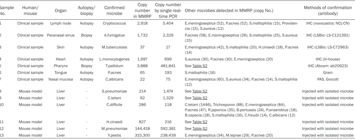
Results
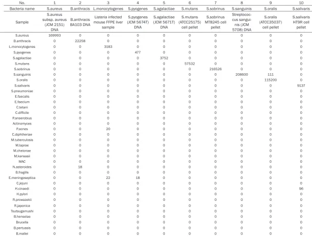
Establishment and validation of
multi-microbi-al remulti-microbi-al-time PCR
To validate the sensitivity and specificity of
each probe and primer set used in the
multi-microbial real-time PCR system, we examined
DNA samples extracted from control bacteria
and fungi (
Figure 1
and Table S2). Each probe
and primer set amplified a gene fragment from
the specific target microbe. The specificity of
each probe-primer set was calculated by
divid-ing the copy number of the target microbe in
the positive control by the sum of all control
samples. With only 9 exceptions, the
probe-primer sets demonstrated more than 95%
specificity. Of the probe-primer sets with low
specificity, some had amplified DNA from
close-ly related bacterial families. For example, the
probe-primer set for
Mycobacterium avium
complex (
MAC
) amplified gene fragments from
nocardia asteroids
and some members of the
Mycobacteriaceae family, including
Myco-
bacterium tuberculosis
,
Mycobacterium
lep-rae
,
Mycobacterium kansasii,
and
Myco-
bacterium avium
, but not
Mycobacterium
che-lonae
. A probe-primer set for
Mycobacterium
kansasii
also reacted with
MAC
. The
probe-primer set for
Neisseria gonorrhoeae
also
amplified a gene fragment of
Neisseria
menin-gitidis
. The probe-primer set for
Providencia
had low specificity (21%) because of
cross-reactions with the enterobacteriae family,
including
Escherichia coli
,
Salmonella enterica
,
Proteus mirabilis
, and
Morganella morganii
;
however, these probe-primer sets failed to
detect
Providencia
spp.
Yersinia pestis
,
Yersinia
enterocolitica
, and
Citrobacter freundii
were
not amplified with the probe-primer set for
Providencia
, although they are members of the
Enterobacteriae family. The probe-primer set
for
Clostridium tetani
cross-reacted with
Helicobacter cinaedi
. The set for
Rickettsia
prowazekii
cross-reacted with
Rickettsia
japonica
. The set for
Burkholderia cepacia
cross-reacted with
Burkholderia mallei
,
although the set for
B.mallei
did not react with
B.cepacia
. Similarly, the set for
E.coli
cross-reacted with
Shigella
spp, although a set for
Shigella
spp. did not react with
E.coli
. All other
probe-primer sets demonstrated high
specifici-ty (>95%), and no, or very low cross-reaction
with off-target bacteria/fungi controls. Some
FFPE samples from a mouse model of bacterial
infection were positive for bacteria other than
the target bacteria (Table S2). The
multi-micro-bial real-time PCR system also detected human
internal control genes, such as
glyceraldehyde-3-phosphate dehydrogenase and beta-actin
(Tables S1 and S2), which were tested to
deter-mine if there was an inhibitory effect in FFPE
samples on the PCR.
Detection of bacteria and fungi in FFPE
sam-ples
To evaluate if the multi-microbial real-time PCR
method is able to detect microbes in
pathologi-cal samples, FFPE samples from patients with
infectious diseases were tested (
Table 2
;
Figure 2
). The multi-microbial real-time PCR
detected at least 7 bacteria/fungi from the
FFPE samples, including
Cryptococcussp
,
monocy-15482
Int J Clin Exp Pathol 2015;8(11):15479-15488
Table 1.
List of Target Bacteria and Fungi
Bacteria
Firmicutes
Bacillales: Staphylococcus aureus, Bacillus anthracis, Listeria monocytogenes.
Lactobacilales: Streptococcus pyogenes, Streptococcus agalactiae, Streptococcus mutans, Streptococcus sobrinus, Streptococcus sanguinis, Streptococcus oralis, Streptococcus salivaris, Streptococcus pneumoniae, Enterococcus faecalis, Enterococcus faecium.
Clostridiales: Clostridium tetani, Clostridium difficile, Peptostreptococcus anaerobius. Actinobacteria
Actinomyces, Propionibacterium acnes.
Corynebacteriales: Corynebacterium diphteriae, Mycobacterium tuberculosis, Mycobacterium laprae, Mycobacterium chelonae, Mycobacte-rium kansasii, MycobacteMycobacte-rium avium complex, Nocardia asteroides.
Bacteroides
Bacteroides fragilis, Elizabethkingia meningoseptica. Proteobacteria
Francisella tularensis, Stenotrophomonas maltophilia, Legionella pneumophila, Aeromonas hydrophila, Haemophilus influenzae. Campylobacteriales: Campylobacter jejuni, Helicobacter cinaedi, Helicobacter pylori.
Rickettsiales: Rickettsia prowazekii, Rickettsia japonica, Orientia tsutsugamushi. Rhizobiales: Bartonella henselae, Brucella.
Bukholderiales: Bordetella pertussis, Burkhoderia mallei, Burkhoderia cepacia. Neisseriales: Neisseria gonorrhoeae, Neisseria meningitidis.
Pseudomonadales: Moraxella catarrhalis, Pseudomonas aeruginosa, Acinetobacter baumannii. Vibrionales: Vibrio cholerae, Vibrio parahaemolyticus, Vibrio vulnificus.
Enterobacteriales: Escherichia coli, Salmonella enterica, Shigella, Klebsiella pneumonia, Yersinia pestis, Yersinia enterocolitica, Citrobacter-freundii, Proteus mirabilis, Morganella morganii, Providencia.
Tenericutes
Mycoplasma pneumoniae. Fusobacteria
Fusobacterium nucleatum. Spirochaetes
Leptospira interrogans, Treponema pallidum. Chlamydiae
Chlamydia psittaci, Chlamydia trachomatis, Chlamydia pneumoniae. Fungi
Aspergillus fumigatus, Aspergillus nigar, Aspergillus flavus, Cryptococcus, Candida albicans, Histoplasma, Trichosporon, Mucor, Coccidioides.
togenes
, and
Treponema pallidum
. The
pres-ence of these microbes in the FFPE samples
was confirmed by immunohistochemistry or
additional methods (
Figure 2
). The copy
num-ber of each microbe was also determined by a
single, real-time PCR specific to each microbe.
Copy numbers calculated using our
multi-microbial real-time PCR were comparable to
those determined by the individual real-time
PCR (
Table 2
). In addition, six microbes were
detected in FFPE samples from mouse models
(
Table 2
, Table S2). Together, the
multi-microbi-al remulti-microbi-al-time PCR successfully detected DNA
from at least 13 microbial species in FFPE
samples.
Detection of bacteria and fungi in the lungs of
AIDS patient autopsy samples
Using the multi-microbial real-time PCR, we
investigated the presence of bacteria and fungi
in 10 lung samples from autopsies of AIDS
patients. The multi-microbial real-time PCR
detected 17 bacterial DNA and one fungal DNA
in 9 cases (
Table 3
). The multi-microbial
real-time PCR also revealed copy numbers of each
bacterium. The most commonly detected
spe-cies was
Staphylococcus aureus
(n=8), but
copy numbers were low in all cases. All samples
positive for
Pseudomonas aeruginosa
(n=6)
showed high bacterial copy numbers.
Ente-
rococcus faecalis
(n=6),
Elizabethkingia me-
ningoseptica
(n=4) and
Candida albicans
(n=4)
were occasionally detected. In some cases, the
microbes detected in the samples correlated
with the patients’ clinical diagnoses. For
exam-ple, a high copy number of
MAC
was detected in
a clinical case of atypical mycobacterial
dis-ease (Case No. 5). In Case No. 9,
P.aeruginosa
15483
Int J Clin Exp Pathol 2015;8(11):15479-15488
Table 2.
Detection of bacteria and fungi in FFPE samples
Sample
No.
Human/
mouse
Organ
Autopsy/
biopsy
Confirmed
microbe
Copy
number
in MMRP
Copy number
by single
real-time PCR
Other microbes detected in MMRP (copy No.)
Methods of confirmation
(antibody)
1 Clinical sample Lymph node Autopsy Cryptococcus 2,918 5,404 E.meningoseptica (52), P.acnes (52), S.maltophilia (15), Providen-cia (15), S.aureus (12)
IHC (novocastra: NCL-CN)
2 Clinical sample Paranasal sinus Biopsy A.fumigatus 1,732 2,329 P.acnes (58), E.meningoseptica (26), S.maltophilia (25), S.aureus
(15) IHC (LSBio: LS-C121391)
3 Clinical sample Skin Autopsy M.tuberculosis 37 17 E.meningoseptica (42), S.maltophilia (20), H.cinaedi (18), P.acnes
(14) IHC (LSBio: LS-C72963)
4 Clinical sample Heart Autopsy L.monocytogenes 1,997 699 S.aureus (30), P.acnes (30), E.meningoseptica (20) IHC (In-house)
5 Clinical sample Pharynx Biopsy T.pallidum 3,888 481,841 See Table S2 IHC (Abcam: ab20923)
6 Clinical sample Tongue Autopsy P.acnes 65 193 S.maltophilia (16) Gram
7 Clinical sample Nasal mucosa Autopsy C.albicans 22 75 E.meningoseptica (60), S.aureus (34), P.acnes (14), S.maltophilia (12)
PAS, Grocott
8 Mouse model Liver - S.pneumoniae 214 1,474 See Table S2 Injected with isolated microbe
9 Mouse model Liver - C.tetani 92 1,329 See Table S2 Injected with isolated microbe
10 Mouse model Liver - C.difficile 286 118 C.tetani (1446), Trichosporon (88), E.meningoseptica (84),
P.acnes (47), R.japonica (35), B.pertussis (24), P.anaerobius (18), B.cepacia (18), S.maltophilia (16), C.freudii (14), C.albicans (12)
Injected with isolated microbe
11 Mouse model Liver - H.cinaedi 827 216 See Table S2 Injected with isolated microbe
12 Mouse model Liver - M.pneumoniae 144,418 592,381 See Table S2 Injected with isolated microbe
15484
Int J Clin Exp Pathol 2015;8(11):15479-15488
cultivated from patient sputum culture. Case
No. 6 was from a patient with sepsis, the cause
of which had been unclear in his lifetime;
how-ever, our system detected a high copy number
of
S.aureus
from lung samples.
E.coli
,
K.pneumoniae
,
and P.aeruginosa
were
detect-ed in case No. 7, a patient whose lung showdetect-ed
organized pneumonia.
Discussion
In the present study, we developed a new
real-time PCR system, designated the
[image:6.629.103.520.84.529.2]“multi-micro-bial real-time PCR”, with the potential to detect
68 bacterial and 9 fungal species
simultane-ously, even in FFPE samples. This system
detected human pathogenic bacteria and fungi
in the lungs of diseased AIDS patients who had
not received any anti-retroviral therapy. The
sensitivity and specificity of the multi-microbial
real-time PCR system are equivalent to those of
standard real-time PCR systems. Moreover,
once the system is established, it follows
sim-ple protocols and is comsim-pleted rapidly,
requir-ing only 2 hours obtainrequir-ing results. The system
15485
Int J Clin Exp Pathol 2015;8(11):15479-15488
is flexible, in that new probe-primer sets can be
incorporated; thus, methods for detecting new
species of microbe can be established quickly.
To detect DNA sequences in FFPE samples,
PCR amplicons should be less than 300 bp,
because DNA is usually fragmented by formalin
fixation [8, 9]. Many probe-primer sets were
designed specifically for this study, because
there are few reports describing real-time PCR
for detection of bacterial/fungal genomes in
FFPE samples. The 16S rRNA gene is encoded
by genomes of almost all bacteria, and variable
regions in this gene have been identified. Thus,
while there are some known shortcomings,
using the 16S rRNA sequence is a promising
strategy to identify bacterial pathogens [10].
The 23S rRNA gene has more sequence
varia-tions between bacterial species than the 16S
[11]. More recently, the locus between the 16S
and 23S regions, the so-called internal
tran-scribed region, has been targeted, because it
contains greater variability than either 16S or
23S rRNA, allowing even better discrimination
of bacterial species [12]. In the present study,
we designed probe-primer sets targeting
main-ly 16S rRNA, 23S rRNA, and the internal
tran-scribed regions. High specificities were
achieved in the cross-reaction analysis, with
few exceptions. Generally, to categorize a
bac-terial strain, it is necessary to amplify and
sequence more than 1 kbp of the 16S rRNA
region [10]. The use of amplicons shorter than
210 bp may be the main reason for the
cross-reactions observed in our system. High copy
numbers in target bacteria will assist
identifica-tion of microbes in cases where cross-reacidentifica-tion
is suspected. Although there is some room to
improve the specificity of the probe-primer sets
for reducing the cross-reactivities, the system
as currently designed will function as a system
for preliminary screening.
Systems for simultaneous molecular detection
of bacteria and fungi DNA in blood samples
have been previously reported [13-15]. Among
them, Septifast® is a commercialized
detec-tion system that can detect 25 pathogens in
clinical samples using real-time PCR [16].
Septifast® is useful for screening blood
sam-ples for bacterial and fungal infections [17-20].
However, there is no report describing the
[image:7.629.99.532.77.344.2]15486
Int J Clin Exp Pathol 2015;8(11):15479-15488
Table 3.
Detection of bacteria/fungi in the lung of AIDS autopsies
Case
No.
Clinical diagnosis
Pathological diagnosis
Detected bacteria by multibacteria real-time PCR
(Copy number)
1 Toxoplasma encephalitis, PCP Blonchial pneumonia, Adrenal CMV infection, Epididymis small abscess
P.aeruginosa (4476), S.aureus (171), C.albicans (66), F.nucleatum (56), S.maltophilia (26)
2 PCP, SLE PCP, CMV pneumonia, SLE S.aureus (44), S.maltophilia (41), P.acnes (20), E.faecium (14)
3 No Data No Data P.aeruginosa (20406), E.faecalis (174), E.meningoseptica
(55), C.albicans (42), S.maltophilia (23) 4 CMV infection, Pulmonary edema, DIC Systemic CMV infection Not detected
5 CMV retinaitis, CMV colitis, Atypical mycobacterial disease, Chronic blonchial pneumonia, Kidney disfarction, Liver mass
Atypical mycobacterial disease, ML,
CMV infection MAC (18721), P.aeruginosa (6746), M.kansasii (4468), S.aureus (167), S.maltophilia (97), Providencia (42), E.meningoseptica (33), M.morganii (32), C.albicans (30), P.acnes (22), E.faecalis (10)
6 ML, Cryptococcus meningitis, CMV reti-naitis, Aspiration pneumonitis
ML, Cryptococcus meningitis, CMV infection, sepsis
S.aureus (38515), E.faecalis (70)
7 PML, Aspiration pneumonitis, Hemophilia A, Oral candidiasis, Chronic hepatitis C
PML, Organized pneumonia E.coli (7217224), Providencia (95254), K.pneumoniae (87912), P.aeruginosa (5784), E.faecalis (352), S.aureus (36), E.meningoseptica (31)
8 PCP, CMV pneumonia Severe pneumonia (PCP and CMV
pneumonia)
M.tuberculosis (125), E.meningosepticum (36), S.aureus (21)
9 Kidney disfunction induced drug, ML, Lung abscess (P.aeruginosa), KS, CMV infection, Amoeba liver abscess, adrenal disfanction
ML, CMV infection, Lung abscess, KS
P.aeruginosa (5891), E.faecalis (3423), S.aureus (45), B.fragills (26)
10 No Data No Data P.aeruginosa (727389), Providencia (65430), E.faecalis
(4093), S.oralis (749), E.faecium (734), C.albicans (202), P.acnes (70), S.maltophilia (52), S.aureus (20) CMV: cytomegalovirus, KS: Kaposi’s sarcoma, ML: malignant lymphoma, PCP: Pneumocystis pneumonia, PML: progressive multifocal leukoencephalopathy, SLE: systemic lu-pus erythematosus; B.fragills: Bacteroides fragills, C.albicans: Candida albicans, E.coli: Escherichia coli, E.faecalis: Enterococcus faecalis E.faecium: Enterococcus faecium, E.meningoseptica: Elizabethkingia meningoseptica, F.nucleatum: Fusobacterium nucleatum, K.pneumoniae: Klebsiella pneumoniae, M.kansasii: Mycobacterium kansasii, M.morganii: Morganella morganii, M.tuberculosis: Mycobacterium tuberculosis, MAC: Mycobacterium avium complex, P.acnes: Propionibacterium acnes, P.aeruginosa: Pseudomonas aeruginosa, S.aureus: Staphylococcus aureus, S.maltophilia: Stenotrophomonas maltophilia, S.oralis: Streptococcus oralis.
ity of Septifast® to detect bacterial DNA in
FFPE samples. Our multi-microbial real-time
PCR can detect a greater number of bacterial/
fungal species than Septifast®, and can detect
DNA from pathogens even in FFPE samples in
which DNA had degraded into fragments of
less than 300 bp. This provides a large
advan-tage in screening for pathogens in pathological
samples. Generally, FFPE samples at autopsy
were fixed in formalin for a longer time than
FFPE biopsy samples, suggesting a greater
degradation of DNA. Our system identified
microbial DNA not only in biopsy FFPE samples,
but also in autopsy FFPE samples. Our system
provides a useful tool for safe detection of
pathogens in FFPE samples, even after long
periods of storage. In addition to FFPE samples,
blood and other fluid samples, such as cerebral
spinal fluid, pharyngeal swab, throat washing,
and pleural and peritoneal effusions may be
also suitable for our system. Various types of
samples will be tested by our system to confirm
its suitability in the future.
Using the multi-microbial real-time PCR, we
detected 17 bacterial and one fungal species
from 10 lung samples from autopsies of AIDS
patients. Basically, a pathogen detected with a
high copy number in Septifast® system is
cor-related with the severity of sepsis [21]. On the
other hand, when small quantities of bacteria
are detected, it is difficult to determine whe-
ther they are associated with disease as a
pathogen or a colonization event. A series of
studies using bacterial DNA sequencing
approaches have shown that healthy lungs
har-bor a unique steady-state microbiota, which is
very different from other organs, such as the
skin and gut [22]. Slight alterations to the
microbiota in the lung could underlie
suscepti-bility to lung disease. Additionally, the detection
of bacteria may be complicated by previous
treatments with antibiotics. Thus, when
bacte-ria are detected in low copy numbers, we need
to consider the whole picture, including the
copy numbers of other bacteria, the patient’s
history of antibiotics usage, and the condition
of host’s immunity.
15487
Int J Clin Exp Pathol 2015;8(11):15479-15488
FFPE samples and preserved frozen samples
from cadavers. Further research is required to
confirm the utility of the system to probe
addi-tional patient samples, such as blood and other
fluids. This system may provide a powerful tool
for screening clinical samples for the presence
of bacteria and fungi, particularly pathological
samples, including FFPE samples from patients
with uncertain diagnoses. In addition, this
sys-tem is inherently flexible, allowing for addition
of additional species of microbe on an
as-need-ed basis. Use of this method in combination
with a previously developed multivirus real-time
PCR system will be considered for generation of
a real-time PCR system detecting almost all
human pathogens, which will compensate for
some of the disadvantages of next generation
sequencing-based assays.
Acknowledgements
The authors thank Drs. Michio Suzuki, Koichi
Imaoka, Shuji Ando, Hideaki Ohno, Masanori
Kai, Tetsu Mukai, Masatomo Morita, Jiro
Mitobe, Hidenobu Senpuku, Hideyuki Taka-
hashi, Noriyo Nagata, Kazunari Kamachi, Keigo
Shibayama, Atsuko Horino, Makoto Ohnishi,
and Haruo Watanabe of the National Institute
of Infectious Diseases for providing DNA
extracted from bacteria/fungi for use as
con-trols. This work was partly supported by Health
and Labor Sciences Research Grants (no.
H24-AIDS-I-003, Shinko-Ippan 015 and
H25-Shinko-Shitei 006 to HK) from the Ministry of
Health, Labor and Welfare, Research Program
on HIV/AIDS Grants, Japan Agency for Medical
Research and Development, and a Grant-in-Aid
for Scientific Research from the Ministry of
Education, Culture, Sports, Science and
Technology of Japan (No. 24659212 to HK).
Disclosure of conflict of interest
None.
Address correspondence to:
Harutaka Katano,
Department of Pathology, National Institute of
Infectious Diseases, 1-23-1 Toyama, Shinjuku-ku,
Tokyo 162-8640, Japan. Tel: 81-3-5285-1111; Fax:
81-3-5285-1189; E-mail: [email protected]
References
[1] Dark P, Blackwood B, Gates S, McAuley D,
Perkins GD, McMullan R, Wilson C, Graham D,
Timms K and Warhurst G. Accuracy of
LightCycler(®) SeptiFast for the detection and
identification of pathogens in the blood of
pa-tients with suspected sepsis: a systematic
re-view and meta-analysis. Intensive Care Med
2015; 41: 21-33.
[2] Onori M, Coltella L, Mancinelli L, Argentieri M,
Menichella D, Villani A, Grandin A, Valentini D,
Raponi M and Russo C. Evaluation of a
multi-plex PCR assay for simultaneous detection of
bacterial and viral enteropathogens in stool
samples of paediatric patients. Diagn Microbiol
Infect Dis 2014; 79: 149-154.
[3] Van Lint P, De Witte E, De Henau H, De Muynck
A, Verstraeten L, Van Herendael B and Weekx
S. Evaluation of a real-time multiplex PCR for
the simultaneous detection of Campylobacter
jejuni, Salmonella spp., Shigella spp./EIEC,
and Yersinia enterocolitica in fecal samples.
Eur J Clin Microbiol Infect Dis 2015; 34:
535-542.
[4] Lee DY, Shannon K and Beaudette LA.
Detection of bacterial pathogens in municipal
wastewater using an oligonucleotide
microar-ray and real-time quantitative PCR. J Microbiol
Methods 2006; 65: 453-467.
[5] Bertelli C and Greub G. Rapid bacterial
ge-nome sequencing: methods and applications
in clinical microbiology. Clin Microbiol Infect
2013; 19: 803-813.
[6] Katano H, Kano M, Nakamura T, Kanno T,
Asanuma H and Sata T. A novel real-time PCR
system for simultaneous detection of human
viruses in clinical samples from patients with
uncertain diagnoses. J Med Virol 2011; 83:
322-330.
[7] Eisen MB, Spellman PT, Brown PO and Botstein
D. Cluster analysis and display of genome-wide
expression patterns. Proc Natl Acad Sci U S A
1998; 95: 14863-14868.
[8] Dietrich D, Uhl B, Sailer V, Holmes EE, Jung M,
Meller S and Kristiansen G. Improved PCR
per-formance using template DNA from
formalin-fixed and paraffin-embedded tissues by
over-coming PCR inhibition. PLoS One 2013; 8:
e77771.
[9] van Dongen JJ, Langerak AW, Bruggemann M,
Evans PA, Hummel M, Lavender FL, Delabesse
E, Davi F, Schuuring E, Garcia-Sanz R, van
Krieken JH, Droese J, Gonzalez D, Bastard C,
White HE, Spaargaren M, Gonzalez M, Parreira
A, Smith JL, Morgan GJ, Kneba M and
Macintyre EA. Design and standardization of
PCR primers and protocols for detection of
clonal immunoglobulin and T-cell receptor
gene recombinations in suspect
lymphoprolif-erations: report of the BIOMED-2 Concerted
Action BMH4-CT98-3936. Leukemia 2003; 17:
2257-2317.
di-15488
Int J Clin Exp Pathol 2015;8(11):15479-15488
agnostic laboratory: pluses, perils, and pitfalls.
J Clin Microbiol 2007; 45: 2761-2764.
[11] Rantakokko-Jalava K, Nikkari S, Jalava J,
Eerola E, Skurnik M, Meurman O, Ruuskanen
O, Alanen A, Kotilainen E, Toivanen P and
Kotilainen P. Direct amplification of rRNA
genes in diagnosis of bacterial infections. J
Clin Microbiol 2000; 38: 32-39.
[12] Lehmann LE, Hunfeld KP, Emrich T,
Haberhausen G, Wissing H, Hoeft A and Stüber
F. A multiplex real-time PCR assay for rapid
de-tection and differentiation of 25 bacterial and
fungal pathogens from whole blood samples.
Med Microbiol Immunol 2008; 197: 313-324.
[13] Altun O, Almuhayawi M, Ullberg M and Ozenci
V. Clinical evaluation of the FilmArray blood
culture identification panel in identification of
bacteria and yeasts from positive blood culture
bottles. J Clin Microbiol 2013; 51: 4130-4136.
[14] Wellinghausen N, Kochem AJ, Disque C, Muhl
H, Gebert S, Winter J, Matten J and Sakka SG.
Diagnosis of bacteremia in whole-blood
sam-ples by use of a commercial universal 16S
rRNA gene-based PCR and sequence analysis.
J Clin Microbiol 2009; 47: 2759-2765.
[15] Liesenfeld O, Lehman L, Hunfeld KP and Kost
G. Molecular diagnosis of sepsis: New aspects
and recent developments. Eur J Microbiol
Immunol (Bp) 2014; 4: 1-25.
[16] Chang SS, Hsieh WH, Liu TS, Lee SH, Wang CH,
Chou HC, Yeo YH, Tseng CP and Lee CC.
Multiplex PCR system for rapid detection of
pathogens in patients with presumed sepsis -
a systemic review and meta-analysis. PLoS
One 2013; 8: e62323.
[17] Gosiewski T, Flis A, Sroka A, Kedzierska A,
Pietrzyk A, Kedzierska J, Drwila R and Bulanda
M. Comparison of nested, multiplex, qPCR;
FISH; SeptiFast and blood culture methods in
detection and identification of bacteria and
fungi in blood of patients with sepsis. BMC
Microbiol 2014; 14: 313.
[18] Burdino E, Ruggiero T, Allice T, Milia MG,
Gregori G, Milano R, Cerutti F, De Rosa FG,
Manno E, Caramello P, Di Perri G and Ghisetti
V. Combination of conventional blood cultures
and the SeptiFast molecular test in patients
with suspected sepsis for the identification of
bloodstream pathogens. Diagn Microbiol Infect
Dis 2014; 79: 287-292.
[19] Herne V, Nelovkov A, Kutt M and Ivanova M.
Diagnostic performance and therapeutic
im-pact of LightCycler SeptiFast assay in patients
with suspected sepsis. Eur J Microbiol Immunol
(Bp) 2013; 3: 68-76.
[20] Dierkes C, Ehrenstein B, Siebig S, Linde HJ,
Reischl U and Salzberger B. Clinical impact of
a commercially available multiplex PCR system
for rapid detection of pathogens in patients
with presumed sepsis. BMC Infect Dis 2009;
9: 126.
[21] Ziegler I, Josefson P, Olcen P, Molling P and
Stralin K. Quantitative data from the SeptiFast
real-time PCR is associated with disease
se-verity in patients with sepsis. BMC Infect Dis
2014; 14: 155.
1
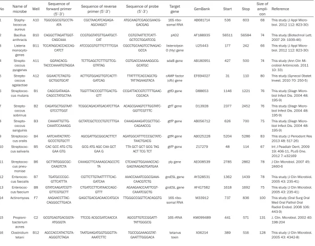
Table S1.
List of probe and primer sequences
No
Name of
microbe
Well
forward primer
Sequence of
(5’-3’)
Sequence of reverse
primer (5’-3’)
Sequence of probe
(5’-3’)
Target
gene
GenBank
Start
Stop
Size of
ampli-con
Reference
1 Staphy-lococcus aureus A10 TGGCGGCGTGCCTA-ATA CGCTAACATCAGAGA-AGCAAGCT ATGCAAGTCGAGCGAACG-GACGAG 16S ribo-somal RNAAB681714 536 603 68 This study (J Appl
Micro-biol. 2012 112: 823-30)
2 Bacillus
anthracis B10 CAGGCTTAGATTGGT-CAGCAAA CCGTGGTATGTTGAATGCT-CAT CGTGTAATTCTCATT-GCTCCTGGATCCG pXO2 AF188935 56511 56584 74 This study (Biotechnol Lett. 2007 29: 1939-46) 3 Listeria monocyto-genes B11 TCCATAGCACCACCAG-CATCT ATCCGCGTGTTTCTTTTCGA CGCCTGCAAGTCCTAAGAC-GCCA listeriolysin O (hly) gene
U25443 177 242 66 This study (J Appl
Micro-biol. 2012 112: 823-30)
4
Strepto-coccus pyogenes
A11
GGRACACG-TACCCAAAATGTAGGA TCTTGAGCTCTTTGTTCG-GTRTAG CGTGACCAAAAAGGCG-GCATGC sdaB gene AB180951 427 500 74 This study (Ann Clin Mi-crobiol Antimicrob. 2011 10: 33)
5
Strepto-coccus agalactiae
A12
GGAACTCTAGTG-GCTGGTGCAT ACTTGTGGAGTTGTCACTT-GATCAG TTATTTTCACCAGCTG-TATTAGAAGTACA cAMP factor (cfb) gene EF694027 31 110 80 This study (Gynecol Obstet Invest. 2010 70: 250-5)
6 Streptococ-cus mutans B1 CAGCGATAAGA-CAGCCTATGCTAA TGGTTTACCCGTTTGACTG-GTT CCGATTACCGTCTTTTGAAC-CGCACA
gtfD gene D88653 1146 1221 76 This study (Diagn
Micro-biol Infect Dis. 2004 48: 195-9) 7 Strepto-coccus sobrinus B2 CAGATGCTGGTAAT-GTCCTTGGT TCGGCAGACATGACATCTTGA ACAGCGAAGTCTTGGTATC-GGTTCGTTTC
gtfT gene D13928 2377 2452 76 This study (Diagn
Micro-biol Infect Dis. 2004 48: 195-9)
8
Strepto-coccus sanguinis
B3
CAAAATTGTTG-CAAATCCAAAGG GCTATCGCTCCCTGTCTTTGA CAAAGAAAGATCGCTTGC-CAGAACCG gtfP gene AB056712 626 700 75 This study (Diagn Micro-biol Infect Dis. 2004 48: 195-9)
9
Streptococ-cus oralis B4 AATCAATACTATC-GCCCTGTGCTT AGCGATTGCGGCACTTCT AGATGGCATTTCCCGCTATC-TAACTGACG gtfR gene AB025228 5204 5286 83 This study (J Periodont Res 2013 48: 517-26) 10
Streptococ-cus salivaris B5 CAC GCC ATG CTG GAA GTG GCG ATG AGC CAA GCT GAA G TTA GCT GCT GCG TAG ACT TCG TCT gtfP gene Z17279 48 114 67 Int J Peadiatr Dent. 2009 19: 406-11, PLoS One. 2012 7: e32169 11
Streptococ-cus pneu-moniae
B6
GCTTATGGGCGC-CAAGTCTA CAAAGCTTCAAAAGCAGCCTC TA CTCAAGTTGGAAACCAC-GAGTAAGAGTGATGAA ply gene AE008539 2785 2862 78 J Clin Microbiol. 2007 45: 2460-6
12 Enterococ-cus faecalis B7 TGATGCCCGC-GTTCATTTA CGTTCTTGTAATTTTTCAC-GATCAA AAACCAAATCGGCGAAA-CAACGTCTG
groESL gene AY328531 1362 1439 78 This study (J Clin Microbiol. 2005 43: 235-41) 13
Enterococ-cus faecium B8 GTATCAAGATCGTT-GTTCGTGCTT CTGATCCTTCATAACCAGC-GTTT AGAAGAACCAATTCGT-CAAATCGCTG groESL gene AF417582 1618 1692 75 This study (J Clin Microbiol. 2005 43: 235-41) 14 Actinomyces F7
AAGAACCTTAC-CAGGGCTTGACA GAGCTGACGACAACCATGCA TTGGGCCGGTTCACAGGTG somal RNA16S ribo- M33912 737 836 100 This study (Oral Surg Oral Med Oral Pathol Oral Radiol Endod. 2008 106: 443-9) 15 Propioni-bacterium acnes C2 GCGTGAGTGACGGTA-ATGGGTA
TTCCG ACGCGATCAACCA AGCGTTGTCCGGATT-TATTGGGCG
16S rRNA KM099489 441 571 131 J. Clin. Microbiol. 2002 40:
198-204
16 Clostridium
2
17 Clostridium
difficile C1 AAGGGTATTGCTC-TACTGGCATTT CCTCATGGTCTTCAGAA-CAAGCT TGGGCGTGTTTTTTG-GCAATATATCCTCA tcdC gene HQ596359 369 450 82 This study (J Clin Microbiol. 2002 40: 3470-5) 18
Peptostrep-tococcus anaerobius
E8
ACACCATGGGAGTCG-GAAAC ACGCCACCTTCGACGACTT CCGAAGCCGATTATCCAAC-CGCA somal RNA 16S ribo-gene
L04168 1381 1450 70 This study (Microbiology.
2003 149: 1719-27)
19 Mycoplasma
pneumoniae F11 GCCACACCAATGC-CATCA GAGGGAGGAAAAGCTC-GTGTT CCGCGCTTAACCCCGT-GAACGT P1gene M18639 328 395 68 This study (J Clin Microbiol. 2009 47: 2269-71)
20
Coryne-bacterium diphtheriae
B9 TTCTCCTGGCGTG-TACTTTGATC
CACCGCGGGAAGGAATG CCGAGCGTCCACTG-CACTCCG
rpo B gene BX248355 61792 61877 86 This study (Diagn Micro-biol Infect Dis. 2012 73: 111-20)
21 Mycobacte-rium tuber-culosis
F2 TCGACGCGATCGAG-CAA
GGATAACGTCTTTCAGGTC-GAGTAC
ATCTGGACCCGCCAA-CAAGAAGG
IS986 X52471 939 1007 69 This study (J Clin Microbiol.
2012 50: 646-50)
22
Mycobacte-rium laprae F3 CGTTCTGGTTCGCATG-GTT CCCTGGCGATAGCCGTAGT ATGGCGACGGCCTACCTG-GT pra gene X65546 836 899 64 This study (Med Microbiol Immunol. 2004 193: 189-93)
23
Mycobac-terium chelonae
F4
GGTTACTCGCTTGGT-GAATATGTTT CGACGTTTTGCCGACTACCT AAATCCTGTCCACCCC-GTGGA ITS1 gene AM709729 121 190 70 This study (J Clin Microbiol. 2009 47: 1497-502)
24
Mycobac-terium kansasii
F5 GGCGAACGGGTGAG-TAACA
ACCCAGTTTCCCAGGCTTATC TGGGCAATCTGCCCTGCAC 16 SrRNA gene
X15916 59 124 66 This study (J Mol Diagn.
2009 11: 42-8)
25 Mycobacte-rium avium
complex
F6 GCATCGAGAGCCGAT-GAAG
GGATCAATGCTCGGTTGACA ACGTGGGAGGCTGC-GATATGC
24S rRNA gene
X74494 337 407 71 This study (J Clin Microbiol. 2000 38: 4080-5)
26 Nocardia
asteroides F8 GCCTTCGGGTTGTA-AACCTCTT GCCGGTGCTTCTTCTACAG-GTA ACAGGGACGAAGCG-CAAGTGACG 16S rRNA gene AF430019 382 452 71 This study (J Clin Microbiol. 2010 48: 503-11)
27
Fusobac-terium nucleatum
E10 CAAGCGGTGGAG-CATGTG
CTAAGATGTCAAACGCTG-GTAAGG
TTAATTCGACGCAACGC-GAGGA
16S rRNA gene
JQ001893 572 638 67 This study (J Clin
Periodon-tol. 2006 33: 427-33)
28 Leptospira
interrogans F10 ACGATCTCTCAATCTA-ACGGAGAATAC GGTTCCGGTTCCTGTGAAAG TTTTACCTGGACGGTC-GCTGGTCAA lfB1 gene HQ328633 165 239 75 This study (J Clin Microbiol. 2011 49: 2154-60) 29 Treponema
pallidum
F9 GAAGCAGTCGAGGGT-GCAGTA
CGCTGTCAACCATGAAGGAA CGTTGGCGGATCGCGCG 47kDa anti-gen anti-gene
M88769 466 526 61 This study (Sex Transm
Infect. 2011 87: 479-85)
29 Treponema
pallidum H3 TAGAAGGGAGGGC- CAGG-TAGTACA
CAGACACAGCACTC-GTCTTCAAC CGGAGGTGAACTCCGTATT-GAAGTCGG pol A gene U57757 1574 1649 76 This study (J Clin Microbiol. 2010 48: 497-502)
30 Chlamydia
psittaci G3 GGGAAGGTGCTTCAG-GAGATC TCCTGCGCGGATGCTAAT TCCTTGCGCTACTTGGTGT-GACGC ompA gene AB468956 188 258 71 This study (Vet J. 2009 181: 145-50) 31 Chlamydia
trachomatis G4 AACGACATTTCTTGCT-GCAAAG TCAGGACATTTTGCGGATAGG TTACCCATGAAATCCCTCGT-GATATA somal RNA 23S ribo-gene
M19487 1441 1521 81 This study (J Med
Micro-biol. 2006 55: 1667-74)
32 Chlamydia
3
33 Bacteroides
fragills E9 CACTTGACTGTTGTAG-ATAAAGC CATCTTCATTGCAGCATTATCC TGTGCTTGCTTCCAGTC-GTCTATG leuB gene CR626927 3844464 3844598 135 J. Clin. Microbiol. 2013 51: 1593 34
Elizabeth-kingia
meningo-septica
E2
CCAGCAGCCGCG-GTAAT CGGACCCTTTAAACCCAATA-AAT CGGAGGGTGCAAGCGT-TATCC 16 SrRNA gene HM748601 432 495 64 This study
35 Campylo-bacter jejuni D3 TGCTTCTTTACTTGTT-GTGGCTTT TGCTTACAACTGCT-GAATTTTGG CAAAGCATAGTATCTCG-CAATGTTGAT
hipOgene Z36940 1964 2040 77 This study (BMC Microbiol.
2011 11: 113)
36 Helicobacter
cinaedi E12 GAATACGTTCCC-GGGTCTTGT TCCCGACTTAAGGCGAATACA CTCACCGCCCGTCACACCAT 16S rRNA gene AF426158 1309 1377 69 This study (BMC Infect Dis. 2006 6: 86)
37 Helicobacter
pylori F1 TTCCATAGGCTATAAT-GTGATCCAAA GCGCATGTCTTCGGTTAAAAA AGGGCCTATGCCTACCCCT-GCGA phosphoglu-cosamine mutase (glmM) gene
JN390578 78 153 76 This study (J. Clin.
Micro-biol. 2012 50: 3233-7)
38 Rickettsia
prowazekii F12 ATGAGCAGAATGCTTC-TACTTCAACA CCAGTGCTAATACATG-CAAAAGGAT CGGATTGCTGGCTCATCAG-GAGCT gltA gene M17149 785 862 78 This study (Am J Trop Med Hyg. 2005 73: 1083-5)
39 Rickettsia japonica G1 CCGAATTGCCG-GCTCAT GTGAGGCAATACCCGTGCTAA CGGAGCTAACCCTTTT-GCTTGT
gltA gene AY743327732 732 793 62 This study (Jpn J Infect Dis. 2010 63: 353-4) 40 Orientia tsutsuga-mushi G2 AACTGATTT- TATTCAAACTAATGCT-GCT TATGCCTGAGTAAGATACRT-GAATRGAATT TGGGTAGCTTTGGTGGAC-CGATGTTTAATCT 47kDa outer membrane protein gene
L31934 630 747 118 Am J Trop Med Hyg. 2004
70: 351-6
41 Bartonella
henselae C12 GATCCCAAGCCTTCTG- AGATGAT-GCG
GGATRAAYYRGWAAACCT-TYMYCGG CCACCGTGGGCTTT-GAAAAACGCT ITS region L35101 321 421 101 Emerg Infect Dis. 2005 11: 1894-8
42 Brucella D10
CGCCAAGATGTG-CAACGA GGTCGCACCGGCTTTCTA AAATCGCGGCAACCG-GCCTC chromo-some2 NC_010740 308676 308734 59 This study (J Clin Microbiol. 2010 48: 697-702) 43 Bordetella
pertussis C7 GGCATCGACCCCAC-CAA GGTTGTATTCGTCCAGGTT-GAGT TCGGGCGCGCTG-TACCCATCT IS481 gene AB473880 966 1034 69 This study (BMC Res Notes. 2011 4: 11) 44 Burkhold-eria mallei D12 CGAGCGCATCG-TACTCGTA CAAGTCGTGGATGCGCATTA 5’CTgAAACgCgCAgCg 3’MGB YD repeat protein
CP000525.1 267228 267300 73 PLoS One. 2010 5: e15413 45 Burkhold-eria cepacia E1 TCAAGAAGAACGAC-GAGGTGATC CGGCGGCGACACCTT AAACCCGCGTGAAGGTC-GTCAAGA
rec A gene D90120 914 984 71 This study (J Clin Microbiol.
2001 39: 4247-55) 46 Neisseria
gonor-rhoeae
C3
AACGCCACGACG-GTATGC CCGCTGAAACCGGAAAATC TTTCCGTGCGTTAC-GATTCCCCC porA pseu-dogene AJ223446 662 725 64 This study (Sex Transm Infect. 2010 86: 207-11)
47 Neisseria meningitidis C4 TGTTTACCCCTGATT-TACAAGGATT GGCTCACAGCTTGGGTTTTC AGCGAAGGCTTACATG-GTTTCCACATCC
sodC gene AJ001313 1879 1954 76 This study (PLoS One.
2011 6: e19361) 48 Francisella
tularensis D8 CCATATCACTGGCTTT-GCTAGACTAGT TGTTGGCAAAAGCTA-AAGAGTCTAAA CAAACCCAGACCTTCAAAC- AAATTATAAAAC-CACA
hypothetical
protein CP003048 453602 453708 107 This study
49 Stenotro-phomonas maltophilia E11 GCC-GAAAGCCCAAGGTTT CGACTTTCGTCCTCGCCTTA CGCAACGTTAATCGGCG-CAGG 23S rRNA gene
AF273255 1278 1354 77 This study (J Appl Micro-biol. 2011 111: 1185-93)
50 Legionella pneumoph-ila D11 TGCCAAGTGGTTTG-CAATACAA CTCGACAGTGACTGTATC-CGATTT ATCAATGCTGGAAATGGTGT-TAAACCCGG
mip gene AM051115 408 487 80 This study (Eur J Clin
4
51 Moraxella
catarrhalis C5 GGTGAGTGCCGCTTT-TACAAC TGTATCGCCTGCCAAGACAA TGCTTTTGCAGCTGTTAGC-CAGCCTAAG copB outer membrane protein gene
U69982 49 121 73 This study (J Clin Microbiol.
2003 41: 1386-90)
52 Pseudo-monas aeruginosa D9 AGCCTTCCTG-GTCCCCTTAC CCTAATGAACCCCAGTGTATA-AGTTTG TGAACTGACGGTCGC-CAACGGTT
oprL gene Z50191 131 202 72 This study (J Clin Microbiol.
1997 35: 1295-9)
53 Acineto-bacter baumannii E7 GCTAATGCTGGCGTAA-CAGTTACTC TTTACCGCCATTGTTGTGTTG TGCTTGGTTACACTTTC-CAAGACAG
omp gene AY485227 253 330 78 This study (J Clin Microbiol.
2012 50: 1412-4)
54 Aeromonas
hydrophia D7 TGGAACCACACCTTC-GTCATC TGTAACGCTTGTCCCACTG-GTA CCGTACAAGGACAAGGC-GAGCAGC hemolysin gene CO000462 467593 467668 76 This study (Forensic Sci-ence International. 2012 222: 11-26)
55 Vibrio
chol-erae D4 CATAGAGCTTGGATCCGGAG- TCGATGATCTTGGAGCATTCC TCATCATGCACCGCC-GGGTTG ctxA gene HM042644 558 637 80 This study (J Clin Microbiol 1992 30: 2118-21)
56 Vibrio
parahaemo-lyticus
D5
ACTCAACACAAGAAGA-GATCGACAA GATGAGCGGTTGATGTCCAA CGCTCGCGTTCACGAAAC-CGT thermolabile hemolysin CP006005.1 682659 682866 208 Appl. Environ. Microbiol. 2007 73: 5840-7
57 Vibrio
vulni-ficus D6 TGCGGGTGGTTCG-GTTAA TTCTTCTTTATCTAG-GCCCCAAACT AGCTGTCACGGCAGTTG-GAACC vvhA gene M34670 1833 1905 73 This study (J Clin Microbiol. 1998 36: 2887-92)
58
Hae-mophilus influenzae
C6
AGCGGCTTG-TAGTTCCTCTAACA CAACAGAGTATCCGC-CAAAAGTT CGATGCTGCAGGCAATG-GTGCT omp P6 M19391 118 191 74 This study (Diagn Micro-biol Infect Dis. 2009 64: 366-73)
59 Escherichia
coli C8 CGGCGTGGTGTAGAG-CATT GCAGTCTTACTTCCAT-GATTTCTTTAAC CGCTGCGATGGATCCCGG beta-D-gluc-uronidase (uid A gene)
U00096.3 1695532 1695601 70 This study (water research 2009 43: 3019-28)
60 Salmonella enterica C9 AATGGCGGCGAAT-TACGA CGGATCCCTTTGCGAATAAC CAGTAATGGTATCTGCT-GAAGTTGAGGA
InvA gene U43272 1732 1799 68 This study (J Appl
Micro-biol. 2012 112: 823-30) 61 Shigella C10
AACTGCCTGTGTTCG-GTCTTCT CATCTGGGATAAGATTATCACCG- TGCAGCACCTTCCTTG-GCCCA ipaH gene AE005674 749519 749587 69 This study (J Appl Micro-biol. 2012 112: 823-30) 62 Klebsiella
pneumoniae C11 CGGGCGTAGCGC-GTAA GATACCCGCATTCACAT-TAAACAG CCCGGCATGGATCGTTC-CGA mdh gene AM051115 140 201 62 This study (Plos One 2008 3: e3701) 63 Yersinia pestis D1 TTTCCATCCTGAGAAG-TAAATGTTAAGTA GGAACCACTAGCACATCTGT-TAACTTT TGGGATCACCCGCG-GCATCT
caf1 gene CP001595 67282 67361 80 This study (J Forensic Sci. 2006 51: 548-8) 64 Yersinia
en-terocolitica
D2 TCACGGAAAGGTTA-AGTCATCTGTAT
TGCCCCGTATGCCATTG TGATGGGTCAGTCAGTA-CAAGTAAGACG
ail gene AJ605740 9405 9477 73 This study (Appl Environ
Microbiol. 2008 74: 6060-7) 65 Citrobacter freundii E3 TGGCACGAGCGCTT-TAATC TGAAGGTGGCGGAATAACG AGCATGGCCGGAGCTGT-CATC
cfa gene U09771 92 152 61 This study (Curr Microbiol.
2005 51: 229-32) 66 Proteus mirabilis E4 TGTGGCGGGTACTA-ATGCAA CGCCTCTAACATGCGGTACA CACAGTTACCCCCGG-TATTTGGAA
ureC gene M31834 2406 2472 67 This study (Curr Microbiol.
2012 65: 44-53) 67 Morganella
morganii E5 GTGAAATCCCC-GGGCTTAA ACCCCCCTCTACAAGACTC-TAGCT CCGGGAATTGCATCT-GATACTGG 16S rRNA gene DQ358146 524 592 69 This study (Lett Appl Micro-biol. 2012 54: 292-8) 68 Providencia E6
GGCGGCAGGCCTAA-CAC CGTCAGCGAGAAGCAAGCT TGCAAGTCGAGCGGTAA-CAGGG 16s RNA gene AY994312 31 91 61 This study (Jpn J Infect Dis. 2012 65: 545-7) 69 Aspergillus fumigatus G6 GCCGAAGACCCCAA-CATG CGGAACCAAGAGATCCGTTGT ACGCTGTTCTGAAAGTATG-CAGTCTG
ITS1 gene JQ080317 95 191 97 This study (J Clin Microbiol. 2009 47: 379-84) 70 Aspergillus nigar G7 ACCCCAACACGAA-CACTGTCT CAAGAGATCCATTGTT-GAAAGTTTTAAC AAAGCGTGCAGTCTGAGTT-GATTGAATGC
ITS 1/2 GU256739 102 185 84 This study (J Clin Microbiol.
5
71 Aspergillus
flavus G8 TACCTTAGTTGCTTCCGTGTTTACTG- TACAATCAACTCAGACTTCAC-TAGATCAGA CGCCCGCCGGAGACACCA ITS1 gene HQ026744 37 168 132 This study (J Clin Microbiol. 2011 49: 4150-7) 72
Cryptococ-cus
G9 GCCTTAAATGTGT-TAGTGGGAAGGT
CCATAGGCCCAGCGAAACT TTACCTGTCAGCCCGGC-GTAAT
ITS2 gene AJ876523 391 459 69 This study (Diagn Microbiol
Infect Dis. 2009 65: 69-72)
73 Candida
albicans G10 GGTTTGGTGTTGAG-CAATACGA AAGCGATCCCGCCTTACC TGGGTTTGCTTGAAAGACG-GTAG somal RNA 5.8S ribo- EF065152 96 161 66 This study (Eur J Clin Microbiol Infect Dis. 2012 31: 2237-45)
74 Histoplasma G11 CAGCCAGCCAGGAG-CAAT
CCCCCAATTATCGTTCACTGA TGGTGCGAGAGGATG-CAAGGTT
GAPDH gene AF273703 652 717 66 This study (J Clin Microbiol. 2011 49: 3204-8) 75
Trichospo-ron G12 GCTTGGGTCTGTATGGCAG- AGGCTCACCAGCACT-CATACTTG CGGTGAGCATACTAG-GAGCTGCAAAG spacer (IGS) intergenic 1 region
AB439003 345 415 71 This study (Microbiol
Im-munol. 2011 55: 483-8)
76 Mucor H1
CGACTAGAGA-TTGGGCGTGTTT
CCCCCCAGAACCCAAAAA TATGACTCGCTCAGCATCT-TAGCG
18S ribo-somal RNA
gene
AF113440 1003 1077 75 This study (J Clin Microbiol. 2011 49: 2151-3)
77 Coccidioi-des
H2 CTGGACAATGCCAT-GCTATACTG
TGTGTCCCCACGCATCTGTA AAGGCCACACGTCCCTT-GTCG
RS hypothet-ical protein
mRNA
XM_001245957 1262 1332 71 This study (Med Mycol. 2010 48: 466-9)
78 Beta-actin H4
TGAGCGCGGCTA-CAGCTT TCCTTAATGTCACGCACGATTT ACCACCACGGCCGAGCGG beta-actin NM_001101 655 714 60 Clin Cancer Res. 2006 12: 29-33
79 Human
GAPDH-DNA
H5 GCTCCCTCTTTCTTTG-CAGCAAT
TACCATGAGTCCTTCCAC-GATAC
TCCTGCACCACCAACTGCT-TAGCACC
hGAPDH-DNA
NW_001838050.1 1055926 1056029 104 J Infect Dis. 2006 193: 773-82
80 Control
6
Table S2.
Validation of the sensitivity and specificity of each probe and primer set in the multi-microbial real-time PCR system
No.
1
2
3
4
5
6
7
8
9
10
Bacteria name
S.aureus
B.anthracis L.monocytogenes S.pyogenes
S.agalactiae
S.mutans
S.sobrinus
S.sanguinis
S.oralis
S.salivaris
Sample
S.aureus
subsp. aureus
(JCM 2151)
DNA
B.anthracis
BA103 DNA
Listeria infected
mouse FFPE liver
sample
S.pyogenes
(JCM 5674T)
DNA
S.agalactiae
(JCM 5671T)
DNA
S.mutans
(ATCC25175)
cell pellet
S.sobrinus
MT8245 cell
pellet
Streptococ-cus
sangui-nis (JCM
5708) DNA
S.oralis
(ATCC35037)
cell pellet
S.salivaris
HT9R cell
pellet
1 S.aureus 169993 0 0 0 0 0 0 0 0 0
2 B.anthracis 0 22258 0 0 0 0 0 0 0 0
3 L.monocytogenes 0 0 3183 0 0 0 0 0 0 0
4 S.pyogenes 0 0 0 477 0 0 0 0 0 0
5 S.agalactiae 0 0 0 0 3752 0 0 0 0 0
6 S.mutans 0 0 0 0 0 57532 0 0 0 0
7 S.sobrinus 0 0 0 0 0 0 216526 0 0 0
8 S.sanguinis 0 0 0 0 0 0 0 208600 111 0
9 S.oralis 0 0 0 0 0 0 0 0 115200 0
10 S.salivaris 0 0 0 0 0 0 0 0 0 9137
11 S.pneumoniae 0 0 0 0 0 0 0 0 0 0
12 E.faecalis 0 0 0 0 0 0 0 0 0 0
13 E.faecium 0 0 0 0 0 0 0 0 0 0
14 C.tetani 0 0 0 0 0 0 0 0 0 0
15 C.difficile 0 0 0 0 0 0 0 0 0 0
16 P.anaerobius 0 0 0 0 0 0 0 0 0 0
17 Actinomyces 0 0 0 0 0 0 0 0 0 0
18 P.acnes 0 0 20 0 0 0 0 0 0 0
19 C.diphtheriae 0 0 0 0 0 0 0 0 0 0
20 M.tuberculosis 0 0 0 0 0 0 0 0 0 0
21 M.laprae 0 0 0 0 0 0 0 0 0 0
22 M.chelonae 0 0 0 0 0 0 0 0 0 0
23 M.kansasii 0 0 0 0 0 0 0 0 0 0
24 MAC 0 0 0 0 0 0 0 0 0 0
25 N.asteroides 0 18 0 0 0 0 0 0 0 0
26 B.fragills 0 0 0 0 0 0 0 0 0 0
27 E.meningoseptica 0 0 22 18 0 0 0 0 0 0
28 C.jejuni 0 0 0 0 0 0 0 0 0 0
29 H.cinaedi 0 0 0 0 0 0 0 0 0 96
30 H.pylori 0 0 0 0 0 0 0 0 0 0
31 R.prowazekii 0 0 0 0 0 0 0 0 0 0
32 R.japonica 0 0 0 0 0 0 0 0 0 0
33 Tsutsugamushi 0 0 0 0 0 0 0 0 0 0
34 B.henselae 0 0 0 0 0 0 0 0 0 0
35 Brucella 0 0 0 0 0 0 0 0 0 0
36 B.pertussis 0 0 0 0 0 0 0 0 0 0
7
38 B.cepacia 0 0 0 0 0 0 0 0 0 0
39 N.gonorrhoeae 0 0 0 0 0 0 0 0 0 0
40 N.meningitidis 0 0 0 0 0 0 0 0 0 0
41 F.tularensis 0 0 0 0 0 0 0 0 0 0
42 S.maltophilia 0 0 0 0 0 0 0 0 0 0
43 L.pneumophila 0 0 0 0 0 0 0 0 0 0
44 M.catarrhalis 0 0 0 0 0 0 0 0 0 0
45 P.aeruginosa 0 0 0 0 0 0 0 0 0 0
46 A.baumannii 0 0 0 0 0 0 0 0 0 0
47 A.hydrophia 0 0 0 0 0 0 0 0 0 0
48 V.cholerae 0 0 0 0 0 0 0 0 0 0
49 V.parahaemolyticus 27 0 0 0 0 0 0 0 0 0
50 V.vulnificus 0 0 0 0 0 0 0 0 0 0
51 H.influenzae 0 0 0 0 0 0 0 0 0 0
52 E.coli 0 0 0 0 26 21 23 0 0 0
53 S.enterica 0 0 0 0 0 0 0 0 0 0
54 Shigella 0 0 0 0 0 0 0 0 0 0
55 K.pneumoniae 0 0 0 0 0 0 0 0 0 0
56 Y.pestis 0 0 0 0 0 0 0 0 0 0
57 Y.enterocolitica 0 0 0 0 0 0 0 0 0 0
58 C.freundii 0 0 0 0 0 0 0 0 0 0
59 P.mirabilis 0 0 0 0 0 0 0 0 0 0
60 M.morganii 0 0 0 0 0 0 0 0 0 0
61 Providencia 0 13 0 0 0 0 0 0 0 0
62 M.pneumoniae 0 0 0 0 0 0 0 0 0 0
63 F.nucleatum 0 0 0 0 0 0 0 0 0 0
64 L.interrogans 0 0 0 0 0 0 0 0 0 0
65 T.pallidum466 0 0 0 0 0 0 0 0 0 0
65 T.pallidum1 0 0 0 0 0 0 0 0 0 0
66 C.psittaci 0 0 0 0 0 0 0 0 0 0
67 C.trachomatis 0 0 0 0 0 0 0 0 0 0
68 C.pneumoniae 0 0 0 0 0 0 0 0 0 0
69 A.fumigatus 0 0 0 0 0 0 0 0 0 0
70 A.nigar 0 0 0 0 0 0 0 0 0 0
71 A.flavus 0 0 0 0 0 0 0 0 0 0
72 Cryptococcus 0 0 0 0 0 0 0 0 0 0
73 C.albicans 0 0 0 0 0 0 0 0 0 0
74 Histoplasma 0 0 0 0 0 0 0 0 0 0
75 Trichosporon 0 0 0 0 0 0 0 0 0 0
76 Mucor 0 0 0 0 0 0 0 0 0 0
77 Coccidioides 0 0 0 0 0 0 0 0 0 0
78 b-actin 0 0 0 54 0 0 0 0 0 0
8
No.
11
12
13
14
15
16
17
18
19
20
Bacteria name
S.pneumoniae E.faecalis
E.faecium
C.tetani
C.difficile
P.anaerobius Actinomyces
P.acnes
C.diphtheriae
M.tuberculosis
Sample
S.pneumoniae
infected
mouse FFPE
liver sample
E.faecalis
(MRY04-402) DNA
E.faecium
(MRY04-403)
DNA
C.tetani
in-fected mouse
FFPE liver
sample
C.difficile
(JCM 1296T)
DNA
P.anaerobius
(JCM 6477)
DNA
A.israelii
(JCM
12964T) DNA
P.acnes (JCM
6425) DNA
(JCM 1310) DNA
C.diphtheriae
M.tuberculosis
H37RV DNA
1 S.aureus 0 0 0 0 0 0 5 0 0 0
2 B.anthracis 0 0 0 0 0 0 0 0 0 0
3 L.monocytogenes 0 0 0 0 0 0 0 0 0 0
4 S.pyogenes 0 0 0 0 0 0 0 0 0 0
5 S.agalactiae 0 0 0 0 0 0 0 0 0 0
6 S.mutans 0 0 0 0 0 0 0 0 0 0
7 S.sobrinus 0 0 0 0 0 0 0 0 0 0
8 S.sanguinis 0 0 0 0 0 0 0 0 0 0
9 S.oralis 0 0 0 0 0 0 0 0 0 0
10 S.salivaris 0 0 0 0 0 0 0 0 0 0
11 S.pneumoniae 215 0 0 0 0 0 6 0 0 0
12 E.faecalis 0 4760 0 0 0 0 0 0 0 0
13 E.faecium 0 0 13179 0 0 0 0 0 0 0
14 C.tetani 0 0 0 92 0 0 0 0 0 0
15 C.difficile 0 0 0 0 16282 0 0 0 0 0
16 P.anaerobius 0 0 0 0 0 168084 0 0 0 0
17 Actinomyces 0 0 0 0 0 0 549 3952 0 0
18 P.acnes 0 0 0 0 0 0 3 134900 27 0
19 C.diphtheriae 0 0 0 0 0 0 0 0 1493000 0
20 M.tuberculosis 0 0 0 7 0 0 0 0 0 23650000
21 M.laprae 0 0 0 0 0 0 0 0 0 0
22 M.chelonae 0 0 0 0 0 0 0 0 0 0
23 M.kansasii 0 0 0 0 0 0 0 0 0 0
24 MAC 0 0 0 17 0 0 0 0 0 3336000
25 N.asteroides 0 0 0 0 0 23 0 0 0 0
26 B.fragills 0 0 0 0 0 0 0 0 0 0
27 E.meningoseptica 13 0 0 24 0 0 0 0 0 0
28 C.jejuni 0 0 0 0 0 0 0 0 0 0
29 H.cinaedi 0 0 0 0 0 0 0 0 0 0
30 H.pylori 0 0 0 0 0 0 0 0 0 0
31 R.prowazekii 0 0 0 0 0 0 0 0 0 0
32 R.japonica 0 0 0 0 0 0 0 0 0 0
33 Tsutsugamushi 0 0 0 0 0 0 0 0 0 0
34 B.henselae 0 0 0 0 0 0 0 0 0 0
35 Brucella 0 0 0 0 0 0 0 0 0 0
36 B.pertussis 0 0 0 0 0 0 0 0 0 0
9
38 B.cepacia 0 0 0 0 0 0 0 0 0 0
39 N.gonorrhoeae 0 0 0 0 0 0 0 0 0 0
40 N.meningitidis 0 0 0 0 0 0 0 0 0 0
41 F.tularensis 0 0 0 0 0 0 0 0 0 0
42 S.maltophilia 0 0 0 0 0 0 0 0 0 0
43 L.pneumophila 0 0 0 0 0 0 0 0 0 0
44 M.catarrhalis 0 0 0 0 0 0 0 0 0 0
45 P.aeruginosa 0 0 0 0 0 0 0 0 0 0
46 A.baumannii 0 0 0 6 0 0 0 0 0 0
47 A.hydrophia 0 0 0 0 0 0 0 0 0 0
48 V.cholerae 0 0 0 0 0 0 0 0 0 0
49 V.parahaemolyticus 0 0 0 0 0 0 0 0 0 0
50 V.vulnificus 0 0 0 0 0 0 0 0 0 0
51 H.influenzae 0 0 0 0 0 0 0 0 0 0
52 E.coli 0 0 0 0 84 10 0 26 0 0
53 S.enterica 0 0 0 0 0 0 0 0 0 0
54 Shigella 0 0 0 0 0 0 0 0 0 0
55 K.pneumonia 0 0 0 0 0 0 0 0 0 0
56 Y.pestis 0 0 0 0 0 0 0 0 0 0
57 Y.enterocolitica 0 0 0 0 0 0 0 0 0 0
58 C.freundii 0 0 0 0 0 0 0 0 0 0
59 P.mirabilis 0 0 0 0 0 0 0 0 0 0
60 M.morganii 0 0 0 0 0 0 0 0 0 0
61 Providencia 0 0 0 0 0 0 0 0 0 0
62 M.pneumoniae 0 0 0 0 0 0 0 0 0 0
63 F.nucleatum 0 0 0 0 0 0 0 0 0 0
64 L.interrogans 0 0 0 0 0 0 0 0 0 0
65 T.pallidum466 0 0 0 0 0 0 0 0 0 0
65 T.pallidum1 0 0 0 0 0 0 0 0 0 0
66 C.psittaci 0 0 0 0 0 0 0 0 0 0
67 C.trachomatis 0 0 0 0 0 0 0 0 0 0
68 C.pneumoniae 0 0 0 0 0 0 0 0 0 0
69 A.fumigatus 0 0 0 0 0 0 0 0 0 0
70 A.nigar 0 0 0 0 0 0 0 0 0 0
71 A.flavus 0 0 0 0 0 0 0 0 0 0
72 Cryptococcus 0 0 0 0 0 0 0 0 0 0
73 C.albicans 0 0 0 6 0 0 0 0 0 0
74 Histoplasma 0 0 0 0 0 0 0 0 0 0
75 Trichosporon 0 0 0 0 0 0 0 0 0 0
76 Mucor 0 0 0 0 0 0 0 0 0 0
77 Coccidioides 0 0 0 0 0 0 0 0 0 0
78 b-actin 5880 0 0 3430 65 0 0 0 0 0
10
No.
21
22
23
24
25
26
27
28
29
30
Bacteria name
M.laprae
M.chelonae
M.kansassi
MAC
N.asteroides
B.fragills
E.meningose-
pticum
C.jejuni
H.cinaedi
H.pylori
Sample
Thai-53 DNA
M.laprae
M.chelonae
type
090312
DNA
M.kansasii
KK21-01 DNA
M.avium JATA
51-01 DNA
N.asteroides
(JCM 3384T)
DNA
B.fragills
(JCM 11019)
DNA
E.meningose-
ptica (JCM
21065) DNA
C.jejuni
subsp.Jejuni
(JCM2013)
DNA
H.cinaedi 1296
strain infected
mouse FFPE
liver sample
H.pylori (JCM
12095) DNA
1 S.aureus 0 0 0 0 18 0 0 0 0 0
2 B.anthracis 0 0 0 0 0 0 0 0 0 0
3 L.monocytogenes 0 0 0 0 0 0 0 0 0 0
4 S.pyogenes 0 0 0 0 0 0 0 0 0 0
5 S.agalactiae 0 0 0 0 0 0 0 0 0 0
6 S.mutans 0 0 0 0 0 0 0 0 0 0
7 S.sobrinus 0 0 0 0 0 0 0 0 0 0
8 S.sanguinis 0 0 0 0 0 0 0 0 0 0
9 S.oralis 0 0 0 0 0 0 0 0 0 0
10 S.salivaris 0 0 0 0 0 0 0 0 0 0
11 S.pneumoniae 0 0 0 0 0 0 0 0 0 0
12 E.faecalis 0 0 0 0 0 0 0 0 0 0
13 E.faecium 0 0 0 0 0 0 0 0 0 0
14 C.tetani 0 0 0 0 0 0 0 0 748 0
15 C.difficile 0 0 0 0 0 0 0 0 21 0
16 P.anaerobius 0 0 0 0 0 0 0 0 9 0
17 Actinomyces 0 0 0 0 0 0 0 0 0 0
18 P.acnes 0 0 23 0 0 0 0 0 32 5
19 C.diphtheriae 0 0 0 0 0 0 0 0 0 0
20 M.tuberculosis 133 0 0 0 0 0 0 0 0 0
21 M.laprae 73022 0 0 0 0 0 0 0 0 0
22 M.chelonae 0 878050 0 0 0 0 0 0 0 0
23 M.kansasii 0 0 102208 109000 0 0 0 0 0 0
24 MAC 567158 0 140892 1016000 126 0 0 0 0 0
25 N.asteroides 0 0 0 0 10790 0 0 0 0 0
26 B.fragills 0 0 0 0 0 147426 0 0 0 0
27 E.meningoseptica 59 0 0 0 20 0 271535 0 33 0
28 C.jejuni 0 0 0 0 0 0 0 9378 0 0
29 H.cinaedi 0 0 0 0 0 0 0 0 828 8
30 H.pylori 0 0 0 0 0 0 0 0 0 5745
31 R.prowazekii 0 0 0 0 0 0 0 0 0 0
32 R.japonica 0 0 0 0 0 0 0 0 6 0
33 Tsutsugamushi 0 0 0 0 0 0 0 0 0 0
34 B.henselae 0 0 0 0 0 0 0 0 0 0
35 Brucella 0 0 0 0 0 0 0 0 0 0
36 B.pertussis 0 0 0 0 0 0 0 0 0 0
11
38 B.cepacia 0 0 0 0 0 0 0 0 0 0
39 N.gonorrhoeae 0 0 0 0 0 0 0 0 0 0
40 N.meningitidis 0 0 0 0 0 0 0 0 0 0
41 F.tularensis 0 0 0 0 0 0 0 0 0 0
42 S.maltophilia 34 0 0 22 13 0 0 0 19 0
43 L.pneumophila 0 0 0 0 0 0 0 0 9 0
44 M.catarrhalis 0 0 0 0 0 0 0 0 0 0
45 P.aeruginosa 0 0 0 0 0 0 0 0 0 0
46 A.baumannii 0 0 0 0 0 0 0 0 0 0
47 A.hydrophia 0 0 0 0 0 0 0 0 0 0
48 V.cholerae 0 0 0 0 0 0 0 0 0 0
49 V.parahaemolyticus 0 0 0 0 0 0 0 0 0 0
50 V.vulnificus 0 0 0 0 0 0 0 0 0 0
51 H.influenzae 0 0 0 0 0 0 0 0 0 0
52 E.coli 0 0 0 0 0 34 0 0 0 0
53 S.enterica 0 0 0 0 0 0 0 0 0 0
54 Shigella 0 0 0 0 0 0 0 0 0 0
55 K.pneumonia 0 0 0 0 0 0 0 0 0 0
56 Y.pestis 0 0 0 0 0 0 0 0 0 0
57 Y.enterocolitica 0 0 0 0 0 0 0 0 0 0
58 C.freundii 0 0 0 0 0 0 0 0 21 0
59 P.mirabilis 0 0 0 0 0 0 0 0 0 0
60 M.morganii 0 0 0 0 0 0 0 0 0 0
61 Providencia 0 0 0 0 0 0 0 0 0 0
62 M.pneumoniae 0 0 0 0 0 0 0 0 0 0
63 F.nucleatum 0 0 0 0 0 0 0 0 0 0
64 L.interrogans 0 0 0 0 0 0 0 0 0 0
65 T.pallidum466 0 0 0 0 0 0 0 0 0 0
65 T.pallidum1 0 0 0 0 0 0 0 0 0 0
66 C.psittaci 0 0 0 0 0 0 0 0 0 0
67 C.trachomatis 0 0 0 0 0 0 0 0 0 0
68 C.pneumoniae 0 0 0 0 0 0 0 0 0 0
69 A.fumigatus 0 0 0 0 0 0 0 0 6 0
70 A.nigar 0 0 0 0 0 0 0 0 0 0
71 A.flavus 0 0 0 0 0 0 0 0 0 0
72 Cryptococcus 0 0 0 0 0 0 0 0 0 0
73 C.albicans 0 0 0 0 0 0 0 0 0 0
74 Histoplasma 0 0 0 0 0 0 0 0 0 0
75 Trichosporon 0 0 0 0 0 0 0 0 21 0
76 Mucor 0 0 0 0 0 0 0 0 0 0
77 Coccidioides 0 0 0 0 0 0 0 0 0 0
78 b-actin 0 0 0 0 0 0 0 0 15571 0
12
No.
31
32
33
34
35
36
37
38
39
40
Bacteria name
R.prowazekii
R.japonica
Tsutsugamushi
B.henselae
Brucella
B.pertussis
B.mallei
B.cepacia
N.gonorrhoeae
N.meningitidis
Sample
R.typhi DNA
R.japonica
DNA
O.tsutsugamushi
Kuroki DNA
B.henselae
(ATCC
49882D-5)
DNA
B.abortus
S99 strain
DNA
B.pertussus
tohama DNA B.mallei DNA
B.cepacia
DNA
N.gonorrhoeae
(Zopf) Trevisan
(ATCC
700825D-5) DNA
N.meningitidis
DNA
1 S.aureus 26 6 0 0 0 0 0 0 0 0
2 B.anthracis 0 0 0 0 0 0 0 0 0 0
3 L.monocytogenes 0 0 0 0 0 0 0 0 0 0
4 S.pyogenes 0 0 0 0 0 0 0 0 0 0
5 S.agalactiae 0 0 0 0 0 0 0 0 0 0
6 S.mutans 0 0 0 0 0 0 0 0 0 0
7 S.sobrinus 0 0 0 0 0 0 0 0 0 0
8 S.sanguinis 0 0 0 0 0 0 0 0 0 0
9 S.oralis 0 0 0 0 0 0 0 0 0 0
10 S.salivaris 0 0 0 0 0 0 0 0 0 0
11 S.pneumoniae 0 0 0 0 0 0 0 0 0 0
12 E.faecalis 0 0 0 0 0 0 0 0 0 0
13 E.faecium 0 0 0 0 0 0 0 0 0 0
14 C.tetani 0 0 0 0 0 0 0 0 0 0
15 C.difficile 0 0 0 0 0 0 0 0 0 0
16 P.anaerobius 0 0 0 0 0 0 0 0 0 0
17 Actinomyces 0 0 0 0 0 0 0 0 0 0
18 P.acnes 26 0 0 35 10 0 0 0 0 0
19 C.diphtheriae 0 0 0 0 0 0 0 0 0 0
20 M.tuberculosis 0 0 0 0 0 0 0 0 0 0
21 M.laprae 0 0 0 0 0 0 0 0 0 0
22 M.chelonae 0 0 0 0 0 0 0 0 0 0
23 M.kansasii 0 0 0 0 0 0 0 0 0 0
24 MAC 0 0 0 0 0 0 0 0 0 0
25 N.asteroides 0 7 0 0 0 0 0 0 0 0
26 B.fragills 0 0 0 0 0 0 0 0 0 0
27 E.meningoseptica 21 0 11 0 0 0 0 0 0 0
28 C.jejuni 0 0 0 0 0 0 0 0 0 0
29 H.cinaedi 0 0 0 0 0 0 0 0 0 0
30 H.pylori 0 0 0 0 0 0 0 0 0 0
31 R.prowazekii 481 118 0 0 0 0 0 0 0 0
32 R.japonica 0 4448 0 0 0 0 0 0 0 0
33 Tsutsugamushi 0 0 5162 0 0 0 0 0 0 0
34 B.henselae 0 0 0 459 0 0 0 0 0 0
35 Brucella 0 0 0 0 44 0 0 0 0 0
36 B.pertussis 0 0 0 0 0 444016 0 0 0 0
13
38 B.cepacia 0 0 0 0 0 0 7235 4210 0 0
39 N.gonorrhoeae 0 0 0 0 0 0 0 0 54922 2825
40 N.meningitidis 0 0 0 0 0 0 0 0 0 869245
41 F.tularensis 0 0 0 0 0 0 0 0 0 0
42 S.maltophilia 15 0 0 0 0 0 0 0 0 0
43 L.pneumophila 0 0 0 0 0 0 0 0 0 0
44 M.catarrhalis 0 0 0 0 0 0 0 0 0 0
45 P.aeruginosa 0 0 0 0 0 0 0 0 0 12
46 A.baumannii 0 0 0 0 0 0 0 0 0 0
47 A.hydrophia 0 0 0 0 0 0 0 0 0 0
48 V.cholerae 0 0 0 0 0 0 0 0 0 0
49 V.parahaemolyticus 0 0 0 0 0 0 0 0 0 0
50 V.vulnificus 0 0 0 0 0 0 0 0 0 0
51 H.influenzae 0 0 0 0 0 0 0 0 0 0
52 E.coli 0 0 0 30 0 0 19 0 0 10
53 S.enterica 0 0 0 0 0 0 0 0 0 0
54 Shigella 0 0 0 0 0 0 0 0 0 0
55 K.pneumonia 0 0 0 0 0 0 0 0 0 0
56 Y.pestis 0 0 0 0 0 0 0 0 0 0
57 Y.enterocolitica 0 0 0 0 0 0 0 0 0 0
58 C.freundii 0 0 0 0 0 0 0 0 0 0
59 P.mirabilis 0 0 0 0 0 0 0 0 0 0
60 M.morganii 0 0 0 0 0 0 0 0 0 0
61 Providencia 0 0 0 17 0 0 13 0 21 0
62 M.pneumoniae 0 0 0 0 0 0 0 0 0 0
63 F.nucleatum 0 0 0 0 0 0 0 0 0 0
64 L.interrogans 0 0 0 0 0 0 0 0 0 0
65 T.pallidum466 0 0 0 0 0 0 0 0 0 0
65 T.pallidum1 0 0 0 0 0 0 0 0 0 0
66 C.psittaci 0 0 0 0 0 0 0 0 0 0
67 C.trachomatis 0 0 0 0 0 0 0 0 0 0
68 C.pneumoniae 0 0 0 0 0 0 0 0 0 0
69 A.fumigatus 0 0 0 0 0 0 0 0 0 0
70 A.nigar 0 0 0 0 0 0 0 0 0 0
71 A.flavus 0 0 0 0 0 0 0 0 0 0
72 Cryptococcus 0 0 0 0 0 0 0 0 0 0
73 C.albicans 0 0 0 0 0 0 0 0 0 0
74 Histoplasma 0 0 0 0 0 0 0 0 0 0
75 Trichosporon 0 0 0 0 0 0 0 0 0 0
76 Mucor 0 0 0 0 0 0 0 0 0 0
77 Coccidioides 0 0 0 0 0 0 0 0 0 0
78 b-actin 0 0 0 0 0 0 0 0 0 0
14
No.
41
42
43
44
45
46
47
48
49
50
Bacteria name
F.tularensis
S.malto-
philia
L.pneumophila
M.catarrhalis P.aeruginosa A.baumannii A.hydrophia
V.cholerae
emolyticus
V.paraha-
V.vulnificus
Sample
clinical pus-
F.tularensis
tule
S.malto-
philia (JCM
1975T)
DNA
L.pneumophila
serum type 1
80-045 DNA
M.catarrhalis
DNA
P.aeruginosa
(MRY06-352)
DNA
A.baumannii
(MRY09-642) DNA
A.hydrophia
(ATCC7966)
cell pellet
V.cholerae
N16961cell
pellet
V.paraha-
emolyticus
(RIMD2210633)
cell pellet
V.vulnificus
(ATCC27562)
cell pellet
1 S.aureus 0 0 0 0 0 0 0 0 0 0
2 B.anthracis 0 0 0 0 0 0 0 0 0 0
3 L.monocytogenes 0 0 0 0 0 0 0 0 0 0
4 S.pyogenes 0 0 0 0 0 0 0 0 0 0
5 S.agalactiae 0 0 0 0 0 0 0 0 0 0
6 S.mutans 0 0 0