Int J Clin Exp Pathol 2013;6(11):2560-2568
www.ijcep.com
/ISSN:1936-2625/IJCEP1309012
Case Report
Occurrence of anaplastic large cell lymphoma following
IgG4-related autoimmune pancreatitis and cholecystitis
and diffuse large B-cell lymphoma
Mitsuaki Ishida
1,2, Keiko Hodohara
3, Keiko Yoshida
2, Akiko Kagotani
2, Muneo Iwai
2, Miyuki Yoshii
1, Hiroko
Okuno
1, Akiko Horinouchi
1, Ryota Nakanishi
1, Ayumi Harada
1, Takashi Yoshida
1, Hidetoshi Okabe
1,21Department of Clinical Laboratory Medicine, 2Division of Diagnostic Pathology, 3Department of Hematology, Shiga University of Medical Science, Shiga, Japan
Received September 3, 2013; Accepted October 2, 2013; Epub October 15, 2013; Published November 1, 2013
Abstract: IgG4-related sclerosing disease is an established disease entity with characteristic clinicopathological features. Recently, the association between IgG4-related sclerosing disease and the risk of malignancies has been suggested. IgG4-related autoimmune pancreatitis with pancreatic cancer has been reported. Further, a few cases of extraocular malignant lymphoma in patients with IgG4-related sclerosing disease have also been documented.
Herein, we describe the first documented case of anaplastic large cell lymphoma (ALCL) following IgG4-related au
-toimmune pancreatitis and cholecystitis and diffuse large B-cell lymphoma (DLBCL). A 61-year-old Japanese male, with a past history of DLBCL, was detected with swelling of the pancreas and tumorous lesions in the gallbladder. Histopathological study of the resected gallbladder specimen revealed diffuse lymphoplasmacytic infiltration with fibrosclerosis in the entire gallbladder wall. Eosinophilic infiltration and obliterative phlebitis were also noted. Immu
-nohistochemically, many IgG4-positive plasma cells had infiltrated into the lesion, and the ratio of IgG4/IgG-positive
plasma cells was 71.6%. Accordingly, a diagnosis of IgG4-related cholecystitis was made. Seven months later, he presented with a painful tumor in his left parotid gland. Histopathological study demonstrated diffuse or cohesive sheet-like proliferation of large-sized lymphoid cells with rich slightly eosinophilic cytoplasm and irregular-shaped large nuclei. These lymphoid cells were positive for CD30, CD4, and cytotoxic markers, but negative for CD3 and
ALK. Therefore, a diagnosis of ALK-negative ALCL was made. It has been suggested that the incidence of malignant
lymphoma may be high in patients with IgG4-related sclerosing disease, therefore, intense medical follow-up is important in patients with this disorder.
Keywords: IgG4-related sclerosing disease, cholecystitis, malignant lymphoma, anaplastic large cell lymphoma
Introduction
IgG4-related sclerosing disease is an
estab-lished disease entity showing characteristic
clinicopathological features [1-3]. This disease
concept was first established in sclerosing pan
-creatitis (autoimmune pan-creatitis) in 2001 [1].
Since then, it has been recognized that this
dis-order can involve various organs, such as liver,
bile duct, gallbladder, nasal cavity, salivary
gland, lacrimal gland, lung, aorta, kidney,
pitu-itary gland, and retroperitoneum [4-21].
IgG4-related sclerosing disease is a systemic fibroin
-flammatory disease characterized clinically by
the formation of tumor-like lesions and
elevat-ed serum IgG4 concentration [1, 3], and
histo-pathologically by the presence of fibrosclerosis
and dense lymphoplasmacytic infiltration with
abundant IgG4-positive plasma cells and high
IgG4/IgG-positive plasma cell ratio
accompa-nied by eosinophilic infiltration and obliterative
phlebitis [2-5].
ALCL following IgG4-related sclerosing disease
B-cell lymphoma and IgG4-related
dacryoade-nitis has been recognized [32-36]. Herein, we
describe the first documented case of anaplas
-tic large cell lymphoma (ALCL) following
IgG4-related autoimmune pancreatitis and
cholecys-titis and diffuse large B-cell lymphoma (DLBCL).
Case report
A 61-year-old Japanese male was detected with
swelling of the pancreas, dilatation of the
pan-creatic duct, and tumorous lesions in the
gall-bladder by magnetic resonance imaging (
Figure
1A
). He had been diagnosed with eosinophilic
granuloma of the nasal cavity at the age of 53.
At the age of 56, he presented with abdominal
pain, and abdominal CT demonstrated multiple
lymph nodes swelling in his abdominal cavity. A
biopsy from the abdominal lymph node revealed
DLBCL (Stage IIIb). Eight cycles of R-CHOP
(rituximab, cyclophosphamide,
hydroxydauno-rubicin, oncovin, and prednisolone) therapy
were performed, and he was free from tumor
recurrence for 5 years.
Laboratory tests revealed elevated eosinophils
and serum IgG and IgG4 concentrations.
Laparoscopic cholecystectomy was performed.
Subsequently, he was administered
predniso-lone (35 mg/day) under a diagnosis of
IgG4-related autoimmune pancreatitis and
cholecys-titis. Three months later, computed tomography
showed a tumorous lesion in his right nasal
cavity and maxillary sinus, and a biopsy
speci-men revealed IgG4-related sclerosing lesion of
the nasal cavity. Seven months after the
chole-cystectomy, he presented with a painful tumor
in his left cervical region, and computed
tomog-raphy demonstrated a large tumorous lesion in
his left parotid gland (
Figure 1B
). A biopsy from
the left parotid gland tumor was performed.
After a diagnosis of ALCL, he received four
cycles of DeVIC (dexamethasone, etoposide,
ifosfamide, and carboplatin) therapy, and
sub-sequently, autologous peripheral blood stem
cell transplantation was performed. However,
he succumbed to respiratory failure and was
suspected with malignant lymphoma invasion
to the brain 16 months after the diagnosis of
[image:2.612.91.520.71.248.2]ALCL.
Figure 1. A: Magnetic resonance imaging showing tumorous lesions in the gallbladder (arrows). B: Computed tomog-raphy demonstrating a left parotid gland tumor.
[image:2.612.90.290.303.451.2]ALCL following IgG4-related sclerosing disease
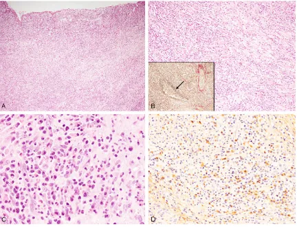
Abdominal lymph node
Diffuse proliferation of large-sized lymphoid
cells and destruction of normal architecture of
the lymph node were observed (
Figure 2
).
These lymphoid cells had large cleaved nuclei
with conspicuous nucleoli, and mitotic figures
and apoptotic bodies were scattered.
Immunohistochemical and
in situ
hybridization
studies were performed using an autostainer
(Ventana) by the same method as previously
reported [37-41]. The large-sized lymphoid cells
were diffusely positive for CD20 (
Figure 2
,
inset) and bcl-2, but negative for CD3, CD10,
CD15, CD30, bcl-6, cyclin D1, and anaplastic
lymphoma kinase (ALK). Moreover,
in situ
hybridization revealed no positive signal for
Epstein-Barr virus-encoded small RNA (EBER).
Accordingly, a diagnosis of DLBCL was made.
Gallbladder
The wall of the gallbladder was thickened, and
diffuse lymphoplasmacytic infiltration with
fibrosclerosis was observed in the entire gall
-bladder wall (
Figure 3A
,
3B
). Infiltrating lympho
-cytes were small in size and had small round
nuclei, and plasma cells were also bland in
appearance (
Figure 3B
,
3C
). Eosinophilic infil
-tration was also observed (
Figure 3C
), but no
neutrophils were seen. Obliterative phlebitis
was also noted by elastica van Gieson staining
(
Figure 3B
, inset).
Immunohistochemical study revealed that
many IgG4-positive plasma cells had infiltrated
into the lesion (73/high-power field) (
Figure
3D
), and the ratio of IgG4/IgG-positive plasmas
was 71.6%. Mild CD3-positive T-lymphocytic
infiltration was observed, and only a few
CD20-Figure 3. Histopathological and immunohistochemical features of the gallbladder. A: Dense lymphoplasmacytic infil -tration is observed in the entire gallbladder wall, HE, x 40. B: Dense lymphoplasmacytic infiltration with fibrosclerotic
change is noted, HE, x 100. Obliterative phlebitis (arrow), elastica van Gieson staining x 100. C: Lymphocytes and
plasma cells appear mature and are without atypia. Eosinophils are also observed, HE, x 400. D: Many IgG4-positive
[image:3.612.92.524.71.401.2]ALCL following IgG4-related sclerosing disease
positive B-lymphocytes had infiltrated into the
lesion.
Kappa
- and
lambda
chain-positive
plas-ma cells were evenly distributed as determined
by
in situ
hybridization.
According to these results, a diagnosis of
IgG4-positive cholecystitis was made. Although a
biopsy from the pancreas was not performed,
swelling of the pancreas was suspected to be
due to autoimmune pancreatitis because the
swelling subsided after administration of
prednisolone.
Nasal cavity
Dense lymphoplasmacytic infiltration with mild
fibrosis was observed under the ciliated epithe
-lium and around the nasal glands (
Figure 4A
,
4B
). Lymphocytes and plasma cells appeared
mature and without atypia (
Figure 4B
).
Obliterative phlebitis was noted (
Figure 4B
,
inset).
Immunohistochemical study revealed that
many IgG4-positive plasma cells had infiltrated
into the lesion (68/high-power field) (
Figure
4C
), and the ratio of IgG4/IgG-positive plasma
cells was 79.1%.
Kappa
- and
lambda
chain-positive plasma cells were evenly distributed as
assessed by
in situ
hybridization.
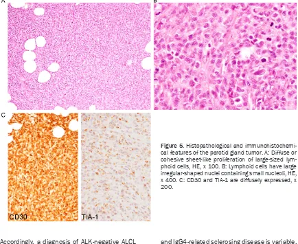
Parotid gland
Diffuse or cohesive sheet-like proliferation of
large-sized lymphoid cells was observed, and
no salivary gland component was present
(
Figure 5A
). Lymphoid cells had moderate
amount of slightly eosinophilic cytoplasm and
irregular-shaped large nuclei containing small
nucleoli, and mitotic figures were scattered
(
Figure 5B
).
Immunohistochemically, these lymphoid cells
were diffusely positive for CD30 and CD4
(
Figure 5C
), and some of these cells were also
positive for epithelial membrane antigen. TIA-1
was expressed in most of the lymphoid cells
(
Figure 5C
), and granzyme B was also expressed
in some of these cells. CD3, CD8, CD10, CD15,
CD20, CD79a, and ALK were not expressed.
[image:4.612.89.524.74.414.2]Moreover,
in situ
hybridization revealed no
posi-tive signal for EBER.
Figure 4. Histopathological and immunohistochemi-cal features of the nasal tumor. A: Dense
lympho-plasmacytic infiltration is observed under the ciliated
epithelium, HE, x 100. B: Dense lymphoplasmacytic
infiltration is also observed around the nasal glands. Lymphocytes and plasma cells are without atypia.
Obliterative phlebitis (arrow, inset), HE, x 200. C:
Abundant IgG4-positive plasma cells have infiltrated
ALCL following IgG4-related sclerosing disease
Accordingly, a diagnosis of ALK-negative ALCL
was made.
Discussion
In this report, we described the first document
-ed case of ALCL following IgG4-relat-ed autoim
-mune pancreatitis and cholecystitis and
DLBCL. Only 7 cases of extraocular malignant
lymphoma occurring in patients with
IgG4-related sclerosing disease with detailed
clinico-pathological features have been reported in
the English literature [27-31].
Table 1
summa-rizes the clinicopathological features of the
pre-viously reported cases as well as the present
one. This condition mainly affects middle-aged
to elderly males (average age 63.6 years (range
41-76), male/female 7/1). The
histopathologi-cal subtypes of malignant lymphoma were
DLBCL (5 cases including the present case), fol
-licular lymphoma (1 case), small lymphocytic
lymphoma (1 case), B-cell lymphoma (subtype
was not available in 1 case), and ALCL (1 case).
The occurrence in extranodal sites is common.
The relationship between malignant lymphoma
and IgG4-related sclerosing disease is variable.
In two of 8 cases, IgG4-related sclerosing
dis-ease developed after the occurrence of
malig-nant lymphoma [27, 28]. In the other 4 cases,
malignant lymphoma occurred during the
medi-cal follow-up of IgG4-related sclerosing disease
[29, 30]. Moreover, a case showing concurrent
occurrence of malignant lymphoma and
IgG4-related autoimmune pancreatitis has been
reported [31]. The present case is very unique
because we show for the first time that
IgG4-related sclerosing diseases (cholecystitis,
auto-immune pancreatitis, and rhinitis) developed
after DLBCL, which resulted in death by ALCL.
This is the first documented case of two differ
-ent histopathological subtype of malignant
lym-phoma occurring before and after IgG4-related
sclerosing disease. These results suggest that
malignant lymphoma can develop both before
and after the occurrence of IgG4-related
scle-rosing disease.
[image:5.612.90.524.76.431.2]Takahashi
et al
. analyzed the incidence of
malignant lymphoma in patients with
IgG4-related sclerosing disease [30]. In 111 patients
Figure 5. Histopathological and immunohistochemi-cal features of the parotid gland tumor. A: Diffuse or cohesive sheet-like proliferation of large-sized lym-phoid cells, HE, x 100. B: Lymphoid cells have largeALCL following IgG4-related sclerosing disease
with IgG4-related sclerosing disease, three
cases developed malignant lymphoma [30].
They concluded that patients with IgG4-related
sclerosing disease may be at an increased risk
of developing malignant lymphoma [30].
Although the mechanism of development of
malignant lymphoma in patients with
IgG4-related sclerosing disease remains unresolved,
an association between autoimmune diseases,
such as Sjögren syndrome, and the
develop-ment of malignant lymphoma is well recognized
[42]. It has been speculated that dysregulation
of B lymphocytes associated with autoimmune
disease leads to abnormal B lymphocytes
pro-liferation, resulting in the occurrence of
malig-nant B-cell lymphoma [43]. Moreover, an
asso-ciation between ocular adnexal marginal zone
B-cell lymphoma and IgG4-related
dacryoade-nitis has recently demonstrated [31-35]. In
addition, Mitsui
et al
. reported a very
interest-ing case of IgG4-related sclerosinterest-ing disease 10
years after chemotherapy for DLBCL [27].
Retrospective analysis of the lymph node
biop-sy specimen diagnosed as DLBCL revealed that
many IgG4-positive plasma cells had infiltrated
outside of the DLBCL lesion, therefore, they
speculated that subclinical IgG4-related
dis-ease was present before development of
DLBCL, and non-neoplastic IgG4-producing
plasma cells survived after chemotherapy,
which led to the development of IgG4-related
sclerosing disease [27]. Therefore, these
results suggest that a history of IgG4-related
sclerosing disease may be a predisposing
con-dition for the development of malignant
lym-phoma [28], and clinical follow-up is important
in patients with IgG4-related sclerosing disea-
se.
ALK-negative ALCL is included in the recent
World Health Organization Classification as a
provisional entity, and is defined as a
CD30-positive T-cell neoplasm that is not reproducibly
distinguishable on morphological grounds from
ALK-positive ALCL, although it lacks ALK pro
-tein [44]. This type of malignant lymphoma
mainly affects middle-aged persons in contrast
to ALK-positive ALCL, which occurs most com
-monly in children and young adults, and shows
[image:6.612.97.523.97.377.2]more aggressive clinical course than
ALK-positive ALCL [44, 45]. Histopathologically,
ALK-negative ALCL is characterized by solid,
Table 1.
Clinicopathological features of malignant lymphoma in patients with IgG4-related sclerosing
disease
Case
No. Age/Gen-der Histological type of ML (location) - Relationship between ML and IgG4-related sclerosing disease Reference Case 1 59/Male Diffuse large B-cell
lympho-ma (supraclavicular lymph node)
Submandibular lymph node biopsy revealed IgG4-related sclerosing disease 10 years after
ML.
[27]
Case 2 41/Male Follicular lymphoma (colon) Renal biopsy revealed IgG4-related
tubulointer-stitial nephritis 14 years after ML. [28]
Case 3 66/Male Diffuse large B-cell lympho-ma (lung, stolympho-mach, ileum, pancreas)
Autopsy revealed DLBCL after 11-year history of Mikulicz’s disease and 8-year history of inflam -matory pseudotumor of the renal pelvis.
[29]
Case 4 65/Female B-cell lymphoma (liver) B-cell lymphoma developed 4 years after a
diagnosis of autoimmune pancreatitis. [30] Case 5 72/Male Diffuse large B-cell
lympho-ma (adrenal gland) Diffuse large B-cell lymphoma developed 5 years after a diagnosis of autoimmune pancre-atitis.
[30]
Case 6 69/Male Diffuse large B-cell
lympho-ma (kidney) Diffuse large B-cell lymphoma developed 3 years after a diagnosis of chronic parotitis. [30] Case 7 76/Male Small lymphocytic lymphoma
(retroperitoneal lymph node) Concurrent autoimmune pancreatitis and small lymphocytic lymphoma. [31] Present
Case 61/Male Diffuse large B-cell lympho-ma (abdominal lymph node) and anaplastic large cell lymphoma (parotid gland)
Autoimmune pancreatitis, IgG4-related cho-lecystitis and rhinitis occurred 5 years after a diagnosis of diffuse large B-cell lymphoma. After one year, anaplastic large cell lymphoma developed.
ALCL following IgG4-related sclerosing disease
cohesive sheets of large-sized neoplastic cells
with abundant eosinophilic to clear cytoplasm
and large irregular-shaped nuclei, sometimes
containing prominent nucleoli [44]. Immuno-
histochemically, these neoplastic cells are
strongly positive for CD30, and CD15 is not
expressed. Loss of T-cell markers can occur,
however, more than half of cases express one
or more T-cell markers. CD4 is positive in a
sig-nificant proportion of cases, whereas
CD8-positive cases are rare [44]. A substantial
minority of cases is positive for epithelial
mem-brane antigen [44]. Moreover, many cases
express cytotoxic markers, such was TIA-1 and
granzyme B [44]. In the present case, the
neo-plastic cells of the parotid gland tumor were
large-sized and had irregular-shaped large
nuclei. Immunohistochemically, these
neoplas-tic cells were positive for CD30 and CD4, and
some of these cells were also positive for
epi-thelial membrane antigen. Moreover, cytotoxic
markers were expressed in most of these cells,
and ALK was negative. These features corre
-sponded to ALK-negative ALCL. Further, this
case had a fatal outcome.
In conclusion, we report the first documented
case of ALK-negative ALCL following
IgG4-related autoimmune pancreatitis and
cholecys-titis and DLBCL. It has been suggested that the
incidence of malignant lymphoma may be high
in patients with IgG4-related sclerosing
dis-ease, therefore, intense medical follow-up is
important in patients with IgG4-related
scleros-ing disease.
Address correspondence to: Dr. Mitsuaki Ishida,
Department of Clinical Laboratory Medicine and
Division of Diagnostic Pathology, Shiga University of Medical Science, Tsukinowa-cho, Seta, Otsu, Shiga, 520-2192, Japan. Tel: 77-548-2603; Fax: +81-77-548-2407; E-mail: mitsuaki@belle.shiga-med. ac.jp
References
[1] Hamano H, Kawa S, Horiuchi A, Unno H, Furuya N, Akamatsu T, Fukushima M, Nikaido T, Na-kayama K, Usuda N, Kiyosawa K. High serum IgG4 concentrations in patients with sclerosing pancreatitis. N Engl J Med 2001; 344: 732-738.
[2] Sato Y, Notohara K, Kojima M, Takata K, Ma-saki Y, Yoshino T. IgG4-related disease: histori-cal overview and pathology of hematologihistori-cal disorders. Pathol Int 2010; 60: 247-258.
[3] Stone JH, Zen Y, Deshpande V. IgG4-related disease. N Engl J Med 2012; 366: 539-551. [4] Zen Y, Nakanuma Y. IgG4-related disease. A
cross-sectional study of 114 cases. Am J Surg Pathol 2010; 34: 1812-1819.
[5] Zen Y, Nakanuma Y. Pathogenesis of IgG4-re-lated disease. Curr Opin Rheumatol 2011; 23: 114-118.
[6] Kamisawa T, Okamoto A. IgG4-related scleros-ing disease. World J Gastroenterol 2008; 14: 3948-3955.
[7] Zen Y, Harada K, Sasaki M, Sato Y, Tsuneyama K, Haratake J, Kurumaya H, Katayanagi K, Ma-suda S, Niwa H, Morimoto H, Miwa A, Uchiya-ma A, PortUchiya-mann BC, NakanuUchiya-ma Y. IgG4-related sclerosing cholangitis with and without hepatic
inflammatory pseudotumor, and sclerosing
pancreatitis-associated sclerosing cholangitis: do they belong to a spectrum of sclerosing pancreatitis? Am J Surg Pathol 2004; 28: 1193-1203.
[8] Hamano H, Kawa S, Ochi Y, Unno H, Shiba N, Wajiki M, Nakazawa K, Shimojo H, Kiyosawa K. Hydronephrosis associated with
retroperitone-al fibrosis and sclerosing pancreatitis. Lancet
2002; 359: 1403-1404.
[9] Zen Y, Kitagawa S, Minato H, Kurumaya H, Ka-tayanagi K, Masuda S, Niwa H, Fujimura M, Nakanuma Y. IgG4-positive plasma cells in
in-flammatory pseudotumor (plasma cell granu -loma) of the lung. Hum Pathol 2005; 36: 710-717.
[10] Kitagawa S, Zen Y, Harada K, Sasaki M, Sato Y, Minato H, Watanabe K, Kurumaya H, Katay-anagi K, Masuda S, Niwa H, Tsuneyama K, Saito K, Haratake J, Takagawa K, Nakanuma Y. Abundant IgG4-positive plasma cell infiltration
characterizes chronic sclerosing sialadenitis (Küttner’s tumor). Am J Surg Pathol 2005; 29: 783-791.
[11] Yamamoto M, Takahashi H, Ohara M, Suzuki C, Naishiro Y, Yamamoto H, Shinomura Y, Imai K. A new conceptualization for Mikulicz’s disease as an IgG4-related plasmacytic disease. Mod Rheumatol 2006; 16: 335-340.
[12] Kasashima S, Zen Y, Kawashima A, Konishi K, Sasaki H, Endo M, Matsumoto Y, Kawakami K, Kasashima F, Moriya M, Kimura K, Ohtake H,
Nakanuma Y. Inflammatory abdominal aortic
aneurysm: close relationship to IgG4-related periaortitis. Am J Surg Pathol 2008; 32: 197-204.
[13] Kasashima S, Zen Y, Kawashima A, Endo M, Matsumoto Y, Kasashima F. A new
clinicopath-ological entity of IgG4-related inflammatory
abdominal aortic aneurysm. J Vasc Surg 2009; 49: 1264-1271.
[14] Ishida M, Hotta M, Kushima R, Asai T, Okabe H.
IgG4-related inflammatory aneurysm of the
ALCL following IgG4-related sclerosing disease
[15] Kasashima S, Zen Y, Kawashima A, Endo M, Matsumoto Y, Kasashima F, Ohtake H, Na-kanuma Y. A clinicopathologic study of immu-noglobulin G4-related sclerosing disease of the thoracic aorta. J Vasc Surg 2010; 52: 1587-1595.
[16] Kasashima S, Zen Y. IgG4-related inflammato -ry abdominal aortic aneu-rysm. Curr Opin Rheumatol 2011; 23: 18-23.
[17] Zen Y, Kasashima S, Inoue D. Retroperitoneal and aortic manifestations of immunoglobulin G4-related disease. Semin Diagn Pathol 2012; 29: 212-218.
[18] Kawano M, Mizushima I, Yamaguchi Y, Imai N, Nakashima H, Nishi S, Hisano S, Yamanaka N, Yamamoto M, Takahashi H, Umehara H, Saito T, Saeki T. Immunohistochemical characteris-tics of IgG4-related tubulointerstitial nephritis: Detailed analysis of 20 Japanese cases. Int J Rheumatol 2012; 2012: 609795.
[19] Wong S, Lam WY, Wong WK, Lee KC. Hypophy
-sitis presented as inflammatory pseudotumor
in immunoglobulin G4-related systemic dis-ease. Hum Pathol 2007; 38: 1720-1723. [20] Cheuk W, Yuen HK, Chan JK. Chronic
scleros-ing dacryoadenitis: Part of the spectrum of IgG4-related sclerosing disease? Am J Surg Pathol 2007; 31: 643-645.
[21] Ishida M, Hotta M, Kushima R, Shibayama M, Shimizu T, Okabe H. Multiple IgG4-related scle-rosing lesions in the maxillary sinus, parotid gland and nasal septum. Pathol Int 2009; 59: 670-675.
[22] Yamamoto M, Takahashi H, Tabeya T, Suzuki C, Naishiro Y, Ishigami K, Yajima H, Shimizu Y, Obara M, Yamamoto H, Himi T, Imai K, Shino-mura Y. Risk of malignancies in IgG4-related disease. Mod Rheumatol 2012; 22: 414-418. [23] Kamisawa T, Chen PY, Tu Y, Nakajima H, Egawa
N, Tsuruta K, Okamoto A, Hishima T. Pancreat-ic cancer with a high serum IgG4 concentra-tion. World J Gastroenterol 2006; 12: 6225-6228.
[24] Fukui T, Mitsuyama T, Takaoka M, Uchida K, Matsushita M, Okazaki K. Pacnreatic cancer assocated with autoimmune pancreatitis in re-mission. Intern Med 2008; 47: 151-155. [25] Ghazale A, Chari S. Is autoimmune
pancreati-tis a risk factor for pancreatic cancer? Pancre-as 2007; 35: 376.
[26] Loos M, Esposito I, Hedderich DM, Ludwig L,
Fingerle A, Friess H, Klöppel G, Büchler P. Auto-immune pancreatitis complicated by carcino-ma of the pancreatobiliary system: a case re-port and review of the literature. Pancreas 2011; 40: 151-154.
[27] Mitsui T, Yokohama A, Koiso H, Ishizaki T, Uchi-umi H, Saitoh T, Handa H, Hirato J, Karasawa M, Murakami H, Kojima M, Nojima Y,
Tsuka-moto N. Development of IgG4-related disease 10 years after chemotherapy for diffuse large B cell lymphoma and longstanding bronchial asthma. Int J Hematol 2013; 98: 122-128. [28] Oshima Y, Usui R, Manabe S, Hasegawa N,
Ka-kuta Y, Nitta K, Hatano M. IgG4-related tubu-lointerstitial nephritis and lymphadenopathy after therapy for malignant lymphoma. Intern Med 2012; 51: 1221-1226.
[29] Uehara T, Ikeda S, Hamano H, Kawa S, Moteki H, Matsuda K, Kaneko Y, Hara E. A case of Mi-kulicz’s disease complicated by malignant
lym-phoma: a postmortem histopathological find -ing. Intern Med 2012; 51: 419-423.
[30] Takahashi N, Ghazale AH, Smyrk TC, Mandrek-ar JN, ChMandrek-ari ST. Possible association between IgG4-associated systemic disease with or with-out autoimmune pancreatitis and non-Hodgkin lymphoma. Pancreas 2009; 38: 523-526. [31] Kim T, Grobmyer SR, Dixon LR, Allan RW, Hoch
-wald SN. Autoimmune pancreatitis and con-current small lymphocytic lymphoma: not just a coincidence? J Gastrointest Surg 2008; 12: 1566-1570.
[32] Matsuo T, Ichimura K, Yoshino T. Local recur -rence as immunoglobulin G4 (IgG4)-related disease 10 years after radiotherapy to ocular adnexal extranodal marginal zone B-cell lym-phoma of mucosa-assocaited lymphoid tissue. J Clin Exp Hematopathol 2011; 51: 125-133. [33] Go H, Kim JE, Kim YA, Chung HK, Khwarg SI,
Kim CW, Jeon YK. Ocular adnexal IgG4-related disease: comparative analysis with mucosa-associated lymphoid tissue lymphoma and
other chronic inflammatory conditions. Histo -pathology 2012; 60: 296-312.
[34] Kubota T, Moritani S, Yoshino T, Nagai H, Tera-saki H. Ocular adnexal marginal zone B cell
lymphoma infiltrated by IgG4-positive plasma
cells. J Clin Pathol 2010; 63: 1059-1065. [35] Sato Y, Ohshima K, Ichimura K, Sato M,
Yama-dori I, Tanaka T, Takata K, Morito T, Kondo E, Yoshino T. Ocular adnexal IgG4-related disease has uniform clinicopathology. Pathol Int 2008; 58: 465-470.
[36] Cheuk W, Yuen HK, Chan AC, Shih LY, Kuo TT, Ma MW, Lo YF, Chan WK, Chan JK. Ocular ad -nexal lymphoma associated with IgG4+ chronic
sclerosing dacryoadenitis: a previously unde-scribed complication of IgG4-related scleros-ing disease. Am J Surg Pathol 2008; 32: 1159-1167.
[37] Ishida M, Iwai M, Yoshida K, Kagotani A, Okabe H. Sarcomatoid carcinoma with small cell car-cinoma component of the urinary bladder: A case report with review of the literature. Int J Clin Exp Pathol 2013; 6: 1671-1676.
ALCL following IgG4-related sclerosing disease
Okabe H. Pleomorphic lobular carcinoma in a male breast: case report with review of the lit-erature. Int J Clin Exp Pathol 2013; 6: 1441-1444.
[39] Nakanishi R, Ishida M, Hodohara K, Yoshida T, Yoshii M, Okuno H, Horinouchi A, Iwai M, Yo-shida K, Kagotani A, Okabe H. Prominent ge-latinous bone marrow transformation present-ing prior to myelodysplastic syndrome: a case report with review of the literature. Int J Clin Exp Pathol 2013; 6: 1677-1682.
[40] Ishida M, Mochizuki Y, Iwai M, Yoshida K, Kago-tani A, Okabe H. Pigmented squamous intraep-ithelial neoplasia of the esophagus. Int J Clin Exp Pathol 2013; 6: 1868-73.
[41] Yoshii M, Ishida M, Hodohara K, Okuno Y, Na-kanishi R, Yoshida T, Okabe H. Systemic Ep-stein-Barr virus-positive T-cell lymphoprolifera-tive disease of childhood: Report of a case
with review of the literature. Oncol Lett 2012;
4: 381-384.
[42] Jonsson MV, Theander E, Jonsson R. Predic-tors for the development of non-Hodgkin lym-phoma in primary Sjögren’s syndrome. Presse Med 2012; 41: e511-516.
[43] Hansen A, Lipsky PE, Dörner T. B-cell lymphop
-roliferation in chronic inflammatory rheumatic
diseases. Nat Clin Pract Rheumatol 2007; 3: 561-569.
[44] Mason DY, Harris NL, Delsol G, Stein H, Campo
E, Kinney MC, Jaffe ES, Falini B. Anaplastic
large cell lymphoma, ALK-negative. In: Swerd
-low SH, Campo E, Harris NL, Jaffe ES, Pileri SA,
Stein H, Thiele J, Vardiman JW, eds. World
Health Organization Classification of Tumours of Haematopoietic and Lymphoid Tissues. Lyon: IARC Press, 2008. pp: 317-319.
[45] ten Berge RL, Oudejans JJ, Ossenkoppele GJ, Meijer CJ. ALK-negative systemic anaplastic