MOLECULAR CHARACTERIZATION AND PHYLOGENETIC
ANALYSIS OF CULEX QUINQUEFASCIATUS BY CYTOCHROME C
OXIDASE SUBUNIT II
Sahar Abd1* and Emad Ahmed2
1
*Lecturer Department of Biology, College of Science for Women, University of Baghdad,
Baghdad, Iraq.
2
Prof. Dr. Department of Biology, College of Science for Women, University of Baghdad,
Baghdad, Iraq.
ABSTRACT
Molecular Genetic methods were used in this study to find out the
genetic diversity of Culex collected from Nahrawan region of Baghdad
in Iraq, the amplified segments of mitochondrial DNA which consisted
of the genes COII by using Polymerase Chain Reaction (PCR) with the
existence of pairs from the specific primers. The PCR amplified
fragments in these mosquitoes were approximately 685 base pairs and
the amino acids were more than 95% similar between Culex
quinquefasciatus from USA and C. quinquefosciatus from Baghdad,
and there is a substitution in 31 bases distributed along the nucleotide
sequence, the variations ranged between 16 type of transition while the
12 type Transversion. These segments contain the major amino acid residues of cytochrome c
oxidase included in electron transport and ligand binding.
KEYWORDS: Culex mosquitoes; DNA barcode; cytochrome oxidase II (COII),
Phylogenetic Analysi.
INTRODUCTION
Mosquitoes are responsible for the transmission of parasitic and viral infections to both
humans and livestock, with substantial morbidity and mortality.[1] There are over 3,500 different species of mosquitoes throughout the world.[2] Some diseases transmitted by mosquitoes include malaria, yellow fever, encephalitis and RVF among others.[3] Prevention
Volume 7, Issue 8, 39-45. Research Article ISSN 2277– 7105
Article Received on 23 Feb. 2018,
Revised on 16 March 2018, Accepted on 06 April 2018
DOI: 10.20959/wjpr20188-11684
*Corresponding Author
Sahar Abd
Lecturer Department of
Biology, College of Science
for Women, University of
vaccinations to reduce and/or prevent transmission.[2] Mosquito control is carried out by habitat control, use of insecticides, larvicides and breeding control using sterile males.[4] Most mosquito transmitted organisms have an obligatory developmental stage that takes place in
the mosquito, and in some cases completely rely on the vectors for transmission. The primary
identification of mosquitoes has over time been carried out by morphological
characterization.[5] Mosquitoes are observed under a differentiating microscope and only persons with knowledge of the features that differentiate one species from another have the
capacity to carry out morphological identification. An unambiguous identification of
mosquitoes using morphological characters requires taxonomic experience and specimens as
intact as possible.[6] In addition to the morphological tools, molecular methods for sub species classification have been proposed.[7] These include PCRRFLPs which apply differences in the length of DNA fragments after digestion with restriction endonucleases.[8]
Mitochondrial Cytochrome oxidase C subunit I and II (COI and COII), Cytochrome oxidase
B, 16S rRNA gene are helpful in species identification, phylogenetic analyses and other
related studies.[9] this study used in identification of insect specimens and have proven highly informative for phylogenetic inference.
MATERIALS AND METHODS
Field samples were obtained from different selected locations in Baghdad, Periods of higher
mosquito population densities were targeted. DNA was extracted separately for mosquitoes
from Culex quinquefasciatus by using Genomic DNA Mini Kit Tissue Protocol.
DNA Amplification by (PCR) was used to amplify a segment of the COII gene using the
primer pair table (1) to find the optimal condition has identified for (Initial denaturation and
annealing) after a work several experiments to get for this condition, the temperature has
changed through the work of (Gradient PCR) for all samples to select the optimal condition
table (2), and also changed the concentration for DNA template between (1.5-2µl) where is
considered these two factors from important factors in primer annealing with complement.
Table (2): The optimum condition of detection COII.
No. Phase Tm (ᵒC) Time No. of cycle
1- Initial Denaturation 94ᵒC 5 min.
35cycle
2- Denaturation -2 94ᵒC 40sec.
3- Annealing 62ᵒC 35sec.
4- Extension-1 72ᵒC 35sec.
5- Extension -2 72ᵒC 10 min.
RESULTS AND DISCUSSIONS
Total genomic DNA was extracted from each mosquito using Genomic DNA Mini Kit Tissue
Protocol Figure 1. Samples not analyzed immediately were stored at −20°C. The polymerase
chain reaction (PCR) was used to amplify a segment of the COII gene using the specific
primer 685 bp pair AAGTCGCTAAAGCTCCTACTG / CTTCATCAAAGTCGTCTCCA
and standard assay conditions, find the optimal temperature in ratio of strong fragment, 62°C
were chosen for annealing temperature for the PCR experiment Figure 2.
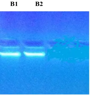
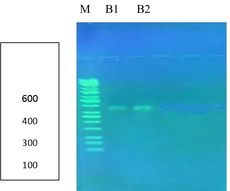
B1 B2
[image:3.595.63.536.89.181.2] [image:3.595.219.378.352.520.2]M B1 B2
Figure 2: Electrophoresis of PCR products of the COII gene using the specific primer
685 bp pair collected from of Iraq. PCR product from Iraq sample, M: Marker; B1 female Baghdad; B2 male Baghdad.
Fig 3 shows the locations of nucleotide substitutions and the sequences showed very low
variation between species on Alignment and phylogenetic analysis, Table (3) shows the
substitutions classified according to the type of base change between these2 species. The
COII gene is 95% similar between Culex quinquefasciatus from USA and C.
quinquefosciatus from Baghdad. The number of transversion was less than that of transitions
In all transversions, the frequency of change between A and T. For a comparison of COII,
amino acid sequences were deduced by using the insect mitochondrial code.685 amino acid
residues are present and similarity is shown in Figure 3 This is similar from comparisons of
mtDNA between closely related mammalian species in which transitions occur more
frequently than transversions.[10] There was also variation between species on phylogenetic analysis Figure 4 the evolutionary history was inferred using the Neigh bour-Joining method.
The percentage of replicate trees in which the associated taxa clustered together in the
bootstrap test are shown next to the branches. The tree is drawn to scale, with branch lengths
in the same units as those of the evolutionary distances used to estimate the phylogenetic tree.
The rate variation among sites was modelled with genetic distances = 0.030. DNA barcode
approach based on DNA sequences of mitochondrial cytochrome oxidase gene sequences
could identify 62 species among these, in confirmation with the conventional taxonomy.[11] 600
400
300
[image:4.595.144.377.70.263.2]Table (3): Type substitutions classified according sequence of partial mitochondrial
COII sequences of Culex quinquefasciatus from Baghdad compared with Cx.
quinquefasciatus from USA.
Sequence ID Range of nucleotide Nucleotide Location Type of substitution sample gb|HQ724617.1| 3124 to 3735
[image:5.595.41.566.133.616.2]Figure 3: Multiple sequence alignment of partial mitochondrial COII sequences of
Culex quinquefasciatus from Baghdad compared with Cx. quinquefasciatus from USA mitochondrion in GenBank, complete genome Sequence ID: gb|HQ724617.1|
seq from Jakarta
seq from Bangladesh
seq from Tunisia
seq from Turkey
seq from USA
seq from Thailand
seq from Baghdad-Iraq
[image:6.595.104.485.69.416.2] [image:6.595.88.513.511.673.2]REFERENCES
1. Davies. (2006). Risk of a rift valley fever epidemic at the haj in Mecca, Saudi Arabia.
Revue scientifique et technique (International Office of Epizootics), 25(1): 137–47.
Retrieved from http://www.ncbi.nlm.nih.gov/pubmed/16796043
2. Tolle, M. a. (2009). Mosquito-borne diseases. Current problems in pediatric and
adolescent health care, 39(4): 97–140. doi:10.1016/j.cppeds.2009.01.001
3. Farajollahi, A., Fonseca, D. M., Kramer, L. D., & Marm Kilpatrick. (2011). “Bird biting”
mosquitoes and human disease: A review of the role of Culex pipiens complex
mosquitoes in epidemiology. Infection, genetics and evolution: journal of molecular
epidemiology and evolutionary genetics in infectious diseases, 11: 1577–1585.
doi:10.1016/j.meegid.2011.08.013
4. Jones, O.M. 2012. The Effects of Spinosad on Culex quinquefasciatus Say (Diptera:
Culicidae) and Non-Targer Insect Species. Thesis. University of the Louisiana State.
5. Service, M. W. (2000). Introduction to mosquitoes (Culicidae) ©. Medical entomology
for students. Retrieved fromhttp://www.cambridge.org/052154775X
6. Hackett, B. J., Gimnig, J., Guelbeogo, W., Costantini, C., Koekemoer, L. L., Coetzee, M.,
Collins, F. H., et al. (2000). Ribosomal DNA internal transcribed spacer (ITS2) sequences
differentiate Anopheles funestus and An. rivulorum and uncover a cryptic taxon. Insect
Molecular Biology, 9(March): 369–374.
7. Caterino, M. S., Cho, S., & Sperling, F. a. (2000). The current state of insect molecular
systematics: a thriving Tower of Babel. Annual review of entomology, 45: 1–54.
doi:10.1146/annurev.ento.45.1.1
8. Walton, C., Sharpe, R. G., Pritchard, S. J., Thelwell, N. J., & Butlin, R. K. (1999).
Molecular identification of mosquito species. Biological journal of the Linnean society,
68: 241–256.
9. Dhananjeyan, K. J., Paramasivan, R., Tewari, S. C., Rajendran, R., Thenmozhi, V., Leo,
S. V. J., Venkatesh, A., et al. (2010). Molecular identification of mosquito vectors using
genomic DNA isolated from eggshells, larval and pupal exuvium. Tropical biomedicine,
27(1): 47–53.
10.Brown WM. 1985. The mitochondrial genome of animals. In Maclntyre RJ, ed.
Molecular evolution genetics New York: Plenum. p 95-130.
11.Kumar, N. P.; Rajavel, A. R.; Natarajan, R.; Jambulingam, P. (2007). DNA Barcodes can


![CAMAC bulletin: A publication of the ESONE Committee Issue #14 December 1975 [last pub of series]](data:image/gif;base64,R0lGODlhAQABAIAAAP///wAAACH5BAEAAAAALAAAAAABAAEAAAICRAEAOw==)