organic papers
o192
GoÈrbitz C9H12NO2+NO3ÿ3C9H11NO2 DOI: 101107/S1600536801000575 Acta Cryst.(2001). E57, o192±o194Acta Crystallographica Section E
Structure Reports Online
ISSN 1600-5368
L
-Phenylalanine nitrate (4/1)
Carl Henrik GoÈrbitz
Department of Chemistry, University of Oslo, PO Box 1033 Blindern, N-0315 Oslo, Norway
Correspondence e-mail: [email protected]
Key indicators
Single-crystal X-ray study
T= 150 K
Mean(C±C) = 0.010 AÊ
Rfactor = 0.113
wRfactor = 0.269 Data-to-parameter ratio = 7.3
For details of how these key indicators were automatically derived from the article, see http://journals.iucr.org/e.
#2001 International Union of Crystallography Printed in Great Britain ± all rights reserved
The asymmetric unit of the title compound,l-phenylalaninium
nitrate tris(l-phenylalanine), C9H12NO2+NO3ÿ3C9H11NO2,
contains a nitrate anion and a peculiar sequence of four l
-phenylalanine molecules. Three of them are present as zwitterions, while the last carries the positive charge and acts
as donor in a ±COOH ÿOOC± hydrogen bond with an
O O distance of 2.443 (9) AÊ.
Comment
The title compound, (I), was studied as part of a search for good crystallization conditions for various peptides. Several nice-looking crystals were obtained, but the diffraction patterns revealed that the structure was probably divided into layers that were misaligned along one axis. This made the unit-cell determination dif®cult, and only after testing several specimens was it possible to ®nd the third axis, collect diffraction data and integrate them successfully.
As a result of the poor crystal quality, the ®nalRfactor is
high for a small molecule structure. Nevertheless, the structure proved to be interesting in having an odd sequence of four phenylalanine molecules in the asymmetric unit in addition to
the nitrate anion (Fig. 1). Three of the phenylalanines (B,C
andD) are in the zwitterionic form. The positive charge most
likely (see below) resides with the last amino acid moleculeA,
which is connected to molecule B by a very short ±
COOH ÿOOC± hydrogen bond (Table 2). The shortness of
this interaction can in part be attributed to the fact that the
carboxylate group of molecule B participates in no other
strong interactions, a most unusual situation. The COOH
group of molecule A is furthermore involved in only one
additional hydrogen bond with an H O distance less than
2.50 AÊ.
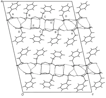
The molecular packing shown in Fig. 2 has, as suspected,
thick hydrophobic layers ofl-phenylalanine side chains, and
also hydrophilic layers composed of two interconnected hydrogen-bonded sheets. This is a very persistent pattern, which occurs not only in the structure of phenylalanine itself
(Weissbuchet al., 1990) and its complexes with mandelic acid
(Okamura et al., 1997), but also in other salts such as l -phenylalanine hydrochloride (Al-Karaghouli & Koetzle, 1975),l-phenylalninel-phenylalaninium formate (GoÈrbitz &
Etter, 1992) andl-phenylalaninel-phenylalaninium
perchlo-rate (Srinivasan & Rajaram, 1997). It is noteworthy that the
latter two structures include onel-phenylalanine zwitterion
and one l-phenylalaninium cation in the asymmetric unit,
which are in both cases connected by a very short hydrogen bond like the one seen in the title structure.
Experimental
l-Phenylalanine was obtained from Sigma and used as received. Crystals in the shape of long ¯at needles were grown by slow evaporation of a dilute nitric acid solution of the amino acid at room temperature.
Crystal data
4C9H12NO2+4NO3ÿ12C9H11NO2
Mr= 723.80
Monoclinic,C2
a= 30.285 (8) AÊ
b= 5.3022 (13) AÊ
c= 22.317 (6) AÊ
= 102.263 (4) V= 3501.9 (15) AÊ3
Z= 4
Dx= 1.373 Mg mÿ3
MoKradiation Cell parameters from 3964
re¯ections
= 1.9±25.0 = 0.10 mmÿ1
T= 150 (2) K Flat needle, colourless 0.550.200.07 mm Data collection
Siemens SMART CCD diffract-ometer
Sets of exposures each taken over 0.3!rotation scans
Absorption correction: empirical (SADABS; Sheldrick, 1996)
Tmin= 0.945,Tmax= 0.993
9411 measured re¯ections
3450 independent re¯ections 2777 re¯ections withI> 2(I)
Rint= 0.100
max= 25.0
h=ÿ36!34
k=ÿ6!6
l=ÿ24!26 Intensity decay: none Re®nement
Re®nement onF2
R[F2> 2(F2)] = 0.113
wR(F2) = 0.269
S= 1.63 3450 re¯ections 474 parameters H atoms constrained
w= 1/[2(F
o2) + (0.098P)2
+ 3.750P]
whereP= (Fo2+ 2Fc2)/3
(/)max= 0.005
max= 0.62 e AÊÿ3
min=ÿ0.54 e AÊÿ3
Extinction correction:SHELXTL
Extinction coef®cient: 0.0053 (14)
Table 1
Selected geometric parameters (AÊ,).
O1AÐC1A 1.219 (10) O2AÐC1A 1.271 (9) N1AÐC2A 1.494 (6) O1BÐC1B 1.247 (10) O2BÐC1B 1.265 (9) N1BÐC2B 1.490 (6)
O1CÐC1C 1.298 (10) O2CÐC1C 1.230 (9) N1CÐC2C 1.489 (6) O1DÐC1D 1.261 (10) O2DÐC1D 1.250 (10) N1DÐC2D 1.491 (6)
O1AÐC1AÐO2A 127.3 (7) O1AÐC1AÐC2A 118.2 (6) O2AÐC1AÐC2A 114.5 (6) O1BÐC1BÐO2B 125.4 (6) O1BÐC1BÐC2B 118.5 (6) O2BÐC1BÐC2B 115.8 (6)
O2CÐC1CÐO1C 124.9 (6) O2CÐC1CÐC2C 118.1 (6) O1CÐC1CÐC2C 117.0 (5) O2DÐC1DÐO1D 125.9 (6) O2DÐC1DÐC2D 117.5 (7) O1DÐC1DÐC2D 116.5 (6)
Figure 1
The structure of the title compound with the atomic numbering indicated. Displacement ellipsoids are shown at the 50% probability level and H atoms are shown as spheres of arbitrary size.
Figure 2
organic papers
o194
GoÈrbitz C9H12NO2+NO3ÿ3C9H11NO2 Acta Cryst.(2001). E57, o192±o194O1AÐC1AÐC2AÐN1A ÿ46.3 (8) N1AÐC2AÐC3AÐC4A 167.2 (6) C2AÐC3AÐC4AÐC5A 61.0 (10) O1BÐC1BÐC2BÐN1B 12.4 (9) N1BÐC2BÐC3BÐC4B 55.4 (7) C2BÐC3BÐC4BÐC5B 83.5 (8)
O1CÐC1CÐC2CÐN1C ÿ10.3 (9) N1CÐC2CÐC3CÐC4C 55.8 (7) C2CÐC3CÐC4CÐC5C 96.2 (8) O1DÐC1DÐC2DÐN1D ÿ0.7 (9) N1DÐC2DÐC3DÐC4D 53.5 (7) C2DÐC3DÐC4DÐC5D 71.5 (10)
Table 2
Hydrogen-bonding geometry (AÊ,).
DÐH A DÐH H A D A DÐH A
O2AÐHO2A O2B 1.00 1.49 2.443 (9) 158 N1AÐH1A O1Di 0.91 2.11 2.858 (8) 138
N1AÐH1A O1Aii 0.91 2.19 2.844 (10) 128
N1AÐH2A O2Diii 0.91 1.97 2.837 (9) 159
N1AÐH3A O1Div 0.91 1.95 2.785 (9) 152
N1AÐH3A O1Av 0.91 2.50 3.071 (8) 121
N1BÐH1B O2Cvi 0.91 2.16 3.053 (10) 167
N1BÐH2B O2C 0.91 2.17 3.012 (10) 154 N1BÐH3B O2Evii 0.91 2.01 2.898 (10) 166
N1CÐH1C O1Evi 0.91 2.19 2.909 (10) 135
N1CÐH2C O1E 0.91 2.04 2.903 (10) 158 N1CÐH3C O1Cvii 0.91 1.86 2.744 (9) 164
N1DÐH1D O3Evi 0.91 2.01 2.886 (10) 161
N1DÐH2D O2Dvi 0.91 2.07 2.896 (9) 151
N1DÐH3D O1Bvii 0.91 2.40 2.954 (9) 119
N1DÐH3D O2Avii 0.91 2.46 3.322 (10) 157
Symmetry codes: (i)x;yÿ1;zÿ1; (ii)x;yÿ1;z; (iii)x;y;zÿ1; (iv)1
2ÿx;yÿ12;1ÿz;
(v)1
2ÿx;yÿ12;ÿz; (vi)x;1y;z; (vii)12ÿx;12y;1ÿz.
Three sets of exposures with the detector set at 2= 29,
crystal-to-detector distance 4.98 cm. Constrained H-atom re®nement withUiso values set to 1.2Ueq of the carrier atom or 1.5Ueqfor the amino groups. The H atom in the shortest hydrogen bond could not be found in difference Fourier maps. It could therefore not be established with certainty whether it is actually bonded to O2Aor O2B(a centered position was considered to be less likely). A combined geometric and force-®eld calculation with the program HYDROGEN (Nardelli, 1999) showed, however, that the H atom is most likely associated with O2A, and it was thus named HO2A. For a 2.44 AÊ O O distance, the
previously observed lengthening of the covalent OÐH bond is about 0.15 AÊ (Steiner & Saenger, 1994). The HO2A position output by
HYDROGENwas consequently adjusted so as to increase the OÐH bond length from 0.85 (default) to 1.00 AÊ. Based on results from a survey of neutron diffraction structures with COOH groups from the Cambridge Structural Database (Allen & Kennard, 1993), the CÐ OÐH angle was furthermore set to 111.5and the the CÐCÐOÐH
torsion angle was increased to 175(GoÈrbitz, 2001).SAMErestraints
were used for the four phenylalanine side chains. The absolute structure could not be determined, and Friedel pairs were merged in the ®nal re®nements.
Data collection:SMART(Bruker, 1998); cell re®nement:SAINT
(Bruker, 1998); data reduction: SAINT; program(s) used to solve structure: SHELXTL (Sheldrick, 1997); program(s) used to re®ne structure:SHELXTL; molecular graphics:SHELXTL; software used to prepare material for publication:SHELXTL.
The author thanks Professor Mario Nardelli for carrying
out the HYDROGEN calculations. The purchase of the
Siemens SMART diffractometer was made possible through support from The Research Council of Norway (NFR).
References
Allen, F. & Kennard, O. (1993).Chem. Des. Autom. News,8, 1, 31±37. Al-Karaghouli, A. R. & Koetzle, T. F. (1975).Acta Cryst.B31, 2461±2465. Bruker (1998).SAINT(Version 6.01) andSMART(Version 5.054). Bruker
AXS Inc., Madison, Wisconsin, USA. GoÈrbitz, C. H. (2001). Unpublished results.
GoÈrbitz, C. H. & Etter, M. C (1992).Acta Cryst.C48, 1317±1320. Nardelli, M. (1999).J. Appl. Cryst.32, 563±571.
Okamura, K., Aoe, K., Hiramatsu, H., Nishimura, N. Sato, T. & Hashimoto, K. (1997).Anal. Sci.13, 315±317.
Sheldrick, G. M. (1996). SADABS. University of GoÈttingen, Germany. Sheldrick, G. M. (1997).SHELXTL.Version 5.10. Bruker AXS Inc., Madison,
Wisconsin, USA.
Srinivasan, N. & Rajaram, R. K. (1997).Acta Cryst.C53, 1711±1713. Steiner, T. & Saenger, W. (1994).Acta Cryst.B50, 348±357.
supporting information
Acta Cryst. (2001). E57, o192–o194 [doi:10.1107/S1600536801000575]
L
-Phenylalanine nitrate (4/1)
Carl Henrik G
ö
rbitz
S1. Comment
The title compound, (I), was studied as part of a search for good crystallization conditions for various peptides. Several
nice-looking crystals were obtained, but the diffraction patterns revealed that the structure was probably divided into
layers that were misaligned along one axis. This made the unit-cell determination difficult, and only after testing several
specimens was it possible to find the third axis, collect diffraction data and integrate them successfully.
As a result of the poor crystal quality, the final R factor is high for a small molecule structure. Nevertheless, the
structure proved to be interesting in having an odd sequence of four phenylalanine molecules in the asymmetric unit in
addition to the nitrate anion (Fig. 1). Three of the phenylalanines (B, C and D) are in the zwitterionic form. The positive
charge most likely (see below) resides with the last amino acid molecule A, which is connected to molecule B by a very
short –COOH···-OOC– hydrogen bond (Table 2). The shortness of this interaction can in part be attributed to the fact that
the carboxylate group of molecule B participates in no other strong interactions, a most unusual incidence. The COOH
group of molecule A is furthermore involved in only one additional hydrogen bond with an H···O distance less than 2.50
Å.
The molecular packing shown in Fig. 2 has, as suspected, thick hydrophobic layers of L-phenylalanine side chains, and
also hydrophilic layers composed of two interconnected hydrogen-bonded sheets. This is a very persistent pattern, which
occurs not only in the structure of phenylalanine itself (Weissbuch et al., 1990) and its complexes with mandelic acid
(Okamura et al., 1997), but also in other salts such as L-phenylalanine hydrochloride (Al-Karaghouli & Koetzle, 1975),
L-phenylalnine L-phenylalaninium formate (Görbitz & Etter, 1992) and L-phenylalanine L-phenylalaninium perchlorate
(Srinivasan & Rajaram, 1997). It is noteworthy that the latter two structures include one L-phenylalanine zwitterion and
one L-phenylalaninium cation in the asymmetric unit, which are in both cases connected by a very short hydrogen bond
like the one seen in the title structure.
S2. Experimental
L-Phenylalanine was obtained from Sigma and used as received. Crystals in the shape of long flat needles were grown by
slow evaporation of a dilute nitric acid solution of the amino acid at room temperature.
S3. Refinement
Three sets of exposures with the detector set at 2θ = 29°, crystal-to-detector distance 4.98 cm. Constrained H-atom
refinement with Uiso values set to 1.2Ueq of the carrier atom or 1.5Ueq for the amino groups. The H atom in the shortest
hydrogen bond could not be found in difference Fourier maps. It could therefore not be established with certainty whether
it is actually bonded to O2A or O2B (a centered position was considered to be less likely). A combined geometric and
force-field calculation with the program HYDROGEN (Nardelli, 1999) showed, however, that the H atom is most likely
supporting information
sup-2
Acta Cryst. (2001). E57, o192–o194
the covalent O—H bond is about 0.15 Å (Steiner & Saenger, 1994). The HO2A position output by HYDROGEN was
consequently adjusted so as to increase the O—H bond length from 0.85 (default) to 1.00 Å. Based on results from a
survey of neutron diffraction structures with COOH groups from the Cambridge Structural Database (Allen & Kennard,
1993), the C—O—H angle was furthermore set to 111.5° and the the C—C—O—H torsion angle was increased to 175°
(Görbitz, 2001). SAME 0.005 0.005 constraints were used for the four phenylalanine side chains. The absolute structure
[image:5.610.126.485.342.676.2]could not be determined, and Friedel pairs were merged in the final refinements.
Figure 1
The structure of the title compound with the atomic numbering indicated. Displacement ellipsoids are shown at the 50%
Figure 2
The unit cell and molecular packing viewed along the a axis. Hydrogen bonds have been dashed.
L-Phenylalanine nitrate (4/1)
Crystal data
C9H12NO2+·NO3−·3C9H11NO2
Mr = 180.95 Monoclinic, C2 a = 30.285 (8) Å b = 5.3022 (13) Å c = 22.317 (6) Å β = 102.263 (4)° V = 3501.9 (15) Å3
Z = 16
F(000) = 1536 Dx = 1.373 Mg m−3
Mo Kα radiation, λ = 0.71073 Å Cell parameters from 3964 reflections θ = 1.9–25.0°
µ = 0.10 mm−1
T = 150 K
Flat needle, colourless 0.55 × 0.20 × 0.07 mm
Data collection
Siemens SMART CCD diffractometer
Radiation source: fine-focus sealed tube Graphite monochromator
Detector resolution: 8.3 pixels mm-1
sets of exposures each taken over 0.3° ω rotation scans
Absorption correction: empirical (using intensity measurements)
(SADABS; Sheldrick, 1996)
Tmin = 0.945, Tmax = 0.993
9411 measured reflections 3450 independent reflections 2777 reflections with I > 2σ(I) Rint = 0.100
θmax = 25.0°, θmin = 1.9°
h = −36→34 k = −6→6 l = −24→26
Refinement
Refinement on F2
Least-squares matrix: full R[F2 > 2σ(F2)] = 0.113
wR(F2) = 0.269
S = 1.63 3450 reflections 474 parameters 543 restraints
Primary atom site location: structure-invariant direct methods
Secondary atom site location: difference Fourier map
Hydrogen site location: inferred from neighbouring sites
H atoms treated by a mixture of independent and constrained refinement
w = 1/[σ2(F
o2) + (0.098P)2 + 3.75P]
where P = (Fo2 + 2Fc2)/3
(Δ/σ)max = 0.005
Δρmax = 0.62 e Å−3
Δρmin = −0.54 e Å−3
Extinction correction: SHELXTL, Fc*=kFc[1+0.001xFc2λ3/sin(2θ)]-1/4
Extinction coefficient: 0.0053 (14)
Special details
Refinement. Refinement of F2 against ALL reflections.
Fractional atomic coordinates and isotropic or equivalent isotropic displacement parameters (Å2)
x y z Uiso*/Ueq
O1A 0.3062 (2) 0.3387 (13) 0.0591 (3) 0.0298 (15) O2A 0.3194 (2) 0.1777 (13) 0.1536 (3) 0.0335 (16)
HO2A 0.3151 0.3546 0.1674 0.050*
N1A 0.29139 (18) −0.1575 (13) 0.0158 (3) 0.0210 (16)
supporting information
sup-4
Acta Cryst. (2001). E57, o192–o194
H2A 0.2935 −0.0477 −0.0149 0.032*
H3A 0.2634 −0.1465 0.0243 0.032*
C1A 0.3157 (2) 0.1668 (13) 0.0959 (3) 0.0178 (17) C2A 0.32619 (19) −0.0934 (13) 0.0718 (3) 0.0236 (19)
H21A 0.3246 −0.2220 0.1040 0.028*
C3A 0.37314 (18) −0.1053 (15) 0.0571 (3) 0.033 (2)
H31A 0.3806 −0.2843 0.0514 0.039*
H32A 0.3721 −0.0177 0.0176 0.039*
C4A 0.4108 (2) 0.0093 (14) 0.1049 (3) 0.029 (2) C5A 0.4215 (3) −0.0852 (15) 0.1646 (3) 0.032 (2)
H51A 0.4056 −0.2277 0.1751 0.038*
C6A 0.4549 (3) 0.0266 (18) 0.2088 (3) 0.042 (3)
H61A 0.4621 −0.0404 0.2492 0.050*
C7A 0.4775 (3) 0.2355 (16) 0.1940 (3) 0.040 (3)
H71A 0.5001 0.3141 0.2243 0.048*
C8A 0.4673 (3) 0.3305 (16) 0.1347 (3) 0.043 (3)
H81A 0.4830 0.4739 0.1246 0.051*
C9A 0.4340 (3) 0.2178 (15) 0.0898 (3) 0.040 (3)
H91A 0.4273 0.2830 0.0492 0.048*
O1B 0.2901 (2) 0.3968 (13) 0.2706 (3) 0.0302 (15) O2B 0.3250 (2) 0.5881 (14) 0.2045 (3) 0.0359 (17) N1B 0.3060 (2) 0.7650 (15) 0.3540 (3) 0.0309 (19)
H1B 0.3121 0.9037 0.3784 0.046*
H2B 0.3223 0.6318 0.3726 0.046*
H3B 0.2760 0.7286 0.3477 0.046*
C1B 0.3115 (3) 0.5761 (14) 0.2543 (3) 0.023 (2) C2B 0.3184 (2) 0.8158 (13) 0.2940 (3) 0.025 (2)
H21B 0.2974 0.9478 0.2724 0.030*
C3B 0.36626 (19) 0.9197 (12) 0.3036 (3) 0.028 (2)
H31B 0.3737 0.9430 0.2628 0.033*
H32B 0.3668 1.0883 0.3227 0.033*
C4B 0.4029 (2) 0.7602 (12) 0.3425 (2) 0.0198 (18) C5B 0.4225 (3) 0.5587 (14) 0.3174 (3) 0.029 (2)
H51B 0.4136 0.5237 0.2748 0.035*
C6B 0.4551 (3) 0.4090 (14) 0.3541 (3) 0.033 (2)
H61B 0.4685 0.2735 0.3366 0.040*
C7B 0.4678 (3) 0.4571 (15) 0.4161 (3) 0.031 (2)
H71B 0.4898 0.3536 0.4414 0.037*
C8B 0.4485 (3) 0.6574 (15) 0.4415 (3) 0.033 (2)
H81B 0.4576 0.6913 0.4841 0.039*
C9B 0.4159 (3) 0.8092 (14) 0.4049 (3) 0.030 (2)
H91B 0.4027 0.9452 0.4226 0.036*
O1C 0.2980 (2) 0.0210 (13) 0.4784 (3) 0.0292 (15) O2C 0.3302 (2) 0.2695 (14) 0.4186 (3) 0.0348 (17) N1C 0.2911 (2) 0.3854 (14) 0.5580 (3) 0.0249 (17)
H1C 0.2992 0.4900 0.5908 0.037*
H2C 0.2966 0.2228 0.5703 0.037*
C1C 0.3156 (3) 0.2316 (14) 0.4652 (3) 0.0215 (19) C2C 0.3179 (2) 0.4485 (13) 0.5112 (3) 0.0220 (19)
H21C 0.3036 0.5995 0.4881 0.026*
C3C 0.3661 (2) 0.5201 (12) 0.5420 (3) 0.026 (2)
H31C 0.3831 0.5539 0.5097 0.031*
H32C 0.3651 0.6793 0.5649 0.031*
C4C 0.3918 (2) 0.3257 (13) 0.5857 (2) 0.027 (2) C5C 0.4210 (3) 0.1543 (14) 0.5665 (3) 0.028 (2)
H51C 0.4253 0.1609 0.5256 0.033*
C6C 0.4437 (3) −0.0257 (15) 0.6068 (3) 0.039 (3)
H61C 0.4622 −0.1473 0.5928 0.047*
C7C 0.4395 (3) −0.0277 (17) 0.6671 (3) 0.043 (3)
H71C 0.4558 −0.1470 0.6950 0.052*
C8C 0.4112 (3) 0.1455 (18) 0.6869 (3) 0.047 (3)
H81C 0.4080 0.1423 0.7283 0.056*
C9C 0.3876 (3) 0.3240 (15) 0.6465 (3) 0.037 (2)
H91C 0.3687 0.4437 0.6604 0.044*
O1D 0.2777 (2) 0.4577 (13) 0.9240 (3) 0.0289 (15) O2D 0.3019 (3) 0.0734 (13) 0.9050 (3) 0.0343 (17) N1D 0.2841 (2) 0.6584 (11) 0.8188 (3) 0.0273 (17)
H1D 0.2920 0.7192 0.7845 0.041*
H2D 0.2977 0.7516 0.8518 0.041*
H3D 0.2536 0.6680 0.8144 0.041*
C1D 0.2928 (3) 0.2989 (15) 0.8913 (3) 0.0235 (19) C2D 0.29870 (19) 0.3903 (12) 0.8283 (3) 0.0209 (18)
H21D 0.2779 0.2876 0.7967 0.025*
C3D 0.34613 (19) 0.3540 (12) 0.8177 (3) 0.027 (2)
H31D 0.3452 0.3784 0.7735 0.033*
H32D 0.3551 0.1769 0.8277 0.033*
C4D 0.3827 (2) 0.5236 (14) 0.8534 (2) 0.025 (2) C5D 0.3988 (3) 0.4946 (18) 0.9164 (3) 0.043 (3)
H51D 0.3859 0.3695 0.9379 0.052*
C6D 0.4334 (3) 0.6461 (19) 0.9480 (3) 0.046 (3)
H61D 0.4443 0.6228 0.9908 0.055*
C7D 0.4519 (3) 0.8308 (17) 0.9173 (3) 0.039 (2)
H71D 0.4753 0.9363 0.9388 0.047*
C8D 0.4361 (3) 0.8612 (17) 0.8546 (3) 0.044 (3)
H81D 0.4484 0.9906 0.8335 0.053*
C9D 0.4025 (3) 0.7039 (15) 0.8222 (2) 0.033 (2)
H91D 0.3931 0.7198 0.7789 0.040*
supporting information
sup-6
Acta Cryst. (2001). E57, o192–o194 Atomic displacement parameters (Å2)
U11 U22 U33 U12 U13 U23
C7D 0.037 (4) 0.027 (5) 0.049 (4) −0.001 (4) 0.002 (4) −0.003 (4) C8D 0.042 (5) 0.035 (5) 0.052 (5) −0.002 (4) 0.006 (4) 0.011 (4) C9D 0.034 (4) 0.025 (4) 0.040 (4) −0.001 (4) 0.004 (3) 0.008 (4) O1E 0.061 (4) 0.016 (3) 0.037 (3) 0.001 (3) 0.009 (3) 0.000 (3) O2E 0.048 (4) 0.028 (4) 0.044 (3) −0.004 (3) 0.008 (3) −0.015 (3) O3E 0.072 (5) 0.046 (4) 0.062 (4) 0.016 (4) 0.015 (3) 0.025 (4) N1E 0.040 (4) 0.019 (4) 0.035 (4) 0.006 (4) 0.007 (3) 0.003 (3)
Geometric parameters (Å, º)
O1A—C1A 1.219 (10) O2C—C1C 1.230 (9)
O2A—C1A 1.271 (9) N1C—C2C 1.489 (6)
O2A—HO2A 1.0042 N1C—H1C 0.9100
N1A—C2A 1.494 (6) N1C—H2C 0.9100
N1A—H1A 0.9100 N1C—H3C 0.9100
N1A—H2A 0.9100 C1C—C2C 1.534 (7)
N1A—H3A 0.9100 C2C—C3C 1.523 (7)
C1A—C2A 1.538 (7) C2C—H21C 1.0000
C2A—C3A 1.527 (7) C3C—C4C 1.516 (6)
C2A—H21A 1.0000 C3C—H31C 0.9900
C3A—C4A 1.515 (6) C3C—H32C 0.9900
C3A—H31A 0.9900 C4C—C9C 1.389 (7)
C3A—H32A 0.9900 C4C—C5C 1.397 (6)
C4A—C9A 1.389 (7) C5C—C6C 1.389 (6)
C4A—C5A 1.395 (6) C5C—H51C 0.9500
C5A—C6A 1.388 (6) C6C—C7C 1.380 (8)
C5A—H51A 0.9500 C6C—H61C 0.9500
C6A—C7A 1.380 (8) C7C—C8C 1.390 (6)
C6A—H61A 0.9500 C7C—H71C 0.9500
C7A—C8A 1.388 (6) C8C—C9C 1.396 (6)
C7A—H71A 0.9500 C8C—H81C 0.9500
C8A—C9A 1.396 (6) C9C—H91C 0.9500
C8A—H81A 0.9500 O1D—C1D 1.261 (10)
C9A—H91A 0.9500 O2D—C1D 1.250 (10)
O1B—C1B 1.247 (10) N1D—C2D 1.491 (6)
O2B—C1B 1.265 (9) N1D—H1D 0.9100
N1B—C2B 1.490 (6) N1D—H2D 0.9100
N1B—H1B 0.9100 N1D—H3D 0.9100
N1B—H2B 0.9100 C1D—C2D 1.535 (6)
N1B—H3B 0.9100 C2D—C3D 1.516 (7)
C1B—C2B 1.538 (7) C2D—H21D 1.0000
C2B—C3B 1.521 (7) C3D—C4D 1.514 (6)
C2B—H21B 1.0000 C3D—H31D 0.9900
C3B—C4B 1.515 (6) C3D—H32D 0.9900
C3B—H31B 0.9900 C4D—C9D 1.390 (7)
C3B—H32B 0.9900 C4D—C5D 1.396 (6)
C4B—C9B 1.389 (7) C5D—C6D 1.389 (6)
supporting information
sup-8
Acta Cryst. (2001). E57, o192–o194
C5B—C6B 1.389 (6) C6D—C7D 1.381 (8)
C5B—H51B 0.9500 C6D—H61D 0.9500
C6B—C7B 1.379 (8) C7D—C8D 1.388 (6)
C6B—H61B 0.9500 C7D—H71D 0.9500
C7B—C8B 1.389 (6) C8D—C9D 1.394 (6)
C7B—H71B 0.9500 C8D—H81D 0.9500
C8B—C9B 1.395 (6) C9D—H91D 0.9500
C8B—H81B 0.9500 O1E—N1E 1.243 (9)
C9B—H91B 0.9500 O2E—N1E 1.272 (11)
O1C—C1C 1.298 (10) O3E—N1E 1.230 (12)
C1A—O2A—HO2A 111.3 C2C—N1C—H2C 109.5
C2A—N1A—H1A 109.5 H1C—N1C—H2C 109.5
C2A—N1A—H2A 109.5 C2C—N1C—H3C 109.5
H1A—N1A—H2A 109.5 H1C—N1C—H3C 109.5
C2A—N1A—H3A 109.5 H2C—N1C—H3C 109.5
H1A—N1A—H3A 109.5 O2C—C1C—O1C 124.9 (6)
H2A—N1A—H3A 109.5 O2C—C1C—C2C 118.1 (6)
O1A—C1A—O2A 127.3 (7) O1C—C1C—C2C 117.0 (5) O1A—C1A—C2A 118.2 (6) N1C—C2C—C3C 110.6 (4) O2A—C1A—C2A 114.5 (6) N1C—C2C—C1C 110.3 (5) N1A—C2A—C3A 109.5 (4) C3C—C2C—C1C 113.0 (4)
N1A—C2A—C1A 109.6 (4) N1C—C2C—H21C 107.6
C3A—C2A—C1A 112.6 (4) C3C—C2C—H21C 107.6
N1A—C2A—H21A 108.4 C1C—C2C—H21C 107.6
C3A—C2A—H21A 108.4 C4C—C3C—C2C 115.7 (4)
C1A—C2A—H21A 108.4 C4C—C3C—H31C 108.4
C4A—C3A—C2A 115.4 (4) C2C—C3C—H31C 108.4
C4A—C3A—H31A 108.4 C4C—C3C—H32C 108.4
C2A—C3A—H31A 108.4 C2C—C3C—H32C 108.4
C4A—C3A—H32A 108.4 H31C—C3C—H32C 107.4
C2A—C3A—H32A 108.4 C9C—C4C—C5C 119.4 (3)
H31A—C3A—H32A 107.5 C9C—C4C—C3C 119.4 (4)
C9A—C4A—C5A 119.5 (3) C5C—C4C—C3C 121.1 (4) C9A—C4A—C3A 119.4 (4) C6C—C5C—C4C 120.5 (4)
C5A—C4A—C3A 121.0 (4) C6C—C5C—H51C 119.8
C6A—C5A—C4A 120.7 (4) C4C—C5C—H51C 119.8
C6A—C5A—H51A 119.6 C7C—C6C—C5C 120.0 (3)
C4A—C5A—H51A 119.6 C7C—C6C—H61C 120.0
C7A—C6A—C5A 119.8 (3) C5C—C6C—H61C 120.0
C7A—C6A—H61A 120.1 C6C—C7C—C8C 119.8 (3)
C5A—C6A—H61A 120.1 C6C—C7C—H71C 120.1
C6A—C7A—C8A 119.8 (3) C8C—C7C—H71C 120.1
C6A—C7A—H71A 120.1 C7C—C8C—C9C 120.6 (4)
C8A—C7A—H71A 120.1 C7C—C8C—H81C 119.7
C7A—C8A—C9A 120.7 (4) C9C—C8C—H81C 119.7
C7A—C8A—H81A 119.6 C4C—C9C—C8C 119.6 (3)
C4A—C9A—C8A 119.4 (3) C8C—C9C—H91C 120.2
C4A—C9A—H91A 120.3 C2D—N1D—H1D 109.5
C8A—C9A—H91A 120.3 C2D—N1D—H2D 109.5
C2B—N1B—H1B 109.5 H1D—N1D—H2D 109.5
C2B—N1B—H2B 109.5 C2D—N1D—H3D 109.5
H1B—N1B—H2B 109.5 H1D—N1D—H3D 109.5
C2B—N1B—H3B 109.5 H2D—N1D—H3D 109.5
H1B—N1B—H3B 109.5 O2D—C1D—O1D 125.9 (6)
H2B—N1B—H3B 109.5 O2D—C1D—C2D 117.5 (7)
O1B—C1B—O2B 125.4 (6) O1D—C1D—C2D 116.5 (6) O1B—C1B—C2B 118.5 (6) N1D—C2D—C3D 111.2 (4) O2B—C1B—C2B 115.8 (6) N1D—C2D—C1D 110.0 (4) N1B—C2B—C3B 110.8 (4) C3D—C2D—C1D 113.7 (4)
N1B—C2B—C1B 109.8 (5) N1D—C2D—H21D 107.2
C3B—C2B—C1B 112.9 (4) C3D—C2D—H21D 107.2
N1B—C2B—H21B 107.7 C1D—C2D—H21D 107.2
C3B—C2B—H21B 107.7 C2D—C3D—C4D 117.0 (4)
C1B—C2B—H21B 107.7 C2D—C3D—H31D 108.0
C4B—C3B—C2B 116.0 (4) C4D—C3D—H31D 108.0
C4B—C3B—H31B 108.3 C2D—C3D—H32D 108.0
C2B—C3B—H31B 108.3 C4D—C3D—H32D 108.0
C4B—C3B—H32B 108.3 H31D—C3D—H32D 107.3
C2B—C3B—H32B 108.3 C9D—C4D—C5D 119.0 (3)
H31B—C3B—H32B 107.4 C9D—C4D—C3D 119.3 (4)
C9B—C4B—C5B 119.4 (3) C5D—C4D—C3D 121.5 (4) C9B—C4B—C3B 119.2 (3) C6D—C5D—C4D 120.9 (4)
C5B—C4B—C3B 121.3 (4) C6D—C5D—H51D 119.6
C6B—C5B—C4B 120.6 (4) C4D—C5D—H51D 119.6
C6B—C5B—H51B 119.7 C7D—C6D—C5D 119.9 (3)
C4B—C5B—H51B 119.7 C7D—C6D—H61D 120.0
C7B—C6B—C5B 119.9 (3) C5D—C6D—H61D 120.0
C7B—C6B—H61B 120.0 C6D—C7D—C8D 119.6 (3)
C5B—C6B—H61B 120.0 C6D—C7D—H71D 120.2
C6B—C7B—C8B 119.8 (3) C8D—C7D—H71D 120.2
C6B—C7B—H71B 120.1 C7D—C8D—C9D 120.7 (4)
C8B—C7B—H71B 120.1 C7D—C8D—H81D 119.6
C7B—C8B—C9B 120.6 (4) C9D—C8D—H81D 119.6
C7B—C8B—H81B 119.7 C4D—C9D—C8D 119.8 (3)
C9B—C8B—H81B 119.7 C4D—C9D—H91D 120.1
C4B—C9B—C8B 119.6 (3) C8D—C9D—H91D 120.1
C4B—C9B—H91B 120.2 O3E—N1E—O1E 121.1 (9)
C8B—C9B—H91B 120.2 O3E—N1E—O2E 120.5 (8)
C2C—N1C—H1C 109.5 O1E—N1E—O2E 118.4 (8)
supporting information
sup-10
Acta Cryst. (2001). E57, o192–o194
N1A—C2A—C3A—C4A 167.2 (6) N1C—C2C—C3C—C4C 55.8 (7) C1A—C2A—C3A—C4A 45.1 (8) C1C—C2C—C3C—C4C −68.4 (6) C2A—C3A—C4A—C9A −116.4 (8) C2C—C3C—C4C—C9C −86.2 (9) C2A—C3A—C4A—C5A 61.0 (10) C2C—C3C—C4C—C5C 96.2 (8) C9A—C4A—C5A—C6A −0.1 (14) C9C—C4C—C5C—C6C 3.5 (13) C3A—C4A—C5A—C6A −177.5 (9) C3C—C4C—C5C—C6C −178.8 (8) C4A—C5A—C6A—C7A 0.8 (16) C4C—C5C—C6C—C7C −3.4 (15) C5A—C6A—C7A—C8A −0.9 (16) C5C—C6C—C7C—C8C 2.1 (16) C6A—C7A—C8A—C9A 0.3 (16) C6C—C7C—C8C—C9C −0.9 (17) C5A—C4A—C9A—C8A −0.6 (15) C5C—C4C—C9C—C8C −2.3 (14) C3A—C4A—C9A—C8A 176.9 (9) C3C—C4C—C9C—C8C −180.0 (9) C7A—C8A—C9A—C4A 0.4 (16) C7C—C8C—C9C—C4C 1.0 (17) O1B—C1B—C2B—N1B 12.4 (9) O2D—C1D—C2D—N1D 177.2 (7) O2B—C1B—C2B—N1B −173.5 (7) O1D—C1D—C2D—N1D −0.7 (9) O1B—C1B—C2B—C3B 136.6 (7) O2D—C1D—C2D—C3D −57.4 (9) O2B—C1B—C2B—C3B −49.3 (9) O1D—C1D—C2D—C3D 124.7 (7) N1B—C2B—C3B—C4B 55.4 (7) N1D—C2D—C3D—C4D 53.5 (7) C1B—C2B—C3B—C4B −68.2 (6) C1D—C2D—C3D—C4D −71.3 (6) C2B—C3B—C4B—C9B −93.8 (8) C2D—C3D—C4D—C9D −112.0 (8) C2B—C3B—C4B—C5B 83.5 (8) C2D—C3D—C4D—C5D 71.5 (10) C9B—C4B—C5B—C6B −0.5 (14) C9D—C4D—C5D—C6D 1.3 (16) C3B—C4B—C5B—C6B −177.8 (8) C3D—C4D—C5D—C6D 177.8 (9) C4B—C5B—C6B—C7B 0.8 (15) C4D—C5D—C6D—C7D 0.9 (18) C5B—C6B—C7B—C8B −0.8 (15) C5D—C6D—C7D—C8D −0.9 (18) C6B—C7B—C8B—C9B 0.7 (15) C6D—C7D—C8D—C9D −1.4 (17) C5B—C4B—C9B—C8B 0.3 (13) C5D—C4D—C9D—C8D −3.5 (15) C3B—C4B—C9B—C8B 177.7 (8) C3D—C4D—C9D—C8D 179.9 (9) C7B—C8B—C9B—C4B −0.4 (15) C7D—C8D—C9D—C4D 3.6 (16)
Hydrogen-bond geometry (Å, º)
D—H···A D—H H···A D···A D—H···A
O2A—HO2A···O2B 1.00 1.49 2.443 (9) 158 N1A—H1A···O1Di 0.91 2.11 2.858 (8) 138
N1A—H1A···O1Aii 0.91 2.19 2.844 (10) 128
N1A—H2A···O2Diii 0.91 1.97 2.837 (9) 159
N1A—H3A···O1Div 0.91 1.95 2.785 (9) 152
N1A—H3A···O1Av 0.91 2.50 3.071 (8) 121
N1B—H1B···O2Cvi 0.91 2.16 3.053 (10) 167
N1B—H2B···O2C 0.91 2.17 3.012 (10) 154 N1B—H3B···O2Evii 0.91 2.01 2.898 (10) 166
N1C—H1C···O1Evi 0.91 2.19 2.909 (10) 135
N1C—H2C···O1E 0.91 2.04 2.903 (10) 158 N1C—H3C···O1Cvii 0.91 1.86 2.744 (9) 164
N1D—H1D···O3Evi 0.91 2.01 2.886 (10) 161
N1D—H3D···O1Bvii 0.91 2.40 2.954 (9) 119
N1D—H3D···O2Avii 0.91 2.46 3.322 (10) 157