Review
Factors influencing bone loss in
anorexia nervosa: assessment and
therapeutic options
Isabelle Legroux , Bernard Cortet
To cite: Legroux i, Cortet B. Factors influencing bone loss in anorexia nervosa: assessment and therapeutic options. RMD Open
2019;5:e001009. doi:10.1136/ rmdopen-2019-001009
Received 13 May 2019 Revised 22 October 2019 Accepted 22 October 2019
Department of Rheumatology and eA 4490, Lille University Hospital and University of Lille, Lille, France
Correspondence to Dr isabelle Legroux; isabelle. legroux@ chru- lille. fr © Author(s) (or their employer(s)) 2019. Re- use permitted under CC BY- NC. No commercial re- use. See rights and permissions. Published by BMJ.
Key messages
What is already know about this subject?
► Bone involvement is a frequent complication of anorexia nervosa leading to an increased risk of fracture.
What does this study add?
► Low body mass index (BMi), amenorrhoea and low iGF-1 level are well recognised factors explaining bone involvement, low leptin level, and high adi-ponectin level have been more recently investigated and seem promising.
How might this impact on clinical practice?
► Management of low bone mass requires an increase of BMi and menstrual cycles recovery.
AbstrAct
Decreased mineral density is one of the major complications of anorexia nervosa. The phenomenon is even more pronounced when the disease occurs during adolescence and when the duration of amenorrhoea is long. The mechanisms underlying bone loss in anorexia are complex. Oestrogen deficiency has long been considered as the main factor, but cannot explain the phenomenon on its own. The essential role of nutrition- related factors— especially leptin and adiponectin—has been reported in recent studies. Therapeutic strategies to mitigate bone involvement in anorexia are still a matter for debate. Although resumption of menses and weight recovery appear to be essential, they are not always accompanied by a total reversal of bone loss. There are no studies in the literature demonstrating that oestrogen treatment is effective, and the best results seem to have been obtained with agents that induce bone formation—such as iGF-1—especially when associated with oestrogen. As such, bone management in anorexia remains difficult, hence, the importance of early detection and multidisciplinary follow- up.
Attitudes to food and eating disorders are of considerable importance in our modern societies on account of their physiological and health implications. The increase in the frequency of eating disorders as a whole is well established epidemiologically, and anorexia nervosa (AN) has become a major public health concern in recent years. Its prevalence is 0.2%–1% as opposed to 2% for bulimia.1 According to the DSM- V, AN is characterised by a refusal to maintain a minimum normal weight, significant weight loss, self- esteem that is excessively dependent on body shape and weight, and a distortion of body image. amenorrhoea is no longer required for the diagnosis of AN based on DSM- V criteria.2 Most of the studies on eating disorders focus on AN—which includes a restricting but also binge- purge subtype and is primarily charac-terised by weight loss—and bulimia nervosa, which is characterised by alternating bouts of uncontrolled binge eating and compensatory
behaviours. In many cases, eating disor-ders are intertwined (eg, anorexia- bulimia) or incomplete, and their impact on weight balance is variable. Some studies on atypical forms of AN—in males, or prepubescent anorexia—can also be found in the literature.
This article focusses on eating disorders, and primarily restricting or binge- purge subtypes of AN in adolescents and young adults. Indeed, in the event of significant weight loss, the evolution of AN is marked by significant morbidity, particularly for bone tissue, and a higher risk of osteoporosis. In a recent meta- analysis, the authors found that spinal bone mineral density (BMD) in normal- weight women with bulimia nervosa was significantly lower than in healthy control women. These data suggest that the adverse effects of an eating disorder on BMD cannot be attributed to weight loss alone.3 The mech-anisms underlying bone loss in these patients are numerous (hormonal, endocrine and nutritional). These negative consequences are further aggravated by the fact that eating disorders usually occur during adolescence, which is a critical period for the acquisition of peak bone mass.
on May 7, 2020 by guest. Protected by copyright.
The purpose of this paper is to assess the extent of bone involvement in AN, in epidemiological, diagnostic and physiopathogenic terms, and to attempt to evaluate its therapeutic consequences. Moreover, this article is a narrative review and not a systematic review.
AssessmenT of bone involvemenT in An bone mineral density
BMD can be assessed non- invasively using dual- energy X- ray absorptiometry. In populations of young women or female adolescents with AN, in which some patients are under 20 years old, it is preferable to use the Z- score—that is, the number of SD between a subject’s value and the mean value of subjects of the same age and sex—rather than the T- score—that is, the number of SD between a subject’s value and the mean value of young subjects of the same sex. Osteoporosis is defined as a Z- score of less than −2 SDs, and osteopenia as a Z- score of between −2 and −1 SDs.
BMD in anorexic patients and healthy subjects has been compared by different authors. Anorexia patients always exhibit lower BMDs: osteoporosis is found in about 20%–30% of the patients,4–6 and osteopenia in 50%–90% of them.
bone remodelling markers
Bone remodelling markers can be used to explore both bone formation and bone resorption. In the literature, the most frequently used markers of bone formation are osteocalcin and bone alkaline phosphatases (BAP). Commonly used bone resorption markers include deoxy-pyridinoline, C- terminal extension peptides (CrossLaps or CTX) or N- terminal extension peptides (NTX), and telopeptides (carboxyl- terminal telopeptide of type one collagen).5 These markers are mainly used in postmeno-pausal women, but their interpretation in young women and adolescent girls is more difficult since growth (model-ling) impacts on their values. In the literature, results vary widely, group sizes are often small, and a distinction needs to be made between those studies involving adoles-cent girls and those involving adult anorexia patients. Like postmenopausal women, anorexia patients exhibit an increase in bone resorption, but studies have reported that they also exhibit a significant decrease in bone forma-tion.6 This results in greater bone loss than in women of the same age with hypogonadism. This decrease in bone formation probably depends on the severity of the disease and the age at onset. The underlying mechanism is probably a decrease in IGF-1, a factor that stimulates osteoblast differentiation and therefore bone formation. However, the decrease in bone formation markers is not found in all of the studies. Indeed, in a study4 comparing young adult anorexia patients (n=113) to controls, we found no significant difference between the groups where bone remodelling markers were concerned. Further-more, some authors have reported low levels of bone formation markers and similar levels of bone resorption
markers in adolescents with AN compared with healthy controls, while in adult women with AN compared with healthy controls, they observed decreased levels of bone formation markers and elevated levels of bone resorption markers. The difference may be due to the fact that phys-iological oestrogen levels are low in prepubertal adoles-cents, whereas most adult women with AN are hypoestro-egenaemic relative to healthy controls.7 8
fractures and An
Only a few studies have investigated the risk of fracture in anorexia patients. However, despite this reservation, all of the studies concur that the risk of fracture is higher in patients with AN. Fractures occur more frequently at the usual sites of osteoporotic fractures, that is, most often the vertebrae, then the radius, and then the upper extremity of the femur.
Grinspoon et al9 found a history of fracture in 26% of patients, 97% of whom had low BMD. The mean dura-tion of the disorder was 5.5 years. In patients who have anorexia for a mean duration of 5.8 years, the risk of frac-ture is seven times higher than in healthy women of the same age.10 The prevalence of fractures in AN and other eating disorders has also been investigated using data from a Danish register.11 A higher incidence of fractures was found in anorexia patients after diagnosis (especially in the first year), with the risk of fracture persisting for up to 10 years thereafter. In patients diagnosed with anorexia after 20 years of age, the risk of fracture was higher than in those diagnosed before the age of 20. In a retrospective study involving 208 anorexic subjects (193 female and 15 male) with 13- year follow- up, Lucas et al12 reported 58 fractures. Compared with the expected number of fractures, the risk was multiplied by three. Fractures occurred more often in patients followed up in a hospital setting than in an outpatient setting, and stress fractures due to bone deficiency were also frequent in this population. The results of this study suggest that the cumulative incidence of fractures within 40 years of being diagnosed with AN is 57%.
The risk of fracture is higher in patients with active AN, and this higher risk persists for as long as 10 years after the disease was diagnosed, especially in patients over 20 years of age. A possible explanation for this could be the absence of bone mass recovery in older anorexia patients, in whom the progression of the disorder is potentially more serious.3 4 6
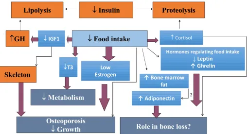
meCHAnisms of bone loss
The pathophysiological mechanisms involved in bone metabolism abnormalities in AN are complex. Various mechanisms have been proposed and are probably inter-related, including oestrogen deficiency, growth hormone and IGF1 metabolism disorders, hypercorticism, vitamin D deficiency, adipose tissue metabolism disorders (leptin, adiponectin), and factors involved in adipocyte/ osteoblast differentiation, such as preadipocyte factor-1
on May 7, 2020 by guest. Protected by copyright.
Figure 1 Main mechanisms of bone involvement in anorexia nervosa.
(Pref-1). Indeed, since osteoblast and adipocyte cells come from the same cell line, the decrease in osteo-blast differentiation probably causes precursors to take the adipocyte pathway. However, while the main biolog-ical pathways are now better understood, the respective weights of the above- mentioned factors in explaining bone impairment remain to be established (figure 1).
sex hormones
A few studies have investigated changes in BMD in AN patient populations.4–11 Women who develop anorexia before the age of 18 have significantly lower BMD than those who develop the disease later, which reflects the impact of the disease on peak bone mass acquisition. Although amenorrhoea is no longer required for the diagnosis of AN based on DSM- V criteria, menstrual dysfunction remains common in this disease. Serum levels of oestrogen and testosterone are lower in adults and adolescents with AN compared with controls, and oestrogen deficiency has been reported to be a signifi-cant etiological factor for bone loss in this population. In anorexia nervosa, the mechanisms underlying oestrogen deficiency are better understood and probably multi-factorial. Other potential factors include hypothalamic dysfunction and weight loss, and the dysregulation of neurohormones, such as GnRH, should also be consid-ered (see below). In the literature, a correlation has been reported between BMD, duration, and age at onset of amenorrhoea.4 5 13 14 Menstrual resumption (associated with weight gain) is often considered a sign of improve-ment and/or recovery in females with AN. Oestrogen is an antiresorptive and reduces osteoclastic activity by
depressing the secretion of RANKL and inflammatory cytokines, and stimulating the secretion of osteoprote-gerin.15 Testosterone also impacts bone, both directly and through its aromatisation from oestradiol. Low testosterone levels have adverse effects on bone, and an increase in testosterone levels following weight gain is a strong predictor of an increase in BMD in girls with AN.16–18
Oestrogen deficiency alone cannot explain bone involvement in anorexic patients, which suggests that other factors are involved. Indeed, in patients who have recovered from anorexia, the gain in bone density precedes the resumption of menses, and oestrogen treat-ment does not prevent bone loss in these patients as indi-cated below.4–6
Hormones and factors involved in regulating food intake
The role of the latter has been substantiated, and a corre-lation between BMD in these patients and nutritional indices such as weight, BMI, lean mass, fat mass, IGF-1, and leptin has been reported in some studies.5 19
In a previous study, we found a correlation between hip BMD and IGF-1 in 113 patients with AN (r=0.4, p<0.0001).4 In another study, we also found that a recovery in weight with a BMI>17 kg/m2 after 1- year follow- up predicted
the recovery in hip BMD at 2 years.13 Other authors5 14 have also reported a positive correlation between bone formation markers (osteocalcin and BAP) and nutrition markers such as BMI, percentage of fat mass, and IGF-1, and a negative correlation between oestradiol and bone resorption markers.
on May 7, 2020 by guest. Protected by copyright.
Alterations in the GH- IGF-1 axis have also been reported in some studies.20 The levels of these hormones increase during puberty and stimulate the proliferation of osteo-blast precursors and their differentiation. IGF-1 is a bone- trophic hormone that stimulates bone formation and growth by acting on osteoblasts and collagen synthesis. In patients with AN, high levels of plasma growth hormone (GH) are associated with low levels of IGF-1, suggesting a resistance to GH. Støving et al20 studied GH secretion over 24 hours in eight anorexic patients and observed an increase in the number of peaks, as well as in their duration and intensity. They also reported an increase in basal secretion (×20, compared with ×4 for pulsatile secretion). The increase in GH peak intensity is probably related to weight loss, and the increase in peak number to oestrogen deficiency. There was no difference in the half- life of GH between patients and healthy controls. Several authors have reported a decrease in IGF-1 levels, but also in IGF-1 carrier proteins—especially IGFBP-3 and IGFBP-2 (and sometimes IGFBP-1)—in anorexic patients.21 22 The decrease in circulating levels of carrier proteins may partly explain the resistance to GH, thus preventing the transfer of IGF-1 to target organs. More-over, IGFBP-3 may be a good predictor of bone loss in anorexic patients, independently of BMI and IGF-1.
The role of leptin in regulating bone mass and density is complex. While it seems to decrease bone density through central action, peripheral leptin seems to increase it. Serum leptin levels are logically lower in AN and have been found to correlate with lower fat mass and bone density measures.6 In a recent study, we investigated factors influencing bone loss in 146 anorexic patients and, in multivariate analysis, we found that leptin level was the main factor (adjusted R2=0.12; p=0.0014) explaining the changes in hip BMD.23
For adiponectin, some authors have reported an increase in serum levels, but results varied depending on the molecular weight fraction of plasma adiponectin that was measured. Some authors only found an increase in the ‘high molecular weight’ fraction. Misra et al24 have reported that higher adiponectin levels corrected for fat mass in girls with AN predict lower spine BMD. We found similar results in a cohort of 80 patients, in which a signif-icant increase in high molecular weight adiponectin was observed only in patients with osteoporosis and BMI
≤17 kg/m2.25 Otherwise, the variance in BMD in AN patients with osteoporosis was explained predominantly by three factors: BMI, leptin and adiponectin. BMI is a reflection of both lean and fat mass, leptin a reflection of subcutaneous and visceral fat, and adiponectin a possible reflection of marrow fat.
Finally, high levels of ghrelin have also been reported in this population. However, some studies have reported that the administration of exogenous ghrelin increases bone mass by altering GH secretion and activating the GH- IGF-1 axis.26 27 Fukushima et al27 also described the expression of ghrelin receptors, and of ghrelin itself, by osteoblasts in culture. The high levels of ghrelin found
in AN patients are probably due to adaptive mechanisms, given its role in food intake, and suggest potential insen-sitivity to this endocrine signal.
Changes in body composition
AN is characterised by marked reductions in fat mass, and less marked but significant reductions in lean mass. Lower lean mass is an important determinant of lower bone density and impaired bone structure in adults and adolescents with AN.6 16 Soyka et al16 have shown that increases in lean mass following weight gains are strongly predictive of coincident increases in bone density in adolescents with AN.
Furthermore, recent imaging studies (using MRI spec-troscopy) have shown an increase in bone marrow fat in women with AN compared with normal- weight controls and women who have recovered from AN, even though patients with anorexia typically exhibit a reduction in subcutaneous and visceral fat. This is associated with an increase in Pref-1, a factor involved in adipocyte/osteoblast differentiation.28 29 These abnormalities can be explained by the common mesenchymal origin of these two cell lines.30 31 Medullary adipocytes are a special population of adipocytes whose secretions in the spinal environment appear to contribute to the decrease in BMD. Further-more, as patients recover from AN, marrow adipose tissue (MAT) tends to return to normal levels. In a study comparing women with AN to women with a history of AN but who had recovered weight and menstrual cyclicity, Fazeli et al found that the women who had recovered from AN had similar MAT levels in the L4 vertebra compared with the normal- weight controls, whereas the women with AN had significantly higher levels of L4 MAT.29 32
stress hormones
The role of hypercortisolism in some patients also remains to be clarified. As is the case with patients taking exogenous corticosteroids, hypercortisolism can lead to low BMD via decreased bone formation- reducing osteo-blast activity and increased bone resorption- promoting osteoclast activity.
High cortisol levels have been reported in this popula-tion, although with normal circadian rhythms. Similarly, an increase in free urinary cortisol is frequently found, with a dexamethasone test suppressing hypercortisolism. This hypercortisolism could be the result of an alteration in hypothalamic function, or promoted by the hyperse-cretion of CRH. In a previous study,4 we reported hyper-cortisolism in only 22% of anorexics with severe bone loss. Audí et al33 found no significant difference in free urinary cortisol levels between anorexic patients and controls. Alternatively, this increase could be an adap-tive mechanism to maintain a euglycemic state when nutritional intakes are low, but could contribute to the decrease in BMD.
Calcium/vitamin D
Although still unclear, the role of calcium/vitamin D deficiency in bone loss seems to be moderate. Most
on May 7, 2020 by guest. Protected by copyright.
studies have failed to demonstrate a relationship between calcium or vitamin D intake and bone parameters in AN. Most adults and adolescents with AN have a higher calcium and vitamin D intake than a control popula-tion due to greater use of supplements.6 In the study conducted by Audí et al,33 the authors reported vitamin D deficiency (25- OH D3 <30 ng/mL) in 24.6% of anorexic patients. We also observed vitamin D deficiency in 42% of anorexics, but also in 44% of controls.4 Nevertheless, optimising calcium and vitamin D status is recommended to optimise bone status, especially in young, growing adolescent patients (as well as in the general population of the same age).
evoluTion of bone DensiTy AfTer WeigHT reCovery or WeigHT gAin
A few studies have evaluated the evolution of BMD in groups of patients with a history of AN, but who had recovered weight. Despite the improvement in BMD when weight returned to normal, some studies reported persistently low bone mass. In a study with 12- month to 24- month follow- up, Bachrach et al34 did not report significant changes in spine, hip and whole- body BMD. In another cohort of 51 patients followed up for 11.7 years after diagnosis of AN, bone mass remained low despite weight recovery.35 Hartman et al36 also reported a decrease in BMD in former anorexia patients who had recovered and maintained a satisfactory BMI for an average of 21 years, compared with a control population matched for age and sex. The authors did not report any fracture events. In a study conducted by Zipfel et al,10 monitoring of spine BMD showed bone gain after weight recovery, with a decrease in the percentage of osteopenic and osteoporotic patients (35 with a 13% gain, and 54 with a 21% gain, respectively). Recently, Jáuregui- Lobera et al37 showed that, despite the efficacy of treatment (in terms of weight recovery and return of regular menses), bone condition varied slightly after a follow- up of 11 months. Indeed, in 80% of their patients, bone mass as measured by quantitative CT remained at a low level. Recovery of BMD in AN seems to be a slow process. On the other hand, they noted a correlation between final BMI and final BMD in patients for whom the duration of follow- up was >11 months, but not when follow- up was <11 months.
These studies suggest that bone mass recovery is not complete, despite weight recovery and recovery from the disease. However, these results have not always been confirmed, and some authors have not found signifi-cant long- term differences in BMD between patients and controls.38 In a recent systematic review of 19 studies, the authors confirmed that weight gain and/ or weight restoration in adolescent females with AN is associated with BMD stabilisation during the first year of follow- up, and that significant improvements can be achieved in the long term (after 16- month follow- up). However, more research is needed to confirm the
apparent association between long- term (30 months) normal- weight maintenance and menses resumption and 80%–100% normalisation of the BMD of the lumbar spine and whole body, respectively.39
TreATmenT non-drug treatment
Weight recovery and resumption of menses
Weight recovery seems to be crucial for increasing bone mass, but is not always sufficient for total recovery of the latter, as previously mentioned. In a previous study,13 we found that resumption of menses was a strong predictor of bone mass gain in the spine, but not in the hip, where weight recovery appeared to be the main predictor. Patients who regained weight and resumed menses after 1 year of follow- up had the highest increase in spine and hip BMD at 2 years. On the other hand, in those whose weight remained low and who were still amenorrhoeic, bone loss persisted (spine: 4%±6.3 vs −1.9%±5.6; p=0.008; hip: 3%±7.1 vs −3.7%±10; p=0.04).
In a 3- month refeeding programme involving 55 patients with AN, the authors reported an increase in hip and spine BMD of +2.6%±3.5% and +1.1±3.6%, respec-tively, with BMI >17.5 kg/m2 in all patients. Twenty- five
of the patients were followed up for 1 year. In those patients whose BMI fell below 17.5 kg/m2, hip BMD
decreased significantly, while in those who maintained a BMI >17.5 kg/m2, spine and hip BMD increased over 15
months (+4.8%±6.2% and +7.1±12.1%, respectively).8 Weight regain is accompanied by a significant increase in bone formation markers (osteocalcin, bone alkaline phosphatase, P1NP) and the normalisation of bone resorption marker (NTX, CTX) levels. Hotta et al14 also reported that BMI >16.4 kg/m2 correlated positively
with an increase in BMD. Castro et al40 also described a decrease in spine and femur BMD of 2.1% and 1.3%, respectively in anorexic patients with BMI <19 kg/m2 and
amenorrhoea. At the same time, in patients with BMI >19 kg/m2 and resumption of menses, spine and femur
BMD increased significantly. Physical activity
The findings in the literature are contradictory. While physical activity is necessary to acquire and main-tain peak bone mass in adults, its protective role against osteoporosis in anorexic patients is still a matter of debate. Assessing physical activity is also very difficult in this population, in which hyperactivity is not rare. For some authors,41 physical activity increases cortical BMD, while for others16 it has no effect, regardless of level of physical activity, and could even have a dele-terious effect. In another study by Bolton et al,42 the authors reported that exercise intensity, rather than duration, had an effect on spine BMD, and that intense activity led to a decrease in spine BMD compared with moderate or no activity. Of course, in the most serious cases, high levels of physical activity are not recommended.
on May 7, 2020 by guest. Protected by copyright.
Drug treatment
Calcium and vitamin D
In most studies, no correlation was found between calcium or vitamin D intake and BMD in anorexic patients.4 10 43 While vitamin and calcium supplementa-tion seems essential, especially in deficient patients, it is not sufficient to reverse bone loss.
Oestrogens
Despite the association between anorexia and oestrogen deficiency, and the strong correlation between bone loss and duration of amenorrhoea, the efficacy of oestrogen- progestin (OP) therapy on bone mass has generally not been demonstrated in the literature. It is worth pointing out that, in most studies, oestrogen intake was achieved by OP contraceptive treatment. Seeman et al44 reported an improvement in lumbar spine BMD after 30 months of OP therapy in pill form compared with a control group, but the values were still significantly lower than in the control group. Moreover, no effect on femoral neck BMD was reported. In a randomised study, Klibanski et al45 investigated the efficacy of oral OP therapy associated with calcium supplementation in 48 anorexic patients. In the treatment group (n=22), 16 patients received hormone replacement therapy (Premarin and Provera), and six patients received an contraceptive pill/oral contraceptives (35 µg ethinyl oestradiol). After 1.5 years of follow- up, no significant difference in BMD was found between the treatment and control groups. Golden et al46 studied changes in bone mass in 50 female adolescents aged 16.8 years after 23 months of OP therapy in pill form (20–35 µg ethinyl oestradiol). They did not find a significant increase in spine and femoral neck BMD at 1- year follow- up, despite the weight gain. In a previous study involving 45 anorexic patients—12 of whom had densitometric osteoporosis—who had received hormone replacement therapy (1 mg oestradiol per os, or an equiv-alent transdermal dose in combination with a continuous daily dose of 100 mg micronised progesterone), we found no evidence of bone loss prevention after 2 years of treat-ment.13 In a recent, randomised, placebo- controlled study, Misra et al47 investigated the efficacy of transdermal oestrogen on bone mass and bone remodelling markers in 110 anorexic patients. The patients were divided into two groups based on bone age (mature ≥15 years, imma-ture <15 years). The maimma-ture group was randomised to receive 100 µg of transdermal 17β-oestradiol with a cyclic dose of progesterone or placebo. The immature group was randomised to receive increasing oral doses of ethinyl- oestradiol (to mimic the increase in oestrogen at puberty) or placebo. The study lasted 18 months and is the first study to have reported a significant increase in spine and hip BMD Z- scores compared with placebo. The results remained significant even after adjustment for age, height, and duration of amenorrhoea. The authors of the study cited the absence of suppression of endog-enous IGF-1 secretion as the reason for this beneficial effect.
The failure of oestrogen treatment can be explained by its mode of action, which is essentially based on (1) inhibiting bone resorption by suppressing the secre-tion of certain cytokines, such as interleukin 1 (IL-1), IL-6, TNFα and PGE2, which activate osteoclasts, and (2) increasing TGFβ and osteoprotegerin, which inhibit osteoclast differentiation and activation. However, the role of oestrogens in bone formation is minor, and in AN the main mechanism of bone loss is the uncoupling of bone remodelling, with an increase in bone resorption and, to some extent, a decrease in bone formation. Others have reported the suppression of IGF-1 secretion by high doses of oestrogens contained in oral oestroprogestins.48
Finally, it is important to highlight the compliance and tolerance issues associated with such treatments in these patients, which lead to the administration of highly random doses, which could also explain the failure of these therapies.
Testosterone
Lower levels of testosterone have been described in AN and have been associated with low BMD. However, in a randomised, double- blind study in women with AN, transdermal testosterone was found to have no effect on BMD at a dose targeted to keep testosterone levels within the normal range.49
IGF-1
In anorexic patients, several authors have reported a deficiency in IGF-1, a hormone involved in bone growth through its stimulating effect on osteoblasts. As such, some authors felt that it would be of interest to investigate the impact of IGF-1 treatment on BMD. In a randomised study involving 60 patients, Grinspoon et al43 compared the efficacy of treatment with IGF-1 alone (30 µg/kg subcutaneously two times per day), IGF-1 combined with oestrogens (ethinyl oestradiol), oestrogen treat-ment alone, and no treattreat-ment. The evaluation lasted 9 months. A significant increase in total bone mass was found in the IGF-1 treatment groups compared with placebo (1.1%±0.5% vs −0.6%±0.8%; p=0.05). Only the combined IGF-1 and oestrogen treatment group showed a significant increase in spine BMD compared with the control group (1.8%±0.8% vs −1%±1.3%; p=0.05). However, at other sites, there was no significant increase in BMD in response to IGF-1 treatment, whether alone or combined, compared with placebo. This study there-fore suggests that IGF-1 treatment may play a role in preventing bone loss in these patients, but further work is needed to confirm these data and to specify the doses to be administered.
Bisphosphonates
Bisphosphonate (BP) treatment is not recommended in young women due to its teratogenic side effects in animal models at high doses. They can cross the blood- barrier and lead to foetal hypocalcaemia. In women with the potential to regain fertility, contraception is recom-mended during and after treatment. However, two BPs (alendronate and risedronate) have been approved by the FDA (Food and Drug Administration) for treatment
on May 7, 2020 by guest. Protected by copyright.
of premenopausal osteoporosis in patients with steroid- induced osteoporosis.50 There are some studies on the use of BPs in AN patients in the literature. In 15 female anorexic patients with osteoporosis (mean age: 16.9±1.6 years) (vs 17 controls), alendronate (10 mg/day) combined with vitamin and calcium supplementation resulted in a significant increase in BMD (spine: 3.5±4.6% vs 2.2±6.1%; femoral neck: 4.4±6.4% vs 2.3±6.9%). However, the authors found no additional benefits of this treatment for the patients, who had regained weight and resumed menses.51 Thus, while alendronate treatment did permit recovery of spine and hip BMD, weight recovery seems to be the most determining factor for increasing bone mass. Where risedronate is concerned,52 there is one study involving 10 anorexic and osteopenic patients (mean age: 28.6±2.6 years) treated with 5 mg per day for 9 months, versus 14 controls. While under treatment, the patients showed a significant increase in spine bone mass (4.9%±1% vs −1±1.3%) coupled with a decrease in bone resorption markers, despite not having recovered their weight. At hip, the variations were not significant. BPs are therefore effective in preventing bone loss in anorexic patients. However, most of the studies were conducted on small groups of patients with follow- ups not exceeding 1 year, and the use of these drugs in young female adoles-cents who are still growing is also an issue. Finally, as the teratogenic effect of these treatments on women of child-bearing age is still unknown, they should be used with caution.
Parathormone (PTH)
In a randomised, controlled trial, Fazeli et al53 inves-tigated the effect of PTH 1–34 (20 ug/day SC) versus placebo on BMD, bone remodelling markers and IGF- I in older mature women with AN. They confirmed the findings of Shibli- Rahhal et al54 and found a 6%–10% increase in spine BMD after 6 months of teri-paratide, as well as an increase in serum P1NP levels in the PTH group.
Recombinant human leptin
Since individuals with AN are leptin- deficient, and because leptin has an anabolic effect on bone, treat-ment with recombinant human (rh)- leptin is a possible strategy for improving bone density in AN patients. In a study conducted by Sienkiewicz et al,55 rh- leptin was administered to 11 lean and strenuously exercising hypo-leptinemic females with hypothalamic amenorrhoea over a period of 9 months. The authors reported an increase in bone mineral content and a trend for BMD in compar-ison to the nine controls who had received a placebo. Rh- leptin has not yet been used specifically in AN. Clin-ical trials would need to be conducted to determine the efficacy of rh- leptin (metreleptin) in increasing BMD in patients with hypothalamic amenorrhoea. Currently, metreleptin is only approved by the FDA for the treat-ment of generalised lipodystrophy, and only under strict conditions.56
ConClusion
Bone loss in AN occurs at an early stage, is severe, and carries a non- negligible risk of fracture. Its underlying mechanisms are little known and probably multifacto-rial. Duration of AN and amenorrhoea seem to play an important role. Therapeutic management is difficult and calls for multidisciplinary approaches. Treatment with OP contraceptive pills or hormone replacement therapy seems to be ineffective. Weight recovery and resumption of menses are essential for the proper development of BMD during AN. Although weight restoration may be the most effective treatment for restoring BMD in AN, most of the patients in the studies reported in the literature exhibited non- significant changes in BMI throughout the study duration. This finding confirms the need for alter-native pharmacological interventions in this population, and the importance of early detection and management of these patients.
Contributors iL and BC previously discussed the plan of the manuscript. iL wrote the first draft. BC amended and modified the draft. iL wrote the final version of the draft that was checked by BC.
funding The authors have not declared a specific grant for this research from any funding agency in the public, commercial or not- for- profit sectors.
Competing interests None declared. Patient consent for publication Not required.
Provenance and peer review Commissioned; externally peer reviewed. open access This is an open access article distributed in accordance with the Creative Commons Attribution Non Commercial (CC BY- NC 4.0) license, which permits others to distribute, remix, adapt, build upon this work non- commercially, and license their derivative works on different terms, provided the original work is properly cited, appropriate credit is given, any changes made indicated, and the use is non- commercial. See: http:// creativecommons. org/ licenses/ by- nc/ 4. 0/.
orCiD iD
isabelle Legroux http:// orcid. org/ 0000- 0002- 5754- 6077
RefeRences
1 Smink FRE, van Hoeken D, Hoek HW. Epidemiology, course, and outcome of eating disorders. Curr Opin Psychiatry 2013;26:543–8. 2 American Psychiatric Association. Diagnostic and statistical
manual of mentals disorders. 5th edn. DSM- V. Arlington: American Psychiatric Publishing, 2013.
3 Robinson L, Aldridge V, Clark EM, et al. A systematic review and meta- analysis of the association between eating disorders and bone density. Osteoporos Int 2016;27:1953–66.
4 Legroux- Gérot I, Vignau J, D'Herbomez M, et al. Evaluation of bone loss and its mechanisms in anorexia nervosa. Calcif Tissue Int
2007;81:174–82.
5 Soyka LA, Grinspoon S, Levitsky LL, et al. The effects of anorexia nervosa on bone metabolism in female adolescents. J Clin
Endocrinol Metab 1999;84:4489–96.
6 Misra M, Klibanski A. Anorexia nervosa and bone. J Endocrinol
2014;221:R163–76.
7 Fazeli PK, Klibanski A. Effects of anorexia nervosa on bone metabolism. Endocr Rev 2018;39:895–910.
8 Viapiana O, Gatti D, Dalle Grave R, et al. Marked increases in bone mineral density and biochemical markers of bone turnover in patients with anorexia nervosa gaining weight. Bone
2007;40:1073–7.
9 Grinspoon S, Thomas E, Pitts S, et al. Prevalence and predictive factors for regional osteopenia in women with anorexia nervosa. Ann
Intern Med 2000;133:790–4.
10 Zipfel S, Seibel MJ, Löwe B, et al. Osteoporosis in eating disorders: a follow- up study of patients with anorexia and Bulimia nervosa. J
Clin Endocrinol Metab 2001;86:5227–33.
11 Vestergaard P, Emborg C, Støving RK, et al. Fractures in patients with anorexia nervosa, bulimia nervosa, and other eating disorders--a nationwide register study. Int J Eat Disord 2002;32:301–8.
on May 7, 2020 by guest. Protected by copyright.
12 Lucas AR, Melton LJ, Crowson CS, et al. Long- term fracture risk among women with anorexia nervosa: a population- based cohort study. Mayo Clin Proc 1999;74:972–7.
13 Legroux- Gérot I, Vignau J, Collier F, et al. Factors influencing changes in bone mineral density in patients with anorexia nervosa- related osteoporosis: the effect of hormone replacement therapy.
Calcif Tissue Int 2008;83:315–23.
14 Hotta M, Shibasaki T, Sato K, et al. The importance of body weight history in the occurrence and recovery of osteoporosis in patients with anorexia nervosa: evaluation by dual X- ray absorptiometry and bone metabolic markers. Eur J Endocrinol 1998;139:276–83. 15 Misra M, Klibanski A. Anorexia nervosa and its associated
endocrinopathy in young people. Horm Res Paediatr
2016;85:147–57.
16 Soyka LA, Misra M, Frenchman A, et al. Abnormal bone mineral accrual in adolescent girls with anorexia nervosa. J Clin Endocrinol
Metab 2002;87:4177–85.
17 Baskaran C, Misra M, Klibanski A. Effects of anorexia nervosa on the endocrine system. Pediatr Endocrinol Rev 2017;14:302–11.
18 Drabkin A, Rothman MS, Wassenaar E, et al. Assessment and clinical management of bone disease in adults with eating disorders: a review. J Eat Disord 2017;5:42.
19 Legroux- Gérot I, Vignau J, Biver E, et al. Anorexia nervosa, osteoporosis and circulating leptin: the missing link. Osteoporos Int
2010;21:1715–22.
20 Støving RK, Veldhuis JD, Flyvbjerg A, et al. Jointly amplified basal and pulsatile growth hormone (GH) secretion and increased process irregularity in women with anorexia nervosa: indirect evidence for disruption of feedback regulation within the GH- insulin- like growth factor I axis. J Clin Endocrinol Metab 1999;84:2056–63.
21 Counts DR, Gwirtsman H, Carlsson LM, et al. The effect of anorexia nervosa and refeeding on growth hormone- binding protein, the insulin- like growth factors (IGFs), and the IGF- binding proteins. J
Clin Endocrinol Metab 1992;75:762–7.
22 Hotta M, Fukuda I, Sato K, et al. The relationship between bone turnover and body weight, serum insulin- like growth factor (IGF) I, and serum IGF- binding protein levels in patients with anorexia nervosa. J Clin Endocrinol Metab 2000;85:200–6.
23 Legroux- Gérot I, Vignau J, d’Herbomez M, et al. Predictive factors of change in BMD at 1 and 2 years in women with anorexia nervosa: a study of 146 cases. Osteoporos Int 2012;23:2855–61.
24 Misra M, Miller KK, Cord J, et al. Relationships between serum adipokines, insulin levels, and bone density in girls with anorexia nervosa. J Clin Endocrinol Metab 2007;92:2046–52.
25 Legroux- Gérot I, Vignau J, Viltart O, et al. Adipokines and bone status in a cohort of anorexic patients. Joint Bone Spine
2019;86:95–101.
26 Delhanty PJD, van der Eerden BCJ, van der Velde M, et al. Ghrelin and unacylated ghrelin stimulate human osteoblast growth via mitogen- activated protein kinase (MAPK)/phosphoinositide 3- kinase (PI3K) pathways in the absence of GHS- R1a. J Endocrinol
2006;188:37–47.
27 Fukushima N, Hanada R, Teranishi H, et al. Ghrelin directly regulates bone formation. J Bone Miner Res 2005;20:790–8.
28 Fazeli PK, Bredella MA, Misra M, et al. Preadipocyte factor-1 is associated with marrow adiposity and bone mineral density in women with anorexia nervosa. J Clin Endocrinol Metab
2010;95:407–13.
29 Fazeli PK, Bredella MA, Freedman L, et al. Marrow fat and preadipocyte factor-1 levels decrease with recovery in women with anorexia nervosa. J Bone Miner Res 2012;27:1864–71.
30 Ecklund K, Vajapeyam S, Feldman HA, et al. Bone marrow changes in adolescent girls with anorexia nervosa. J Bone Miner Res
2010;25:298–304.
31 Bredella MA, Fazeli PK, Miller KK, et al. Increased bone marrow fat in anorexia nervosa. J Clin Endocrinol Metab 2009;94:2129–36. 32 Fazeli PK, Klibanski A. The paradox of marrow adipose tissue in
anorexia nervosa. Bone 2019;118:47–52.
33 Audí L, Vargas DM, Gussinyé M, et al. Clinical and biochemical determinants of bone metabolism and bone mass in
adolescent female patients with anorexia nervosa. Pediatr Res
2002;51:497–504.
34 Bachrach LK, Katzman DK, Litt IF, et al. Recovery from osteopenia in adolescent girls with anorexia nervosa. J Clin Endocrinol Metab
1991;72:602–6.
35 Herzog W, Minne H, Deter C, et al. Outcome of bone mineral density in anorexia nervosa patients 11.7 years after first admission. J Bone
Miner Res 1993;8:597–605.
36 Hartman D, Crisp A, Rooney B, et al. Bone density of women who have recovered from anorexia nervosa. Int J Eat Disord
2000;28:107–12.
37 Jáuregui- Lobera I, Bolaños- Ríos P, Sabaté J. Bone mineral density in anorexia nervosa: only weight and menses recovery? Endocrinol Nutr 2016;63:458–65.
38 Wentz E, Mellström D, Gillberg C, et al. Bone density 11 years after anorexia nervosa onset in a controlled study of 39 cases. Int J Eat
Disord 2003;34:314–8.
39 El Ghoch M, Gatti D, Calugi S, et al. The association between weight Gain/Restoration and bone mineral density in adolescents with anorexia nervosa: a systematic review. Nutrients 2016;8:E769. 40 Castro J, Lazaro L, Pons F, et al. Adolescent anorexia nervosa: the
catch- up effect in bone mineral density after recovery. J Am Acad
Child Adolesc Psychiatry 2001;40:1215–21.
41 Rigotti NA, Nussbaum SR, Herzog DB, et al. Osteoporosis in women with anorexia nervosa. N Engl J Med 1984;311:1601–6.
42 Bolton JGF, Patel S, Lacey JH, et al. A prospective study of changes in bone turnover and bone density associated with regaining weight in women with anorexia nervosa. Osteoporos Int 2005;16:1955–62. 43 Grinspoon S, Thomas L, Miller K, et al. Effects of recombinant
human IGF- I and oral contraceptive administration on bone density in anorexia nervosa. J Clin Endocrinol Metab 2002;87:2883–91. 44 Seeman E, Szmukler GI, Formica C, et al. Osteoporosis in anorexia
nervosa: the influence of peak bone density, bone loss, oral contraceptive use, and exercise. J Bone Miner Res 1992;7:1467–74. 45 Klibanski A, Biller BM, Schoenfeld DA, et al. The effects of estrogen
administration on trabecular bone loss in young women with anorexia nervosa. J Clin Endocrinol Metab 1995;80:898–904. 46 Golden NH, Lanzkowsky L, Schebendach J, et al. The effect of
estrogen- progestin treatment on bone mineral density in anorexia nervosa. J Pediatr Adolesc Gynecol 2002;15:135–43.
47 Misra M, Katzman D, Miller KK, et al. Physiologic estrogen
replacement increases bone density in adolescent girls with anorexia nervosa. J Bone Miner Res 2011;26:2430–8.
48 Howgate DJ, Graham SM, Leonidou A, et al. Bone metabolism in anorexia nervosa: molecular pathways and current treatment modalities. Osteoporos Int 2013;24:407–21.
49 Miller KK, Meenaghan E, Lawson EA, et al. Effects of risedronate and low- dose transdermal testosterone on bone mineral density in women with anorexia nervosa: a randomized, placebo- controlled study. J Clin Endocrinol Metab 2011;96:2081–8.
50 Robinson L, Aldridge V, Clark EM, et al. Pharmacological treatment options for low bone mineral density and secondary osteoporosis in anorexia nervosa: a systematic review of the literature. J Psychosom Res 2017;98:87–97.
51 Golden NH, Iglesias EA, Jacobson MS, et al. Alendronate for the treatment of osteopenia in anorexia nervosa: a randomized, double- blind, placebo- controlled trial. J Clin Endocrinol Metab
2005;90:3179–85.
52 Miller KK, Grieco KA, Mulder J, et al. Effects of risedronate on bone density in anorexia nervosa. J Clin Endocrinol Metab
2004;89:3903–6.
53 Fazeli PK, Wang IS, Miller KK, et al. Teriparatide increases bone formation and bone mineral density in adult women with anorexia nervosa. J Clin Endocrinol Metab 2014;99:1322–9.
54 Shibli- Rahhal A, McCormick L. Teriparatide treatment of osteoporosis in a patient with anorexia nervosa. Eat Weight Disord
2013;18:229–31.
55 Sienkiewicz E, Magkos F, Aronis KN, et al. Long- term metreleptin treatment increases bone mineral density and content at the lumbar spine of lean hypoleptinemic women. Metabolism 2011;60:1211–21. 56 Hebebrand J, Milos G, Wabitsch M, et al. Clinical trials required to
assess potential benefits and side effects of treatment of patients with anorexia nervosa with recombinant human leptin. Front Psychol
2019;10:769.
on May 7, 2020 by guest. Protected by copyright.