Original Article
High prevalence of ground-glass opacity in synchronous
multiple primary lung cancer
Wenxin Luo1*, Panwen Tian1*, Rui Zhang1, Ping Zhou2, Yiyun Fu2, Weimin Li1
Departments of 1Respiratory and Critical Care Medicine, 2Pathology, West China Hospital of Sichuan University, Chengdu, China. *Equal contributors.
Received October 31, 2017; Accepted September 10, 2018; Epub October 15, 2018; Published October 30, 2018
Abstract:Objectives: The detection rate of synchronous multiple primary lung cancer (SMPLC) has risen signifi -cantly over the past decades. We herein investigated the clinicopathological, radiological and molecular features of patients with SMPLC. Methods: We retrospectively reviewed and analyzed 97 consecutive patients who were diagnosed with SMPLC at West China Hospital of Sichuan University between 2014 and 2017. The 97 patients were
classified into three groups according to the radiological feature of the tumors: 24 patients (24.7%) in Group A (all tumors being solid lesions), 36 (37.1%) in Group B [solid and ground-glass opacity (GGO) tumors coexisting] and 37 (38.2%) in Group C (all tumors being GGO lesions). Results: Of the 97 patients, 73 (75.3%) harbored at least one GGO tumor and 60.3% (129/214) of their surgically resected tumors were GGO tumors. Subgroup analyses found more females (P=0.046), non-smokers (P=0.013) and patients with three tumors (P=0.005) in Group C than in Group A. Additionally, both the largest tumor dimension and the sum of tumor dimension in Group C were smaller than those in Group A (P<0.001 for both). Concordance between histological subtyping and clinical diagnostic cri
-teria was observed in 93.0% (53/57) of patients, and that between genetic analysis and clinical diagnostic cri-teria was identified in 46.2% (6/13) of patients. Conclusions: GGO tumor was quite common in SMPLC and the clinical characteristics of GGO SMPLC were different from solid SMPLC. Histological subtyping, instead of genotyping, could
be advocated as an additional reference to differentiate SMPLC from intrapulmonary metastases.
Keywords: Multiple primary lung cancers, synchronous, ground-glass opacity, genetic alteration
Introduction
Synchronous multiple primary lung cancer (SMPLC), first reported by Beyreuther in 1924 [1], is defined as the presence of two or more separate primary lung cancers in a single pa- tient at the same period of time [2]. With the aging of population, the prevalence of lung can-cer screening and the advances in detection technique for lung cancer, e.g., high-resolution computed tomography (CT) and positron emis-sion tomography-computed tomography (PET-CT), the detection rate of SMPLC has risen sig-nificantly over the past decades [3-5]. Studies have reported that among the patients who received surgical treatment with lung cancer, 0.2%-8.0% were finally diagnosed as SMPLC [3-8].
Distinguishing SMPLC from intrapulmonary me- tastases remains a clinical challenge, since the treatment strategy and prognosis of the two
diseases are completely different [3, 9-11]. Although synchronous multiple lung cancers are easy to be defined as SMPLC when the tumors are of different histological types [e.g., lung adenocarcinoma (LUAD) and lung squa -mous cell carcinoma (LUSC)], the majority of synchronous multiple lung cancers involve tumors of the same histological type [2, 12]. Thus, it is essential to explore better methods of differential diagnosis for SMPLC and intrapul-monary metastases.
In the present study, we retrospectively ana-lyzed the clinicopathological, radiological and molecular features of patients with SMPLC, in an attempt to improve SMPLC management.
Materials and methods
Patients
Sichuan University between January 2014 and April 2017. For this research the patient inclu-sion criteria were determined according to the clinical diagnostic criteria of SMPLC from Ame- rican College of Chest Physicians (ACCP) guide-line [2] as: a) different histology; or b) same his -tology but with primary tumors in different lobes, and no N2, N3 involvement or systemic metastases. As a result, 97 patients (1.3%) with SMPLC were included in our study. This research was approved and waived for written informed consent by the Institutional Review Board of West China Hospital of Sichuan Uni-versity, China.
Evaluation of tumor CT imaging performance
CT scans were obtained with SIEMENS SO- MATOM® Definition Flash scanners (Munich, Germany). Two chest radiologists, each with more than 2 years of experience in diagnosing thoracic diseases, independently assessed the
tumor imaging performance. The ground-glass opacity (GGO) was defined as a hazy area of the lung with preservation of bronchial and vascu-lar margins [13]. According to the radiological feature of tumors, we classified the 97 patients into three groups: 24 patients (24.7%) in Group A (all tumors being solid lesions), 36 (37.1%) in Group B (solid and GGO tumors coexisting) and 37 (38.2%) in Group C (all tumors being GGO lesions).
Postoperative pathological evaluation and molecular detection
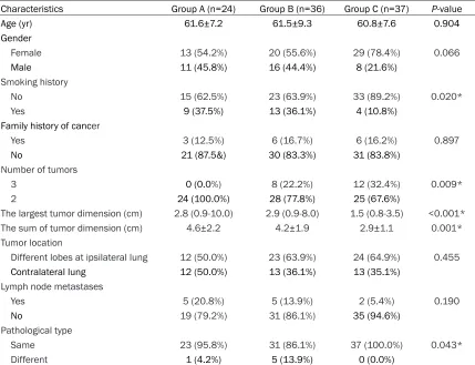
[image:2.612.92.521.96.425.2]The postoperative histology was determined by two experienced lung pathologists. LUAD was classified according to the criteria of IASLC/ ATS/ERS International Multidisciplinary LUAD Classification [14]. For each LUAD tumor, the proportions of the five histological subtypes (acinar, papillary, micropapillary, solid, and lep-idic) were calculated. Epidermal growth factor Table 1. Clinical and pathological characteristics of synchronous multiple primary lung cancer pa-tients classified into three different groups
Characteristics Group A (n=24) Group B (n=36) Group C (n=37) P-value
Age (yr) 61.6±7.2 61.5±9.3 60.8±7.6 0.904
Gender
Female 13 (54.2%) 20 (55.6%) 29 (78.4%) 0.066
Male 11 (45.8%) 16 (44.4%) 8 (21.6%)
Smoking history
No 15 (62.5%) 23 (63.9%) 33 (89.2%) 0.020*
Yes 9 (37.5%) 13 (36.1%) 4 (10.8%)
Family history of cancer
Yes 3 (12.5%) 6 (16.7%) 6 (16.2%) 0.897
No 21 (87.5&) 30 (83.3%) 31 (83.8%)
Number of tumors
3 0 (0.0%) 8 (22.2%) 12 (32.4%) 0.009*
2 24 (100.0%) 28 (77.8%) 25 (67.6%)
The largest tumor dimension (cm) 2.8 (0.9-10.0) 2.9 (0.9-8.0) 1.5 (0.8-3.5) <0.001*
The sum of tumor dimension (cm) 4.6±2.2 4.2±1.9 2.9±1.1 0.001* Tumor location
Different lobes at ipsilateral lung 12 (50.0%) 23 (63.9%) 24 (64.9%) 0.455
Contralateral lung 12 (50.0%) 13 (36.1%) 13 (35.1%)
Lymph node metastases
Yes 5 (20.8%) 5 (13.9%) 2 (5.4%) 0.190
No 19 (79.2%) 31 (86.1%) 35 (94.6%)
Pathological type
Same 23 (95.8%) 31 (86.1%) 37 (100.0%) 0.043*
Different 1 (4.2%) 5 (13.9%) 0 (0.0%)
receptor (EGFR) mutation status was tested by polymerase chain reaction amplification or direct DNA sequencing. Anaplastic Lymphoma kinase (ALK) and C-ros oncogene 1 (ROS1) fusions were detected by immunohistochemis-try method [15].
Statistical analysis
Statistical analyses were performed using SPSS version 17.0 (SPSS Inc., Chicago, IL, USA). Measurement data were expressed as mean ± standard deviation or median (range), and com-parison between groups was performed by one-way ANOVA or nonparametric test. Count data were expressed as frequency or percent -age, and group comparison was performed by Chi-square test or Fisher’s exact test. P-values less than 0.05 were considered statistically significant.
Results
Patient clinicopathological characteristics
There were 97 consecutive patients who were diagnosed with SMPLC at West China Hospital of Sichuan University between January 2014 and April 2017. The clinicopathological charac-teristics of the 97 patients with SMPLC were summarized in Table 1. No mortality occurred during the follow-up period (median 14 months; range 1-45 months). Of the 97 SMPLC patients, 62 (64.0%) were female and 35 (36.0%) were male, with a median age of 64 years (range 30-77 years). Seventy-one patients (73.2%) reported no history of smoking. About the am-
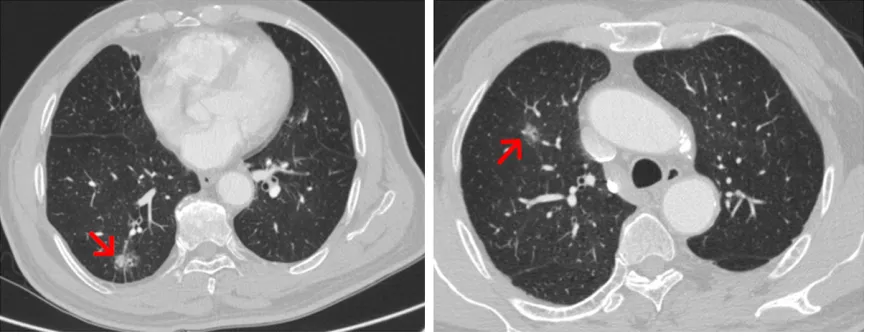
ount of tumors, 77 (79.4%) patients had two primary lung cancers and 20 (20.6%) had th-ree primary lung cancers. The majority (91/97, 93.8%) of patients harbored tumors with the same histological type and the other 6 patients had tumors with different histological types. Notably, 75.3% (73/97) of the patients har -bored at least one GGO tumor. Figure 1 shows a case of SMPLC manifested with two different GGO tumors at the upper lobe and the lower lobe of the right lung, respectively, on the CT scan.
Of all 97 patients, 24 (24.7%) were in Group A, 36 (37.1%) in Group B and 37 (38.2%) in Group C. In the subgroup analyses, the difference of smoking history, number of tumors, the largest tumor dimension, the sum of tumor dimension and pathological type among the three groups was statistically significant (P<0.05), whereas no statistical difference existed in age, gender, family history of cancer, tumor location or lym- ph node metastases. Furthermore, compared with Group A, Group C had more females (P=0.046), more non-smokers (P=0.013) and more patients with three tumors (P=0.005). On the other hand, both the largest tumor dimen-sion (P<0.001) and the sum of tumor dimen -sion in Group C were smaller than those in Group A (P<0.001).
Surgical procedure and postoperative pathol-ogy
[image:3.612.90.525.72.240.2]All the 97 patients received surgery treatment and no mortality occurred during the peropera-tive period (Table 2). The majority (98.4%) of Figure 1. Chest CT showing a case of synchronous multiple primary lung cancer (SMPLC) manifested with two
Table 2. Surgical procedure of the patients
Characteristics tumors (%)Ipsilateral Contralateral tumors (%)
Surgical stage
Single-stage 60 (98.4%) 12 (33.3%)
Two-stage 1 (1.6%) 24 (66.7%)
Surgical approach
Thoracotomy 16 (26.2%) 10 (27.8%)
VATS 45 (73.8%) 20 (55.6%)
Thoracotomy + VATS 0 (0.0%) 6 (16.6%) Surgical resection type
Lobectomy 3 (4.9%) 2 (5.6%) Lobectomy + sublobectomy 39 (63.9%) 23 (63.9%)
Sublobectomy 19 (31.2%) 11 (30.5%)
VATS, video-assisted thoracoscopic surgery.
129/214) were more than solid tumors (39.7%, 85/214).
There were 57 patients for whom all tumors were diagnosed as specific subtypes of LUAD. Fifty-three patients (93.0%) were found to have tumors with different patterns of histological subtypes, i.e., 42 patients (73.7%) had tumors with different predominant histological sub-types, and 11 (19.3%) had tumors with the same predominant histological subtype but with different proportions of other subtypes.
Genetic alterations
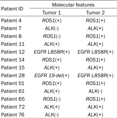
Among the 214 surgically resected patients, 160 were detected with the genetic alterations.
EGFR activating mutations were identified in 50.0% (33/66) tumors, ALK found in 9.4% (15/160) tumors, and ROS1 fusions in 7.8% (11/141) of the tumors. Thirteen patients had all tumors detected and 6 patients showed dif-ferent molecular alteration in separate tumors (Table 4), suggesting a concordance of 46.2% (6/13) between molecular features and clinical diagnostic criteria.
Discussion
Despite the rising incidence of SMPLC, we are lack of a consistent diagnostic standard. Most studies refer to ACCP diagnostic criteria, which emphasize that clinicians should take clinical, imaging and pathological features into consid-eration [2]. In the present study, we analyzed the clinicopathological and radiological charac-teristics of 97 patients with SMPLC and ex-
Table 3. Clinical and pathological characteris-tics of the lesions
Characteristics Number (%)
Total 214 (100%)
Location
Right upper lobe 71 (33.2%)
Right middle lobe 22 (10.3%)
Right lower lobe 51 (23.8%)
Left upper lobe 42 (19.4%)
Left lower lobe 28 (13.2%)
Histological type and subtype
Sum of LUAD 206
AIS 26 (12.7%)
MIA 23 (11.2%)
Lepidic predominant 55 (26.7%)
Acinar predominant 48 (23.3%)
Papillary predominant 19 (14.1%)
Solid predominant 5 (2.4%)
Micropapillary predominant 1 (0.5%)
Not known 29 (7.5%)
LUSC 6
Others 2
Density on CT scan
Solid 85 (39.7%)
Mixed GGO 76 (35.5%)
Pure GGO 53 (24.8%)
ADC, adenocarcinoma; AIS, adenocarcinoma in situ;
LUAD, lung adenocarcinoma; LUSC, lung squamous cell
carcinoma; MIA, minimally invasive adenocarcinoma;
GGO, ground-glass opacity.
those with tumors at the contralateral lung underwent two-stage surgery. Video-assist- ed thoracoscopic surgery (VATS) was the most common approach for both ipsilateral (73.8%) and contralateral tumors (55.6%), and the combination of lobectomy and sub-lobectomy was the predominant resection type for both (63.9% and 63.9%, respe-ctively).
A total of 214 tumors from the 97 patients were radically resected (Table 3). The most common locations were the right upper lobe (33.2%), followed by the right lower lobe (23.8%). The major histological type was LUAD (96.3%, 206/214), with lepidic (26.7%) and acinar (23.3%) as the leading subtypes. It was noteworthy that GGO tumors (60.3%,
[image:4.612.90.289.309.631.2]Table 4. Molecular alterations of thirteen patients with specific genotypes
Patient ID Molecular features
Tumor 1 Tumor 2
Patient 4 ROS1(+) ROS1(+)
Patient 7 ALK(-) ALK(+)
Patient 8 ROS1(-) ROS1(+)
Patient 11 ALK(+) ALK(+)
Patient 12 EGFR L858R(+) EGFR L858R(+)
Patient 14 ROS1(+) ROS1(+)
Patient 15 ALK(+) ALK(+)
Patient 28 EGFR 19-del(+) EGFR L858R(+)
Patient 51 ROS1(+) ROS1(+)
Patient 61 ALK(+) ALK(-)
Patient 65 ROS1(-) ROS1(+)
Patient 72 ALK(+) ALK(+)
Patient 76 ALK(-) ALK(+)
EGFR, epidermal growth factor receptor; ALK, anaplastic
Lymphoma kinase; ROS1, c-ros oncogene 1.
plored the validity of histological subtyping and genotyping in differential diagnosis between SMPLC and intrapulmonary metastases. One important finding of this study was that patients with SMPLC harbored high frequency of GGO tumors. Among the 97 SMPLC patients, 75.3% had at least one GGO tumor and 60.3% of the 214 surgically resected tumors were GGO lesions. Consistently, a previous study sh-owed that GGO lesions accounted for 79.23% of the 833 surgically removed SMPLC tumors [16]. Although some previous studies reported the clincopathological characteristics of SM- PLC, they did not report the difference between GGO SMPLC and solid SMPLC. When compar -ing the clinical characteristics of patients with different imaging features, we found that GGO SMPLC was more common in females (P= 0.046) and non-smokers (P=0.013) than solid SMPLC, with more tri-primary tumors (P= 0.005) and smaller dimensions (P<0.001). These indicated that for patients with syn- chronous multiple lung GGO lesions, clinicians should take SMPLC into consideration, espe-cially in females and non-smokers.
Another major finding of this study was that most (93.8%) of SMPLC patients had tumors with one same histological type, with LUAD being the most frequently observed one (96.3%). Consistent with our finding, Zhang et al. summarized clinicopathological
characteris-tics of 285 patients with SMPLC and found all the tumors of 81.8% of patients were LUAD [10]. Against the traditional difficulty in distin -guishing SMPLC from intrapulmonary metasta-ses in synchronous multiple LUAD tumors, the application of histological subtyping is now offering valuable information [2, 17, 18]. As is known, LUAD is histologically heterogeneous with a mixture of lepidic, acinar, papillary, solid, and micropapillary subtypes [19]. ACCP has suggested synchronous multiple LUAD tumors being defined on the basis of the histological subtyping, i.e., the proportions of different his-tological subtypes [2]. In the present study, we observed the concordance up to 93.0% be-tween histological subtyping and clinical diag-nostic criteria. In a previous study, Murphy et al. also applied histological subtyping for distin-guishing independent primary tumors and me- tastases, and found the concordance of 81.8% between histological subtyping and patterns of DNA rearrangement breakpoints [17]. Thus, histological subtyping could be advocated as an additional reference to differentiate SMPLC from intrapulmonary metastases.
Many studies assessed particular genetic alter-ations to define clonal relalter-ationship of multiple lung cancers, assuming that a match of genetic alterations defines a single clone and metasta -ses, whereas a difference defines separate cancers [5, 20]. We also explored the applica -tion of genetic analysis in diagnosis of SMPLC, and found that the concordance of 46.2% (6/13) between molecular alterations (EGFR
activating mutations, ALK and ROS1 fusions) and clinical diagnostic criteria. Similarly, the accordance in EGFR mutations between sepa-rate primary tumors in patients with SMPLCs was reported to be 35% [21]. In addition, previ -ous studies have revealed that the discordance in genetic alterations (EGFR and KRAS muta-tions) between lung primary tumor and meta-static sites varied from 10% to 85% [22-27]. The heterogeneity of genetic alterations in clearly related lung primary tumor and meta-static site calls for caution. Therefore, it is unclear that either the different genotypes in specific driver genes identify separate primary cancers or that the same genetic alteration defines intrapulmonary metastases.
-quently in females, non-smokers and patients with tri-primary tumors. Our study also indicat-ed that histological subtyping, instead of geno-typing, could be advocated as an additional reference to distinguish SMPLC from intrapul-monary metastases.
Acknowledgements
This work was supported by the Science and Technology Support Program of Sichuan Pro- vince (No.2016CZYD0001 to Weimin Li and No.2016SZ0073 to Panwen Tian).
Disclosure of conflict of interest
None.
Address correspondence to: Dr. Weimin Li, Depart- ment of Respiratory and Critical Care Medicine, West China Hospital of Sichuan University, 37
Guoxue Xiang, Chengdu 610041, Sichuan, China.
Tel: +86-28-85423998; Fax: +86-28-85582944; E-mail: [email protected]
References
[1] Martini N and Melamed MR. Multiple primary lung cancers. J Thorac Cardiovasc Surg 1975; 70: 606-612.
[2] Kozower BD, Larner JM, Detterbeck FC and
Jones DR. Special treatment issues in non-small cell lung cancer: diagnosis and manage-ment of lung cancer, 3rd edition: American col-lege of chest physicians evidence-based clinical practice guidelines. Chest 2013; 143: e369S-e399S.
[3] Ishikawa Y, Nakayama H, Ito H, Yokose T, Tsub-oi M, Nishii T and Masuda M. Surgical treat-ment for synchronous primary lung adeno- carcinomas. Ann Thorac Surg 2014; 98: 1983-1988.
[4] Liu M, He W, Yang J and Jiang G. Surgical treat -ment of synchronous multiple primary lung cancers: a retrospective analysis of 122 pa-tients. J Thorac Dis 2016; 8: 1197-1204.
[5] Detterbeck FC, Franklin WA, Nicholson AG, Gi -rard N, Arenberg DA, Travis WD, Mazzone PJ, Marom EM, Donington JS, Tanoue LT, Rusch VW, Asamura H, Rami-Porta R; IASLC Staging and Prognostic Factors Committee; Advisory
Boards; Multiple Pulmonary Sites Workgroup.
The IASLC lung cancer staging project: back-ground data and proposed criteria to distin-guish separate primary lung cancers from metastatic foci in patients with two lung tu-mors in the forthcoming eighth edition of the
TNM classification for lung cancer. J Thorac
Oncol 2016; 11: 651-665.
[6] Chang YL, Wu CT and Lee YC. Surgical treat-ment of synchronous multiple primary lung cancers: experience of 92 patients. J Thorac Cardiovasc Surg 2007; 134: 630-637.
[7] Mun M and Kohno T. Single-stage surgical
treatment of synchronous bilateral multiple lung cancers. Ann Thorac Surg 2007; 83: 1146-1151.
[8] Trousse D, Barlesi F, Loundou A, Tasei AM, Doddoli C, Giudicelli R, Astoul P, Fuentes P and
Thomas P. Synchronous multiple primary lung cancer: an increasing clinical occurrence
re-quiring multidisciplinary management. J Tho -rac Cardiovasc Surg 2007; 133: 1193-1200.
[9] Shimada Y, Saji H, Otani K, Maehara S, Maeda J, Yoshida K, Kato Y, Hagiwara M, Kakihana M, Kajiwara N, Ohira T, Akata S and Ikeda N. Sur -vival of a surgical series of lung cancer pa-tients with synchronous multiple ground-glass opacities, and the management of their resid-ual lesions. Lung Cancer 2015; 88: 174-180.
[10] Zhang Z, Gao S, Mao Y, Mu J, Xue Q, Feng X
and He J. Surgical outcomes of synchronous multiple primary non-small cell lung cancers. Sci Rep 2016; 6: 23252.
[11] Peng Y, Ren W, Wang H, Li M, Feng Z and Peng Z. Surgical treatment is an effective approach for patients with synchronous multiple primary lung cancers. J Cancer Res Ther 2017; 13: 702-706.
[12] Riquet M, Cazes A, Pfeuty K, Ngabou UD, Fou
-cault C, Dujon A and Banu E. Multiple lung can -cers prognosis: what about histology? Ann Tho-rac Surg 2008; 86: 921-926.
[13] Pedersen JH, Saghir Z, Wille MM, Thomsen LH,
Skov BG and Ashraf H. Ground-glass opacity
lung nodules in the era of lung cancer CT screening: radiology, pathology, and clinical management. Oncology (Williston Park) 2016; 30: 266-274.
[14] Travis WD, Brambilla E, Noguchi M, Nicholson AG, Geisinger KR, Yatabe Y, Beer DG, Powell CA, Riely GJ, Van Schil PE, Garg K, Austin JH, Asamura H, Rusch VW, Hirsch FR, Scagliotti G,
Mitsudomi T, Huber RM, Ishikawa Y, Jett J, San-chez-Cespedes M, Sculier JP, Takahashi T, Tsuboi M, Vansteenkiste J, Wistuba I, Yang PC,
Aberle D, Brambilla C, Flieder D, Franklin W, Gazdar A, Gould M, Hasleton P, Henderson D, Johnson B, Johnson D, Kerr K, Kuriyama K, Lee
JS, Miller VA, Petersen I, Roggli V, Rosell R, Saijo N, Thunnissen E, Tsao M and Yankelewitz D. International association for the study of lung cancer/american thoracic society/euro-pean respiratory society international
multidis-ciplinary classification of lung adenocarcino -ma. J Thorac Oncol 2011; 6: 244-285.
[16] Guo H, Mao F, Zhang H, Qiu Y and Shen-Tu Y.
Analysis on the prognostic and survival factors of synchronous multiple primary lung cancer. Zhongguo Fei Ai Za Zhi 2017; 20: 21-27.
[17] Murphy SJ, Aubry MC, Harris FR, Halling GC, Johnson SH, Terra S, Drucker TM, Asiedu MK, Kipp BR, Yi ES, Peikert T, Yang P, Vasmatzis G and Wigle DA. Identification of independent
primary tumors and intrapulmonary metasta-ses using DNA rearrangements in non-small-cell lung cancer. J Clin Oncol 2014; 32: 4050-4058.
[18] Zhang Y, Hu H, Wang R, Ye T, Pan Y, Wang L, Zhang Y, Li H, Li Y, Shen L, Yu Y, Sun Y, Chen H
and Garfield D. Synchronous non-small cell
lung cancers: diagnostic yield can be improved by histologic and genetic methods. Ann Surg Oncol 2014; 21: 4369-4374.
[19] Travis WD, Brambilla E, Nicholson AG, Yatabe Y, Austin JH, Beasley MB, Chirieac LR, Dacic S, Duhig E, Flieder DB, Geisinger K, Hirsch FR, Ishikawa Y, Kerr KM, Noguchi M, Pelosi G, Pow -ell CA, Tsao MS and Wistuba I. The 2015 world
health organization classification of lung tu -mors: impact of genetic, clinical and radiologic
advances since the 2004 classification. J Tho -rac Oncol 2015; 10: 1243-1260.
[20] Shen C, Wang X, Tian L, Zhou Y, Chen D, Du H, Wang W, Liu L and Che G. “Different trend” in
multiple primary lung cancer and intrapulmo-nary metastasis. Eur J Med Res 2015; 20: 17.
[21] Cheng H, Lei BF, Peng PJ, Lin YJ and Wang XJ.
Histologic lung cancer subtype differentiates synchronous multiple primary lung adenocar-cinomas from intrapulmonary metastases. J Surg Res 2017; 211: 215-222.
[22] Chang YL, Wu CT, Lin SC, Hsiao CF, Jou YS and Lee YC. Clonality and prognostic implications of p53 and epidermal growth factor receptor somatic aberrations in multiple primary lung cancers. Clin Cancer Res 2007; 13: 52-58.
[23] Takamochi K, Oh S, Matsuoka J and Suzuki K.
Clonality status of multifocal lung adenocarci-nomas based on the mutation patterns of
EGFR and K-ras. Lung Cancer 2012; 75:
313-320.
[24] Chung JH, Choe G, Jheon S, Sung SW, Kim TJ, Lee KW, Lee JH and Lee CT. Epidermal growth
factor receptor mutation and pathologic-radio-logic correlation between multiple lung nod-ules with ground-glass opacity differentiat- es multicentric origin from intrapulmonary spread. J Thorac Oncol 2009; 4: 1490-1495.
[25] Warth A, Macher-Goeppinger S, Muley T, Thom -as M, Hoffmann H, Schnabel PA, Penzel R, Schirmacher P and Aulmann S. Clonality of multifocal nonsmall cell lung cancer: implica-tions for staging and therapy. Eur Respir J 2012; 39: 1437-1442.
[26] Wang X, Wang M, MacLennan GT, Abdul-Karim
FW, Eble JN, Jones TD, Olobatuyi F, Eisenberg
R, Cummings OW, Zhang S, Lopez-Beltran A,
Montironi R, Zheng S, Lin H, Davidson DD and Cheng L. Evidence for common clonal origin of multifocal lung cancers. J Natl Cancer Inst 2009; 101: 560-570.
[27] Girard N, Deshpande C, Azzoli CG, Rusch VW,
Travis WD, Ladanyi M and Pao W. Use of
epi-dermal growth factor receptor/Kirsten rat sar -coma 2 viral oncogene homolog mutation
test-ing to define clonal relationships among