RESEARCH ARTICLE
Morphometric models for estimating bite force in
Mus
and
Rattus
:
mandible shape and size perform better than lever-arm ratios
Samuel Ginot1,2,*, Anthony Herrel3, Julien Claude1and Lionel Hautier1ABSTRACT
Morphological traits are frequently used as proxies for functional outputs such as bite force performance. This allows researchers to infer and interpret the impacts of functional variation, notably in adaptive terms. Despite their mechanical bases, the predictive power of these proxies for performance is not always tested. In particular, their accuracy at the intraspecific level is rarely assessed, and they have sometimes been shown to be unreliable. Here, we compared the performance of several morphological proxies in estimatingin vivo
bite force, across five species of murine rodents, at the interspecific and intraspecific levels. Proxies used included the size and shape of the mandible, as well as individual and combined muscular mechanical advantage (temporalis, superficial masseter and deep masseter). Maximum voluntary bite force was measured in all individuals included. To test the accuracy of predictions allowed by the proxies, we combined linear regressions with a leave-one-out approach, estimating an individual’s bite force based on the rest of the dataset. The correlations between estimated values and thein vivo
measurements were tested. At the interspecific and intraspecific levels, size and shape were better estimators than mechanical advantage. Mechanical advantage showed some predictive power at the interspecific level, but generally not within species, except for the deep masseter inRattus. In a few species, size and shape did not allow us to predict bite force. Extrapolations of performance based on mechanical advantage should therefore be used with care, and are mostly unjustified within species. In the latter case, size and shape are preferable.
KEY WORDS: Adaptation, Morphology, Murid, Performance, Rodentia
INTRODUCTION
For decades, deductions of functional outputs from morphology have been routinely used in an adaptationist framework to infer the potential selective advantage of phenotypic variation (Gould and Lewontin, 1979; Mayr, 1983). However, this evaluation was often done without measuring functional performance until the integration of functional morphology within evolutionary biology (Arnold, 1983). Since then, some of the best examples of adaptation and adaptive radiation have been illustrated using this integrative approach (e.g. Grant and Grant, 2002; Herrel et al., 2005, 2009).
Yet, relationships between morphology and function vary at different scales (e.g. interspecific, intraspecific or intrapopulation), and only a precise quantification of the links between morphological and functional variation can avoid the pitfalls of a pan-adaptationist approach (Gould and Lewontin, 1979; Mayr, 1983).
In the diversified clade of rodents, links between skull or mandible morphology and diet or ecology have been reported for several groups and at different taxonomic scales (Michaux et al., 2007; Samuels, 2009; Hautier et al., 2009, 2011, 2012; Cox et al., 2012). The influence of skull and mandible morphology onin vivo bite force performance has also been directly tested, using various anatomical variables (e.g. Freeman and Lemen, 2008; Ginot et al., 2018). The use of biomechanical models [i.e. combining muscle physiological cross-sectional area (PCSA) and lines of action] or mechanical descriptors (combinations of mandibular measurements) in these studies allowed the accurate estimation of bite force (compared with in vivo data) at the interspecific level. However, Ginot et al. (2018) showed that these estimates of bite force were less precise at the intraspecific level. Furthermore, this approach requires the accurate dissection of muscles, to dissolve them and measure fiber length for individual muscle strands, and is therefore time consuming and inapplicable to specimens for which muscles have not been preserved. Therefore, other osteological proxies, such as morphometric data or mechanical advantage (i.e. muscular lever-arm ratios) are often used to estimate bite force (Greaves, 1983; Kiltie, 1984; Thomason, 1991; Christiansen and Adolfssen, 2005; Ellis et al., 2008).
Here, we estimated bite force using osteological proxies in five species of murine rodents [Mus musculus Linnaeus 1758; Mus
cervicolor Hodgson 1845; Mus caroli Bohnote 1902; Rattus
exulans (Peale 1848) andRattus tanezumi Temminck 1844], for
whichin vivomeasurements were also taken. Several osteological proxies were tested and compared: mandible size and shape, the mechanical advantage of the temporalis muscle, the mechanical advantage of the superficial masseter muscle and the mechanical advantage of the deep masseter muscle, as well as the combined mechanical advantage of all three. Although mechanical advantage makes mechanical sense and has previously been used as a proxy for function (e.g. Thorington and Darrow, 1996; Velhagen and Roth, 1997; Swiderski and Zelditch, 2010; Blanco et al., 2013; Casanovas-Vilar and van Dam, 2013; Gomes Rodrigues et al., 2016; Fabre et al., 2017; Renaud et al., 2015, 2018a,b; Parmenter et al., 2019; Souquet et al., 2019), its relationship with bite force has never been formally tested. The mechanical expectation is that individuals with larger values for mechanical advantage will show larger bite forces. The tighter this relationship, the more precise our estimates should be, producing a stronger correlation (i.e. closer to 1) between estimated andin vivobite force. However, shape may be a better predictor because the complexity of shape variation as quantified by geometric morphometrics may be more integrative than the limited number of scalars obtained from lever mechanical
Received 8 April 2019; Accepted 13 May 2019
1Institut des Sciences de l’Évolution de Montpellier (UMR5554), 34095 Montpellier Cedex 5, France.2Institut de Génomique Fonctionnelle de Lyon (UMR5242), 69007 Lyon, France.3Muséum National d’Histoire Naturelle (UMR7179), 75005 Paris, France.
*Author for correspondence (samuel.ginot@ens-lyon.fr)
S.G., 0000-0003-0060-9660; A.H., 0000-0003-0991-4434
Journal
of
Experimental
advantage. Notably, the strong allometric component of shape variation means that shape also integrates a large part of the signal linked to size. Although it is expected that more complex methods, using muscular characteristics to estimate forces, will give much better estimations (see Ginot et al., 2018), the aim of this paper was to verify and compare the validity of osteological proxies that are easy to access (allowing large sample sizes), already in use in the literature and available even when soft tissues are absent (e.g. paleontology).
MATERIALS AND METHODS Specimens
Four species were caught in the wild with no control for age:
M. cervicolor (n=65), M. caroli (n=13), R. exulans (n=42) and
R. tanezumi(n=29). All were caught with live traps (either handmade
local cage traps or Sherman traps) over two field sessions (2015 and 2016) during the dry season (February–March) in several localities across northern, eastern and north-eastern Thailand. Sampled sites were around the towns of Tha Wang Pha, Nan Province; Sakaerat, Nakhon Ratchasima Province; Mahasarakham, Mahasarakham Province; and Sahatsakhan, Kalasin Province. Mus musculus specimens (n=51) were raised in the lab at the University of Montpellier and are descendants from wild ancestors captured in the Orkney Islands (Scotland). These individuals all had their bite forces measured at 68 days and were subsequently killed by CO2inhalation.
Males and females were pooled to improve the sample size on which the predictive bite force models were built (see below).
Bite force measurements
Shortly after their capture, we measured the voluntary bite force at the incisors of each individual using a piezoelectric force transducer (Kistler, type 9203, range 0–500 N, accuracy 0.01–0.1 N; Amherst, NY, USA; calibrated by the constructor at 25°C and 36% humidity) attached to a handheld charge amplifier (Kistler, type 5995; Herrel et al., 1999). The force transducer was mounted between two steel bite plates as described in Herrel et al. (1999). We adjusted the distance between the bite plates by measuring it with a caliper, and by increasing or decreasing it via the micrometer head, so that each individual bit at a consistent gape angle of∼30 deg. All animals bit directly onto steel at the same spot on the plates (i.e. at the tip), to ensure a consistent out-lever length. We recorded three trials in a row for each individual, and the maximal score was used in the analyses. All measurements were taken by one user (S.G.) to avoid inter-user variation.
Animals were treated in accordance with the guidelines of the American Society of Mammalogists, and within the European Union
legislation guidelines (Directive 86/609/EEC). Approval notices for trapping and investigation of rodents in the field (Thailand) were provided by the Ethical Committee of Mahidol University, Bangkok, Thailand (number 0517.1116/661), for the CERoPath protocols ( project ANR 07 BDIV 012). All lab procedures were carried out under approval no. A34-172-042 (Hérault Prefecture).
Morphometric analyses
All mandibles were skeletonized manually, after which they were photographed in a standardized way (camera at a fixed distance, mandible positioned flat with the lingual side down) using a Pentax K200D reflex camera, with a 45 mm focal distance. In total, 17 landmarks were placed on each mandible (Fig. 1) using tpsDig2.x software to represent shape, and to calculate the length of lever arms. Additionally, we computed centroid size of the mandible to be used as our measure of size. The coordinate data were imported in R (http://www.R-project.org/) and scaled, centered and superimposed using Procrustes analyses routines from Claude (2008). In order to avoid possible overparameterization of statistical predictive models, shape data were submitted to principal component analyses for variable reduction, and the principal components (PCs) representing a total of 90% of variation were kept. Mechanical advantage for the temporalis muscle, superficial masseter and deep masseter was obtained by computing the respective in-lever/out-lever ratios (Fig. 1). In the case of the superficial masseter, two different in-lever measurements were used, corresponding to the ventral-most insertion point and posterior-most insertion point (following Velhagen and Roth, 1997; Swiderski and Zelditch, 2010). Only the incisor out-lever was used, as ourin vivomeasurements were restricted to incisive bites.
Bite force estimates
Using log10 mechanical advantage (individually and combined),
log10centroid size or shape (PCs including 90% of shape variation)
data, we fitted linear models of log10in vivobite force either within
species or combining the entire dataset (i.e. using all individual values). The model of combined mechanical advantage was built by using them all as explanatory variables in the same model. Interaction effects were checked and found to be non-significant, and were therefore dropped. To test the precision of these models, we used a leave-one-out validation approach. To do so, we took out one individual from the dataset, fitted the model, then used the‘predict’ function in R to compute a bite force estimate for this individual. After iterating this process for all individuals, we compared estimated and in vivo bite force using one-tailed Pearson’s correlation coefficient in which the alternative hypothesis was that
B
A
1/E
14
13
12
11/D
10/F 9
8 3 2
4
5/C
6 7
[image:2.612.48.414.581.737.2]15
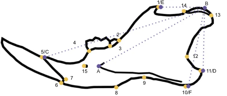
Fig. 1. Outline of the mouse mandible, showing the landmarks used in this study.
Yellow landmarks are those used for shape analysis, and purple landmarks are those used to calculate lever arms. Shape and lever arms were used in separate analyses. A–B: deep masseter in-lever; B–D: superficial masseter in-lever ( posterior-most); B–F: superficial masseter in-lever (ventral-most); B–E: temporalis in-lever; B–C: out-lever.
Journal
of
Experimental
the correlation was greater than 0, because estimations should be positively correlated within vivomeasures. We also computed linear models of estimated againstin vivobite forces, to obtain the adjusted
R̅2values to quantify and compare the precision of the estimations.
Morphological and bite force data are available from the corresponding author on request.
RESULTS
At the interspecific level (Fig. 2A, Table 1), all morphometric estimations of bite force were significantly and positively correlated
with in vivo bite force. The superficial masseter mechanical
advantage was, however, only predictive when using the ventral insertion point (i.e. points B–F in Fig. 1). Considering the correlation coefficients andR̅2values (Table 1), it is clear that the size and shape
of the mandible are better estimators than the individual or combined mechanical advantage. Despite this, the last two do show some predictive power. Shape differences related to bite force variation basically represent differences between small mice, with longer and more slender mandibles, and large rats, with shorter, more robust mandibles, a larger angular process and a posteriorly developed coronoid process (Fig. 2C). It can be seen that intraspecifically (especially in Mus species; Figs 2B and 3), some slopes appear strongly negative, which is necessarily artefactual: if the model (significant or not) has a negative slope, and morphology is less variable than the measured bite force, the leave-one-out prediction based on this model will simply follow the regression line, despite representing very little morphological variation.
Mus musculus
In the lab-reared mice (Fig. 2B), age was controlled, and all specimens in this study were 68 days old. We found a significant positive relationship between shape-estimated bite force andin vivo bite force (r=0.32, t=2.40, d.f.=49, P=0.01). In contrast, the estimations based on mechanical advantage or centroid size were not significantly and positively correlated within vivodata (Table 1).
Mus caroli
In this wild species (Fig. 3A), although we had fewer specimens than in others, we found a significant correlation (but non-significant linear regression) between shape estimates of bite force
and in vivo bite force (r=0.51, t=1.96, d.f.=11, P=0.03). Size
estimations were not significantly related toin vivomeasurements (r=0.47,t=1.75, d.f.=11,P=0.054). However, this may simply be due to the small sample size, and the relationship may in fact be significant with more measurements. Again, the mechanical advantage-based estimations were not significantly related to
in vivobite force data (Table 1).
Mus cervicolor
Our sample was larger than forM. caroliand shape-estimated bite force values were again significantly positively correlated toin vivo data in this wild mouse species (r=0.26,t=2.13, d.f.=63,P=0.019, Fig. 3B). Here, size was a better estimator than shape (r=0.36,
t=3.073, d.f.=63,P=0.0016). However, the mechanical advantage estimates were not correlated toin vivovalues (Table 1, Fig. 3B).
Rattus exulans
ForR. exulans, both size and shape estimations were correlated toin
vivobite force (Fig. 4A), with a stronger correlation for size (r=0.55,
t=4.16, d.f.=40,P<0.001) than for shape estimates (r=0.41,t=2.84, d.f.=40,P=0.0035). In this species, the deep masseter mechanical advantage also had significant predictive power, although less than size or shape (r=0.36, t=2.46, d.f.=40, P=0.0092). Both the superficial masseter and temporalis mechanical advantage estimates of bite fore were not significantly correlated within vivo bite force (Table 1). The combined mechanical advantage yielded a significant correlation between estimated and in vivo bite force, although it was less than that for the deep masseter estimate (r=0.33,
t=2.20, d.f.=40,P=0.017).
Rattus tanezumi
Contrary to findings for all other species, the shape-estimated bite force did not correlate significantly with in vivo data (r=0.16,
t=0.86, d.f.=27, P=0.20; Fig. 4B). However, there were positive correlations between the deep masseter mechanical advantage (r=0.55,t=3.44, d.f.=27,P=0.00095) and size estimates andin vivo bite force (r=0.49, t=2.93, d.f.=27, P=0.0034). The combined mechanical advantage estimations were also correlated to in vivo bite force, although less so than size or deep masseter estimations (r=0.47, t=2.66, d.f.=27, P=0.0065). The other mechanical advantage estimates did not show significant positive correlations
within vivobite force (Table 1).
DISCUSSION
[image:3.612.48.566.606.707.2]Our results show that despite being commonly used as a functional proxy (e.g. Thorington and Darrow, 1996; Velhagen and Roth, 1997; Swiderski and Zelditch, 2010; Blanco et al., 2013; Casanovas-Vilar and van Dam, 2013; Gomes Rodrigues et al., 2016; Fabre et al., 2017; Renaud et al., 2015, 2018a,b; Parmenter et al., 2019; Souquet et al., 2019), mechanical advantage generally does not appear to be an accurate estimator ofin vivobite force, at least for incisor bites, and across our sample of species. In particular, the temporalis mechanical advantage bite force estimates were never significantly related toin vivobite force at the intraspecific level. At the interspecific level, the superficial masseter mechanical
Table 1. Pearson correlation coefficients and adjustedR̅² from the various analyses run in this study
Size Shape
Mechanical advantage
d.f.
SM SM2 DM T SM2+DM+T
R̅² r R̅² r R̅² r R̅² r R̅² r R̅² r R̅² r
Interspecific 0.53 0.73 0.52 0.72 0.00 −0.01 0.17 0.42 0.18 0.42 0.20 0.45 0.32 0.57 1; 198
Mus caroli 0.15 0.47 0.19 0.51 −0.09 0.03 −0.97 0.93 0.14 −0.46 0.61 −0.80 0.72 −0.87 1; 11
Mus cervicolor 0.12 0.36 0.05 0.26 −0.01 0.04 −0.02 −0.01 0.00 0.12 0.69 −0.84 −0.02 0.01 1; 63
Mus musculus −0.01 0.09 0.09 0.32 0.32 −0.58 0.06 −0.28 0.00 0.13 −0.01 −0.15 0.01 0.16 1; 49
Rattus exulans 0.28 0.55 0.15 0.41 −0.02 −0.08 0.00 −0.13 0.11 0.36 −0.02 −0.01 0.09 0.33 1; 40
Rattus tanezumi 0.21 0.49 −0.01 0.16 −0.03 −0.11 0.06 0.31 0.28 0.55 0.04 −0.26 0.18 0.47 1; 27
SM, superficial masseter mechanical advantage (posterior-most insertion); SM2, superficial masseter mechanical advantage (ventral-most insertion); DM, deep masseter mechanical advantage; T, temporalis mechanical advantage.
Bold values denote significance (P<0.05).
Journal
of
Experimental
advantage only allowed us to make correct predictions ofin vivobite force when using the ventral-most insertion, and the temporalis mechanical advantage also showed some predictive power
(although both had lowR̅2values; Table 1). In contrast, the deep
masseter mechanical advantage did better, with significant correlations between estimated andin vivodata at the interspecific 0.8
1.0
Size estimate
Shape estimate
DP
. mass.
estimate
Sp. mass. estimate
Sp. mass. 2 estimate
T
e
mp. estimate
Combined MA
estimate
1.2 1.4
A
B
C
0.90
0.92 0.94 0.96 0.98
0.94 0.95 0.96 0.97
0.92 0.93 0.95
0.94 0.96
0.93 0.94 0.96
0.95 0.97
0.88 0.96
0.92 1.00 0.9
0.8 1.0 1.1 1.2 0.92 0.94 0.96 0.98 1.00
0.4
0.6
0.82
1.2
1.1
1.0
0.9
0.8
0.7
1.1
1.0
0.9
0.8
0.7
0.6
0.5 1.0
In vivo BF
Strong BF
Weak BF
In vivo BF
1.5 0.8 0.9 1.0 1.1
0.8 1.0 1.2 1.4 0.86 0.90 0.94 0.7 0.8 0.9 1.1
1.0 0.6 0.8 1.0 1.4
[image:4.612.49.383.39.702.2]1.2
Fig. 2. Bite force estimates plotted againstin vivo bite force at the interspecific and intraspecific level.Bite force estimates were based on morphological variables and are plotted at the interspecific level (n=200) (A) and intraspecific level
forMus musculus(n=51) raised in the lab (B).
For A, small gray symbols are individual values, while large black symbols are species averages. Lines represent significant (P<0.05) positive linear regressions (based on individual data in A and B). Note the difference in ordinate scales between A and B here and in Figs 3 and 4, reflecting the difference in the amount of variation between interspecific and intraspecific levels. Squares:Rattus exulans; circles:
Rattus tanezumi; plus signs:Mus caroli; crosses:
Mus cervicolor; triangles:Mus musculus. Dp. mass.,
deep masseter mechanical advantage; Sp. mass., superficial masseter mechanical advantage; Temp., temporalis mechanical advantage; MA, mechanical advantage; BF, bite force. (C) Predicted shape differences for maximal (black circles) and minimal (red triangles) bite force at the interspecific level.
Journal
of
Experimental
level, as well as in both rat species studied here (R. exulansand
R. tanezumi). These results may appear, at first sight, surprising
when considering the typical role assigned to individual muscles
during incision in rodents (e.g. Hiiemae, 1971; Cox and Jeffery, 2015). The temporalis and superficial masseter are usually cited as major actors in gnawing (i.e. biting at the incisors), while the deep
0.5 0.6 0.7 0.8
A
B
Size estimate
0.6 0.8 1.0 1.2 1.4
Shape estimate
0.66 0.70 0.74
Dp. mass. estimate
0.50 0.60 0.70
Sp. mass. estimate
0.68 0.70 0.72 0.74
Sp. mass. 2 estimate
0.69 0.70 0.71 0.72 0.73
T
e
mp. estimate
0.5 0.6 0.7 0.8 0.9
0.50 0.60 0.70 0.80
In vivo BF
Combined MA
estimate
0.65 0.70 0.75 0.80 0.85
0.6 0.7 0.8 0.9
0.66 0.70 0.74 0.78
0.70 0.72 0.74 0.76 0.78
0.70 0.72 0.74 0.76 0.78
0.735 0.745 0.755
0.5 0.6 0.7 0.8 0.9 1.0
0.65 0.70 0.75 0.80
[image:5.612.48.413.52.693.2]In vivo BF
Fig. 3. Bite force estimates plotted againstin vivobite force at the intraspecific level for mice.Bite force estimates were based on morphological variables and are plotted for wildMus caroli
(n=13) (A) and wildMus cervicolor(n=65) (B). Lines represent significant (P<0.05) positive linear regressions. Dp. mass., deep masseter mechanical advantage; Sp. mass., superficial masseter mechanical advantage; Temp., temporalis mechanical advantage; MA, mechanical advantage; BF, bite force.
Journal
of
Experimental
0.5 0.6 0.7 0.8 0.9
A
B
Size estimate
0.4 0.6 0.8 1.0 1.2
Shape estimate
0.5 0.6 0.7 0.8 0.9
Dp. mass. estimate
0.75 0.80 0.85 0.90 0.95
Sp. mass. estimate
0.70 0.75 0.80 0.85
Sp. mass. 2 estimate
0.70 0.80 0.90 1.00
T
e
mp. estimate
0.2 0.4 0.6 0.8 1.0 1.2
0.5 0.6 0.7 0.8 0.9 1.0
In vivo BF
Combined MA
estimate
1.1 1.2 1.3 1.4 1.5
1.0 1.2 1.4 1.6
1.0 1.1 1.2 1.3 1.4
1.25 1.30 1.35 1.40 1.45
1.20 1.30 1.40
1.28 1.32 1.36
0.8 1.0 1.2 1.4 1.6
0.9 1.1 1.3 1.5
[image:6.612.47.420.53.725.2]In vivo BF
Fig. 4. Bite force estimates plotted againstin vivobite force at the intraspecific level for rats.Bite force estimates were based on morphological variables and are plotted for wildRattus
exulans(n=42) (A) and wildRattus
tanezumi(n=29) (B). Lines represent
significant (P<0.05) positive linear regressions. Dp. mass., deep masseter mechanical advantage; Sp. mass., superficial masseter mechanical advantage; Temp., temporalis
mechanical advantage; MA, mechanical advantage; BF, bite force.
Journal
of
Experimental
masseter and its different sub-parts are typically associated with chewing (i.e. masticating at the molars), although some authors have also found that it is positively involved in gnawing (Druzinsky, 2010). When taken as a whole, our results seem to suggest that the deep masseter may have a larger impact onin vivobite force than the temporalis or superficial masseter (Table 1). However, it must be kept in mind that our measurements ofin vivobite force represent maximum voluntary bite force, during which all muscles are contracting (McBrayer and White, 2002). Therefore, it cannot be taken to be functionally identical to either chewing or gnawing. Intraspecifically, the temporalis mechanical advantage had little or no predictive power forin vivo bite force, including bite force in both rat species (Fig. 4), and had the lowest predictive power of all studied variables interspecifically. Although the muscular properties of the temporalis may reveal another pattern, our results confirm that this muscle, which is reduced in murids compared with the masseter, and not well positioned to produce high forces at low gapes, does not have a major role in force production at the incisor. The temporalis may therefore be acting more as a control for lateral jaw movements as suggested by some authors (Hiiemae, 1971; Cox and Jeffery, 2015, and references therein). The superficial masseter mechanical advantage was also generally not a great predictor of
in vivobite force, except at the interspecific level when using the
ventral insertion point. This suggests that the expansion of the ventral border of the angular process may be more functionally significant than its posterior tip. However, our results do not contradict its role as the main protractor of the mandible. Its action during gnawing may be more related to the maintenance of the mandible in a forward position, against the posterior reaction forces induced by the bitten material (Hiiemae, 1971). Finally, the deep masseter, despite performing fairly badly, was the best of the mechanical advantage proxies (Table 1). One notable point is that it had (some) predictive power in both rat species as well as interspecifically, but in none of the mice species. Although this may be due to biased sampling in the field and other noise sources, this may also hint at evolutionary differences in anatomy between
RattusandMus(e.g. rats may increase the force output of the deep
masseter by modifying lever arms, while mice may vary more in terms of muscular PCSA). It is also notable that the combined mechanical advantage did not perform better than the deep masseter mechanical advantage in both species of rats, while it did perform better than individual mechanical advantage at the interspecific level (Table 1). Although lever arms and mechanical advantage are often used, probably because of the ease of measuring them, their weak performance as proxies for bite force is not entirely surprising. They are extremely simplified approximations of any muscular system, and notably do not account for the fact that (i) muscles insert on areas rather than on single points, (ii) rodents have multi-layered masticatory muscles, and (iii) muscular action has three dimensions rather than two (the transverse axis is ignored). More difficult to obtain, the moment arms of muscles (i.e. the line running from the joint, perpendicularly to the muscle line of action) may be better proxies, but require the cranium and mandible to be in articulation. Of course, even more precise estimations of bite force can be obtained by using PCSA to calculate muscle forces (Ginot et al., 2018). However, the aim of the paper was specifically to test and compare ‘simplistic’ morphological estimators that are currently used by the community, rather than try to obtain the most precise estimation possible.
Both mandible size and shape appear to be reasonably accurate estimators ofin vivobite force, with a better performance for size in most cases, with the exception of M. caroli and M. musculus
(Table 1). This is not surprising for size, which is generally the major correlate with bite force, including in humans (Raadsheer et al., 1999). Yet, neither of these morphometric estimators was perfect, and both had no predictive power in at least one species of our sample (M. musculus for size and R. tanezumi for shape; Table 1). The lack of predictive power of size inM. musculusmay be explained by the limited size variation as all selected mice were of the same age (68 days). However, the same kind of explanation does not seem to fit for shape inR. tanezumi, as its shape variance was the second highest. At the interspecific level, the shape differences associated with in vivo bite force variation basically reflect differences between a large rat (here R. tanezumi) and a small mouse, with a stronger bite being linked to a shorter mandible with enlarged anterior ramus, ventrally extended angular process, longer masseteric ridge and posteriorly extended coronoid process (Fig. 2A). These shape changes therefore integrate aspects that are also reflected in lever-arm ratios (i.e. mechanical advantage) of the various muscles, alongside multiple morphological parameters, which may explain the more robust and accurate predictions of shape-estimated bite force compared with mechanical advantage-based estimations. One caveat that must be noted is that spurious relationships between bite force and shape may appear due to
‘Pinocchio effects’(i.e. when most shape variation is limited to one or few landmarks), which may not be the case for mechanical advantage (Rohlf and Slice, 1990).
Overall, in most species, it appears that mandible size and shape are betterin vivoincisor bite force estimators than mechanical advantage, with stronger correlations between estimated andin vivobite force. However, our results also suggest that this depends on the group studied, as deep masseter mechanical advantage estimates were related toin vivodata in both rats, but in none of the mice. Although our results partly warrant the use of mandible morphology and mechanical advantage as proxies for performance interspecifically (Fig. 1A, Table 1), for example in reconstructions of (sub) fossil function and ecology, they also reveal important imprecision in the estimated values at the intraspecific level, as was found for estimates based on muscular data (Ginot et al., 2018). The large difference in the amount of variation between the intraspecific and interspecific levels certainly results in weaker correlations within species. Yet, bite force is also clearly under the influence of multiple factors intraspecifically, so that morphological variation may only partly explain performance variation. Among such factors, sex (Ginot et al., 2017), age (which was mostly uncontrolled in our wild species sample), behavior (notably motivational state), hormones, social status, health status, inbreeding or genetics, as well as a general plasticity of in vivo bite force depending on abiotic environmental conditions (e.g. temperature, food availability), might play an important role. Furthermore, many-to-one mapping implies that optimal bite force may be attained by various anatomical configurations (Wainwright et al., 2005); therefore, linear relationships between morphology and performance need not always be assumed.
Our study also suggests that, at least at the intraspecific level, testing the quality of morphological proxies of performance should be a prerequisite before making functional and adaptive inferences based on morphology in order to avoid the pitfalls of a pan-adaptationist approach (Arnold, 1983; Gould and Lewontin, 1979).
Acknowledgements
The authors thank S. Morand for the planning and support in the field, and S. Agret for her invaluable work with the lab mice. We also thank P. Cox and one anonymous reviewer for their constructive comments. This is ISEM contribution
ISEM-2019-093.
Journal
of
Experimental
Competing interests
The authors declare no competing or financial interests.
Author contributions
Conceptualization: S.G., J.C., L.H.; Methodology: S.G., J.C., L.H.; Software: S.G., J.C.; Validation: S.G., A.H., J.C.; Formal analysis: S.G.; Investigation: S.G., J.C.; Resources: A.H., J.C.; Data curation: S.G.; Writing original draft: S.G.; Writing -review & editing: S.G., A.H., J.C., L.H.; Visualization: S.G.; Supervision: A.H., J.C., L.H.; Funding acquisition: J.C.
Funding
This work is part of the ANR projects CERoPath (ANR 07 BDIV 012) and BiodivHealthSEA (ANR 11 CEPL 0002), funded by the French Agence Nationale de la Recherche (PI: S. Morand).
References
Arnold, S. J.(1983). Morphology, performance and fitness.Am. Zool.23, 347-361.
doi:10.1093/icb/23.2.347
Blanco, R. E., Jones, W. W. and Milne, N.(2013). Is the extant southern short tailed
opossum a pigmy sabretooth predator?J. Zool.291, 100-110. doi:10.1111/jzo. 12050
Casanovas-Vilar, I. and van Dam, J.(2013). Conservatism and adaptability during
squirrel radiation: what is mandible shape telling us?PLoS ONE8, e61298. doi:10.1371/journal.pone.0061298
Christiansen, P. and Adolfssen, J. S.(2005). Bite forces, canine strength and skull
allometry in carnivores (Mammalia, Carnivora).J. Zool.266, 133-151. doi:10. 1017/S0952836905006643
Claude, J.(2008).Morphometrics with R. Berlin. Springer Science & Business
Media.
Cox, P. G. and Jeffery, N.(2015). The muscles of mastication in rodents and the
function of the medial pterygoid. In Evolution of the Rodents: Advances in Phylogeny, Functional Morphology and Development(ed. P. Cox and L. Hautier), Chapter 13, pp 350-372. Cambridge University Press.
Cox, P. G., Rayfield, E. J., Fagan, M. J., Herrel, A., Pataky, T. C. and Jeffery, N.
(2012). Functional evolution of the feeding system in rodents.PLoS ONE7, e36299. doi:10.1371/journal.pone.0036299
Druzinsky, R. E.(2010). Functional anatomy of incisal biting inAplodontia rufaand
sciuromorph rodents–Part 2: sciuromorphy is efficacious for production of force at the incisors.Cells Tissues Organs192, 50-63. doi:10.1159/000284930
Ellis, J. L., Thomason, J. J., Kebreab, E. and France, J.(2008). Calibration of
estimated biting forces in domestic canids: comparison of post-mortem and in vivo measurements.J. Anat.212, 769-780. doi:10.1111/j.1469-7580.2008.00911.x
Fabre, P.-H., Herrel, A., Fitriana, Y., Meslin, L. and Hautier, L.(2017). Masticatory
muscle architecture in a water rat from Australasia (Murinae, Hydromys) and its implication for the evolution of carnivory in rodents.J. Anat.231, 380-397. doi:10. 1111/joa.12639
Freeman, P. W. and Lemen, C. A.(2008). A simple morphological predictor of bite
force in rodents.J. Zool.275, 418-422. doi:10.1111/j.1469-7998.2008.00459.x
Ginot, S., Claude, J., Perez, J. and Veyrunes, F.(2017). Sex reversal induces size
and performance differences among females of the African pygmy mouse,Mus minutoides.J. Exp. Biol.220, 1947-1951. doi:10.1242/jeb.157552
Ginot, S., Herrel, A., Claude, J. and Hautier, L. (2018). Skull size and
biomechanics are good estimators ofin vivobite force in murid rodents.Anat. Rec.301, 256-266. doi:10.1002/ar.23711
Gomes Rodrigues, H., Šumbera, R. and Hautier, L.(2016). Life in burrows
channelled the morphological evolution of the skull in rodents: the case of African mole-rats (Bathyergidae, Rodentia).J. Mamm. Evol.23, 175-189. doi:10.1007/ s10914-015-9305-x
Gould, S. J. and Lewontin, R. C.(1979). The spandrels of San Marco and the
Panglossian paradigm: a critique of the adaptationist programme.Proc. R. Soc. B
205, 581-598. doi:10.1098/rspb.1979.0086
Grant, P. R. and Grant, B. R.(2002). Unpredictable evolution in a 30-year study of
Darwin’s finches.Science296, 707-711. doi:10.1126/science.1070315
Greaves, W. S.(1983). A functional analysis of carnassial biting.Biol. J. Linn. Soc.
20, 353-363. doi:10.1111/j.1095-8312.1983.tb01596.x
Hautier, L., Bover, P., Alcover, J. A. and Michaux, J. (2009). Mandible
morphometrics, dental microwear pattern, and paleobiology of the extinct Balearic dormouse Hypnomys morpheus.Acta Palaeontologica Polonica 54, 181-195. doi:10.4202/app.2008.0001
Hautier, L., Lebrun, R., Saksiri, S., Michaux, J., Vianey-Liaud, M. and Marivaux, L.(2011). Hystricognathy vs sciurognathy in the rodent jaw: a new morphometric
assessment of hystricognathy applied to the living fossil Laonastes (Diatomyidae).PloS One6, e18698. doi:10.1371/journal.pone.0018698
Hautier, L., Lebrun, R. and Cox, P. G.(2012). Patterns of covariation in the
masticatory apparatus of hystricognathous rodents: implications for evolution and diversification.J. Morphol.273, 1319-1337. doi:10.1002/jmor.20061
Herrel, A., Spithoven, L., Van Damme, R. and De Vree, F. (1999). Sexual
dimorphism of head size in Gallotia galloti: testing the niche divergence hypothesis by functional analyses.Funct. Ecol. 13, 289-297. doi:10.1046/j. 1365-2435.1999.00305.x
Herrel, A., Podos, J., Huber, S. K. and Hendry, A. P.(2005). Bite performance and
morphology in a population of Darwin’s finches: implications for the evolution of beak shape.Funct. Ecol.19, 43-48. doi:10.1111/j.0269-8463.2005.00923.x
Herrel, A., Podos, J., Vanhooydonck, B. and Hendry, A. P.(2009). Force–velocity
trade off in Darwin’s finch jaw function: a biomechanical basis for ecological speciation?Funct. Ecol.23, 119-125. doi:10.1111/j.1365-2435.2008.01494.x
Hiiemae, K.(1971). The structure and function of the jaw muscles in the rat (Rattus
norvegicusL.) III. The mechanics of the muscles.Zool. J. Linn. Soc.50, 111-132. doi:10.1111/j.1096-3642.1971.tb00754.x
Kiltie, R. A.(1984). Size ratios among sympatric neotropical cats.Oecologia61,
411-416. doi:10.1007/BF00379644
Mayr, E.(1983). How to carry out the adaptationist program?Am. Nat.121, 324-334.
doi:10.1086/284064
McBrayer, L. D. and White, T. D. (2002). Bite force, behavior, and
electromyography in the teiid lizard, Tupinambis teguixin. Copeia 2002, 111-119. doi:10.1643/0045-8511(2002)002[0111:BFBAEI]2.0.CO;2
Michaux, J., Chevret, P. and Renaud, S.(2007). Morphological diversity of Old
World rats and mice (Rodentia, Muridae) mandible in relation with phylogeny and adaptation.J. Zool. Syst. Evol. Res.45, 263-279. doi:10.1111/j.1439-0469.2006. 00390.x
Parmenter, M. D., Nelson, J. P., Weigel, S. E., Gray, M. M., Payseur, B. A.
Vinyard, C. J. (2019). Masticatory apparatus performance and functional
morphology in the extremely large mice from Gough Island. Anat. Rec. (Hoboken). doi:10.1002/ar.24053
Raadsheer, M. C., Van Eijden, T. M. G. J., Van Ginkel, F. C. and Prahl-Andersen, B.(1999). Contribution of jaw muscle size and craniofacial morphology to human bite force magnitude. J. Dent. Res. 78, 31-42. doi:10.1177/ 00220345990780010301
Renaud, S., Gomes Rodrigues, H., Ledevin, R., Pisanu, B., Chapuis, J. L. and
Hardouin, E. A. (2015). Fast evolutionary response of house mice to
anthropogenic disturbance on a Sub-Antarctic island.Biol. J. Linn. Soc.114, 513-526. doi:10.1111/bij.12454
Renaud, S., Ledevin, R., Pisanu, B., Chapuis, J.-L., Quillfeldt, P. and Hardouin,
E. A.(2018a). Divergent in shape and convergent in function: adaptive evolution
of the mandible in Sub-Antarctic mice.Evolution72, 878-892. doi:10.1111/evo. 13467
Renaud, S., Ledevin, R., Souquet, L., Rodrigues, H. G., Ginot, S., Agret, S.,
Claude, J., Herrel, A. and Hautier, L.(2018b). Evolving teeth within a stable
masticatory apparatus in Orkney mice.Evol. Biol. 45, 405-424. doi:10.1007/ s11692-018-9459-6
Rohlf, F. J. and Slice, D.(1990). Extensions of the Procrustes method for the
optimal superimposition of landmarks.Syst. Biol.39, 40-59.
Samuels, J. X.(2009). Cranial morphology and dietary habits of rodents.Zool. J.
Linnean Soc.156, 864-888. doi:10.1111/j.1096-3642.2009.00502.x
Souquet, L., Chevret, P., Ganem, G., Auffray, J.-C., Ledevin, R., Agret, S.,
Hautier, L. and Renaud, S.(2019). Back to the wild: does feralization affect the
mandible of non-commensal house mice (Mus musculus domesticus)? Biol. J. Linn. Soc.126, 471-486. doi:10.1093/biolinnean/bly218
Swiderski, D. L. and Zelditch, M. L. (2010). Morphological diversity despite
isometric scaling of lever arms.Evol. Biol.37, 1-18. doi:10.1007/s11692-010-9081-8
Thomason, J. J.(1991). Cranial strength in relation to estimated biting forces in
some mammals.Can. J. Zool.69, 2326-2333. doi:10.1139/z91-327
Thorington, R. K., , Jr and Darrow, K.(1996). Jaw muscles of Old-World squirrels.
J. Morphol. 230, 145-165. doi:10.1002/(SICI)1097-4687(199611)230:2<145:: AID-JMOR3>3.0.CO;2-G
Velhagen, W. A. and Roth, V. L.(1997). Scaling of the mandible in squirrels.
J. Morphol. 232, 107-132. doi:10.1002/(SICI)1097-4687(199705)232:2<107:: AID-JMOR1>3.0.CO;2-7
Wainwright, P. C., Alfaro, M. E., Bolnick, D. I. and Hulsey, C. D.(2005).
Many-to-one mapping of form to function: a general principle in organismal design?Integr. Comp. Biol.45, 256-262. doi:10.1093/icb/45.2.256