Spectroscopic and Molecular Docking Studies on DNA
Binding Interaction of Podophyllotoxin
B. Heidary Alizadeh
1, Gh. Dehghan
2, V. Derakhsh Ahmadi
3S. Moghimi
4, A. Asadipour
5, and A. Foroumadi
*5,61
Iranian Research Institute of Plant Protection (IRIPP), Tehran, Islamic Republic of Iran 2Department of Biology, Faculty of Natural Science, University of Tabriz, Tabriz, Islamic Republic of
Iran
3Department of Pesticides, Iranian Research Institute of Plant Protection, Arak, Islamic Republic of
Iran 4
Drug Design and Development Research Center, Tehran University of Medical Sciences, Tehran, Islamic Republic of Iran
5Department of Medicinal Chemistry, Faculty of Pharmacy and Neuroscience Research Center,
Institute of Neuropharmacology, Kerman University of Medical Sciences, Kerman, Islamic Republic of Iran
6
Department of Medicinal Chemistry, Faculty of Pharmacy and Pharmaceutical Sciences Research Center, Tehran University of Medical Sciences, Tehran, Islamic Republic of Iran
Received: 9 February 2017 / Revised: 9 April 2017 / Accepted: 14 May 2017
Abstract
The binding interaction of novel podophyllotoxin derivative,
(3R,4R)-4-((benzo[d][1,3]dioxol-5-yl)methyl)-dihydro-3-(hydroxy(3,4-dimethoxyphenyl) methyl)
furan-2(3H)-one (PPT), with calf thymus DNA (ctDNA) has been examined using
UV-Visible
absorption
spectrophotometry,
fluorescence
spectroscopy,
viscosity
measurement and molecular docking studies. UV-Vis absorption results showed
hyperchromic effect and low binding constant value (1.01×10⁴ M
-1), indicating
non-intercalative interaction as a binding mode. The competitive fluorescence study also
confirmed the obtained results from UV-Vis absorption spectra. Small changes in the
viscosity of DNA exhibited that the interaction of PPT with DNA is based on groove
binding mode. Molecular docking study showed minor groove interaction and -7.08
kcal/mol as a calculated energy.
Keywords: Spectroscopy; Podophyllotoxin; DNA; Ccompetitive fluorescence; Groove binding.
*
Corresponding author: Tel: +982166954708; Fax: +982166461178; Email: [email protected]
Introduction
Cancer is a worldwide health problem, emerged by different cancer-causing agents (carcinogens) ranged from environmental pollutants to genetic mutations [1]. DNA is the primary target of anticancer drugs, according to cell biologists. DNA-binding molecules are
The non-covalent mode of drug-DNA binding is classified into three types, intercalation, groove binding and electrostatic binding [5]. Planner heterocyclic compounds act as an intercalator and stack between adjacent DNA base pairs, which led to structural changes in DNA [6, 7]. The groove binding in the DNA structure forms via fitting the binders into grooves, resulted in perturbation in DNA structure. Electrostatic binding also occurs with the negatively charged phosphate back bone [8-10].
Podophyllotoxin, is the most abundant natural product, isolated from different plants of genus Podophyllan [11]. A wide range of pharmacological activities have been reported to date for podophyllotoxin derivatives such as chatartic, antimitotic, antirheumatic and antiviral [12]. However, the anticancer activity of these cycloligands proved to be more promising. Several research groups worldwide are engaged in the structural modification of podophyllotoxin scaffold to modify the anti-cancer activity. These ongoing efforts resulted in the development of clinically approved drugs like etoposide, tentiposide and etoposide-etopophos [13-16], which are currently applied for the treatment of small cell lung cancer, testicular carcinoma, non-Hodgkin’s lymphoma and kapasi sarcoma [17, 18].
In this work, DNA binding properties of new podophyllotoxin derivative, (3R,4R)-4-((benzo[d] [1,3]dioxol-5-yl)methyl)-dihydro-3-(hydroxy(3,4-dimethoxyphenyl) methyl)furan-2(3H)-one (PPT) Figure 1, has been studied by using various spectroscopic and hydrodynamic methods. Furthermore, molecular docking study was also utilized to clarify the binding modes of PPT with ctDNA.
Materials and Methods
Materials
Calf thymus DNA (ctDNA), Tris-HCl, methylene blue (MB) and dimethyl sulfoxide (DMSO) were purchased from Sigma-Aldrich Chemical Co. (St. Louis, MO USA). PPT was prepared according to previously
described method [19].
Stock solution preparation
ctDNA stock solution was prepared by dissolving amount of ctDNA in 10 mM Tris-HCl buffer (pH = 7.4) at 4°C during 24 h with continual stirring to ensure the formation of the homogeneous solution. The concentration of ctDNA solution was estimated spectrophotometrically at 260 nm using molar extinction coefficient ε = 6600 cm-1M-1[20]. The ratio was found to be 1.8 which revealed that purity of DNA without any protein contamination [21, 22]. The PPT stock solution was prepared in 10 mM Tris-HCl buffer containing 0.4% DMSO.
UV-Visible spectroscopy measurement
The absorption spectra was recorded using UV-Vis spectrophotometer (T60, PG Instruments, Leicestershire, UK) and quartz cuvette with a path length of 1 cm. The absorption spectra of PPT in the presence of different amount of ctDNA in the Tris-HCl buffer solution (10 mM, pH = 7.4) was recorded and changing in the absorption spectra of ctDNA in the presence of different amount of PPT also monitored.
Fluorescence measurement
Fluorescence emission study was carried out using JASCO (FP-750) (Tokyo, Japan) spectrofluorimeter by using a quartz cuvette (1 cm). In this competitive fluorescence measurement, different concentration of PPT (24-55 µM) was mixed with constant ctDNA (5×10⁻⁵) and the intercalative DNA probe such as methylene blue (MB) (5×10⁻⁵) was used. Samples were excited at 630 nm and emission spectra were recorded from 650-730 nm.
Viscosity measurements
Viscosity measurements were carried out at 25°C. Digital stopwatch was exerted to measure the flow time while each sample was replicated three times. The plot is presented as (η/η0)1/3 versus ratio of DNA/compound concentrations where η is the viscosity
O O O
O
OH
O O
of DNA and η0 is the DNA-PPT complex. Viscosity measurements were performed by keeping ctDNA (5×10⁻⁵) and different concentration of PPT (1-6×10⁻⁵).
Molecular docking studies
Molecular docking studies were carried out using Auto dock 4.2 program package. The crystal structure of the B-DNA dodecamer d (CGCGAATTCGCG) 2 (1BNA) were obtained from protein data bank (PDB) for performing docking. The structure of drug was drawn and has been optimized employing density functional theory (DFT) in conjugation with B3LYP functional and 6-31G* standard basis set in Gaussian 03 program. The ligand root of drug was detected and rotatable bonds were defined, additionally with Gasteiger charges and polar hydrogen were added into the B-DNA model using Auto Dock Tools (ADT) version 1.5.2. The grid box parameters were set in x × y × z directions, 76 × 78 ×120 and a grid spacing of 0.375
Å. Lamarckian genetic algorithms, as accomplished in Auto Dock, were employed to perform docking calculations. The best optimized model having lowest energy was chosen for further analysis which was best viewed in Chimera 1.10.1 program [32].
Results and Discussion
UV-Visible spectroscopic analysis
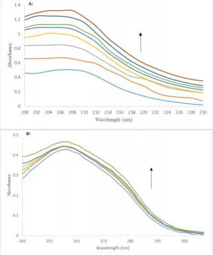
Herein, UV-Visible analysis was applied to study binding modes between ctDNA and PPT. This method is normally utilized to study the interaction of small molecules with DNA [24, 25]. Spectral changes of PPT which occurred via increasing concentration of ctDNA between 3.3–7.8×10⁻⁵ is shown in Figure 2a. PPT exhibited maximum absorption near 206 nm. By increasing in the concentration of ctDNA, hyperchromism is observed with no apparent shift in the peak. In general, the hyperchromic effect is related to
various non-covalent interactions outside of DNA helix [26]. In case of intercalation, a hypochromism along with bathochromic shifts is observed [27]. Therefore, UV-Vis spectroscopy data confirmed the non-intercalative binding of PPT with ctDNA. The binding constant which corresponds to the interaction of PPT, was calculated according to Eq. (1) [28].
= + (
[ ]) Eq. (1)
Where A0 and A are the absorbance of PTT in the
absence and presence of ctDNA, andԑGandԑH-Gare the
absorption coefficients of the PPT and PPT-DNA complex, respectively. K is the binding constant. The binding constant was calculated to be 1.01×10⁴ M-1 from the intercept to slope ratios of versus
1/[ctDNA] plot. This value of binding constant is lower than strong binding constant like EtBr, acridine orange interaction [29], and is close to groove binding compounds. The maximum absorption of ctDNA with different concentrations of PPT (3.7–33 µM) was recorded at 260 nm and depicted in Figure 2b. The hyperchromic effect was observed, demonstrating the non-intercalative binding.
Emission spectroscopic study
A competitive fluorescence measurement was carried out to determine the binding mode of PPT with DNA. Methylene blue (MB) molecules are one of the DNA probes, used to investigate the compound-DNA interactions [30, 31]. The binding of MB molecules to DNA occurred via intercalation mode [32, 33]. Constant DNA was mixed with MB as a probe, then, increasing concentrations of PPT were added to DNA-MB complexes. Interestingly, by adding DNA, emission intensity of MB was quenched. This phenomenon is linked to strong stacking interaction between the adjacent DNA base pairs and MB [34]. The emission spectra of DNA-MB and increasing concentration of PPT are shown in Figure 3. As observed clearly, no changes in the fluorescence intensity of MB was illustrated upon addition of PPT. No release of MB from DNA-MB complex is responsible for this observation and proved the non-intercalative binding mode.
Viscometric study
Changing in DNA viscosity in the presence of the desired compound provides a credible proof for the binding mode [35]. The interaction of small molecules with DNA helix led to increased length of DNA, separation of base pairs and an increase in the viscosity of DNA [36, 37]. In contrast, groove and electrostatic
bindings exhibited no or less change in DNA viscosity [38, 39]. Figure 4 shows the effect of PPT on the viscosity of DNA. There was insignificant change in the viscosity of DNA. Thus, the results was illustrated non-intercalative and groove binding as the most probable modes.
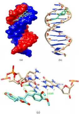
Molecular docking
Molecular docking is an attractive technique to investigate the drug–DNA interactions on the way of rational drug design and discovery [40]. In our experiment, the molecular docking of PPT with B-DNA was performed using Auto Dock 4.2 in order to further clarify the binding mode of PPT with DNA duplex of sequence d(CGCGAATTCGCG)2 dodecamer (PDB ID: 1BNA) and the binding structure of DNA-PPT complex. Generally, more negative binding energy (ΔG), caused by the formation of the most stable complex. As shown
Figure 3. Fluorescence emission spectra of MB-DNA complex in the presence of increasing amount of PPT (24–55 µM).
in Figure 5, the PPT interacts with the O4' atom of the ribose group in the DNA back bone by forming bond with length of 2.06 Å, the PPT also interacts with NH2
(H22 atoms) group of guanine by forming two hydrogen bonds between OH and OMe groups of PPT with 2.03 and 2.62 Å bond lengths, respectively. Conclusively, PPT interacts in the minor groove of the DNA through guanine and ribose of back bone. The estimated free energy of this binding was computed to be -7.08 kcal/mol.
Furthermore, the binding constant (Kb) which was
determined by UV-Vis method was correlated with the free binding energy (ΔG) of the docked model. Basic formula of binding constant and Gibbs free energy is: ΔG =-RT lnKb
Where ΔG is free energy, R is gas constant (1.98 cal/mol/K), T is temperature at which the experiment
was done (25 ◦C i.e. 298 K) and Kb is the binding
constant between PPT and free DNA calculated using UV-Vis spectroscopy 1.01×10⁴.
This docking free energy was compared to experimental UV-Vis free energy value (ΔG), which is -5.44 kcal/mol. These results revealed matches roughly to the free energy calculated by docked drug-DNA model. Therefore, it can be concluded that PPT-DNA docked model is in approximate correlation with our experimental results.
Conclusion
In this work, we investigated the interaction of PPT with ctDNA by using various spectroscopic, viscosity measurement and molecular docking studies. The UV-Vis absorption showed hyperchromicity in spectra and binding constant was calculated to be 1.01 × 10⁴ M-1
that referring to non-intercalative binding between PPT and DNA. Fluorescence emission study was also confirmed the UV-Vis absorption results. Negligible change in viscosity supported previous finding. Molecular docking study showed interaction of PPT via minor groove of DNA. These studies help to understand the mechanism of interaction between target compounds with DNA, and could help to explore new compounds with therapeutic purposes.
Acknowledgement
This work was supported by the research council of Tehran University of Medical Sciences (TUMS) and Iran National Science Foundation (INSF).
References
1. Ducasse H., Arnal A., Vittecoq M., Daoust S.P., Ujvari B., Jacqueline C., Tissot T., Ewald P., Gatenby R.A., King K.C., Bonhomme F., Brodeur J., Renaud F., Solary E., Roche B. and Thomas F. Cancer: an emergent property of disturbed resource-rich environments? Ecology meets personalized medicine. Evol. Appl. 8: 527-540 (2015). 2. Rescifina A., Zagni C., Varrica M.G., Pistarà V. and
Corsaro A. Recent advances in small organic molecules as DNA intercalating agents: Synthesis, activity, and modeling. Eur. J. Med. Chem. 74: 95-115 (2014).
3. Roos W.P. and Kaina B. DNA damage-induced cell death by apoptosis. Trends Mol. Med. 12: 440-450 (2006). 4. Zhang G., Guo J., Zhao N. and Wang J. Study of
interaction between kaempferol-Eu3+ complex and DNA
with the use of the Neutral Red dye as a fluorescence probe. Sensors Actuat. B Chem. 144: 239-246 (2010). 5. Lerman L.S. Structural considerations in the interaction of
DNA and acridines. J. Mol. Biol. 3: 18-30 (1961). 6. Bauer W. and Vinograd J. The interaction of closed
circular DNA with intercalative dyes. 3. Dependence of the buoyant density upon superhelix density and base composition. J. Mol. Biol. 54: 281-298 (1970).
7. Sirajuddin M., Ali S. and Badshah A. Drug-DNA interactions and their study by UV-Visible, fluorescence spectroscopies and cyclic voltammetry. J. Photochem. Photobiol. B. 124: 1-19 (2013).
8. Kikandi S.N., Musah S., Lee K., Hassani J., Rajan S., Zhou A. and Sadik O.A. Comparative Studies of Quercetin Interactions with Monophosphate Nucleotides Using UV-Vis Spectroscopy and Electrochemical Techniques. Electroanalysis 19: 2131-2140 (2007).
9. Silvestri A., Barone G., Ruisi G., Lo Giudice M.T. and Tumminello S. The interaction of native DNA with iron(III)-N,N′-ethylene-bis(salicylideneiminato)-chloride. J. Inorg. Biochem. 98: 589-594 (2004).
10. Wang L., Lin L. and Ye B. Electrochemical studies of the interaction of the anticancer herbal drug emodin with DNA. J. Pharm. Biomed. Anal. 42: 625-629 (2006). 11. Lin H.W., Kwok K.H. and Doran P.M. Production of
podophyllotoxin using cross-species coculture of Linum flavum hairy roots and Podophyllum hexandrum cell suspensions. Biotechnol. Prog. 19: 1417-1426 (2003). 12. Inamori Y., Kubo M., Tsujibo H., Ogawa M., Baba K.,
Kozawa M. and Fujita E. The biological activities of podophyllotoxin compounds. Chem. Pharm. Bull. 34: 3928-3932 (1986).
13. Castro M.A., Del corral J.M., Garcia P.A., Rojo M.V., La Iglesia-Vincete J., Mollinedo F., Cuevas C. and San Feliciano A. Synthesis and biological evaluation of new podophyllic aldehyde derivatives with cytotoxic and apoptosis-inducing activities. J. Med. Chem. 53: 983-993 (2010).
14. Stahelin H.F. and Wartburg A.V. The Chemical and Biological Route from Podophyllotoxin Glucoside to Etoposide: Ninth Cain Memorial Award Lecture. Cancer Res. 51: 5-15 (1991).
15. Baldwin E.L. and Osheroff N. Etoposide, Topoisomerase II and Cancer. Anticancer Agents Med. Chem. 5: 363-372 (2005).
16. Gordaliza M., MaMiguel del Corral J., Castro M.A., Lopez-Vazquez M., Garcia P.A., San Feliciano A. and Garcia-Gravalos M. Selective cytotoxic cyclolignans. Bioorg. Med. Chem. Lett. 5: 2465-2468 (1995).
17. Kamal A., Hssaini S.M.A., Rahim A. and Riyaz S. Podophyllotoxin derivatives: a patent review (2012-2014). Expert Opin. Ther. Pat. 25: 1025-1034 (2015).
18. You Y. Podophyllotoxin derivatives: current synthetic approaches for new anticancer agents. Curr. Pharm. Des. 11: 1695-1717 (2005).
19. Heidary Alizadeh B., Emami S., Dehghan G., Foroumadi A. and Shafiee A. Synthesis of Cytotoxic Isodeoxypodophyllotoxin Analogs. J. Heterocyclic Chem. DOI: 10.1002/jhet.2618 (2016).
20. Dehghan G., Dolatabadi J.E.N., Jouyban A., Zeynali K.A., Ahmadi S.M. and Kashanian S. Spectroscopic Studies on the Interaction of Quercetin–Terbium(III) Complex with Calf Thymus DNA. DNA Cell Biol. 30: 195-201 (2011). 21. Glasel J.A. Validity of nucleic acid purities monitored by
260nm/280nm absorbance ratios. Biotechniques, 18: 62-63 (1995).
22. Subastri A., Ramamurthy C.H., Suyavaran A., Mareeswaran R., Rao P.L., Harikrishna M., Kumar M.S., Sujatha V. and Thirunavukkarasu C. Spectroscopic and molecular docking studies on the interaction of troxerutin with DNA. Int. J. Biol. Macromolec. 78: 122-129 (2015). 23. Pettersen E.F., Goddard T.D., Huang C.C., Couch G.S.,
Greenblatt D.M., Meng E.C. and Ferrin T.E. UCSF Chimera--a visualization system for exploratory research and analysis. J. Comput. Chem. 25: 1605-1612 (2004). 24. Parveen M., Ahmad F., Malla A.M., Sohrab Khan M., Ur
Rehman S., Tabish M., Silva M.R. and Pereira Silva P.S. Structure elucidation and DNA binding specificity of natural compounds from Cassia siamea leaves: A biophysical approach. J. Photochem. Photobiol. B. 159: 218-228 (2016).
25. Jangir D.K., Charak S., Mehrotra R. and Kundu S. FTIR and circular dichroism spectroscopic study of interaction of 5-fluorouracil with DNA. J. Photochem. Photobiol. B. 105: 143-148 (2011).
molecular modeling studies on the interaction of antihypertensive drug; methyldopa with calf thymus DNA. Mol. BioSyst. 10: 338-347 (2014).
27. Tomer E., Goren R. and Monselise S.P. Isolation and identification of seselin in Citrus roots. Phytochemistry, 8: 1315-1316 (1969).
28. Bauri A.K., Foro S., Lindner H.J. and Nayak S.K. Reinvestigation of seselin. Acta Cryst. E. 62: 1340-1341 (2006).
29. Nafisi S., Saboury A.A., Keramat N., Neault J.F. and Tajmir-Riahi H.A. Stability and structural features of DNA intercalation with ethidium bromide, acridine orange and methylene blue. J. Mol. Struct. 827: 35-43 (2007). 30. Kashanian S., Shahabadi N., Roshanfekr H., Shalmashi K.
and Omidfar K. DNA binding studies of PdCl2(LL)(LL =
chelating diamine ligand: N,N-dimethyl trimethylenediamine) complex. Biochemistry, 73: 929-936 (2008).
31. Kashanian S., Javanmardi A., Chitsazan D., Paknejad M. and Omidfar K. Fluorometric study of fluoxetine DNA binding. J. Photochem. Photobiol. B. 113: 1-6 (2012). 32. Douthart R.J., Burnett J.P., Beasley F.W. and Frank B.H.
Binding of ethidium bromide to double-stranded ribonucleic acid. Biochemistry, 12: 214-220 (1973). 33. Shahabadi N., Kashanian S., Khosravi M. and Mahdavi M.
ultispectroscopic DNA interaction studies of a water-soluble nickel(II) complex containing different dinitrogen aromatic ligands. Transit. Metal Chem. 35: 699-705 (2010).
34. Shahabadi N., Kashanian S. and Purfoulad M. DNA interaction studies of a platinum(II) complex, PtCl2(NN)
(NN = 4,7-dimethyl-1,10-phenanthroline), using different instrumental methods. Spectrochim. Acta A. Mol. Biomol. Spectrosc. 72: 757-761 (2009).
35. Zhao T., Bi S., Wang Y., Wang T., Pang B. and Gu T. In vitro studies on the behavior of salmeterol xinafoate and its interaction with calf thymus DNA by multi-spectroscopic techniques. Spectrochim. Acta A. Mol. Biomol. Spectrosc. 132: 198-204 (2014).
36. Husain M.A., Sarwar T., Rehman S.U., Ishqi H.M. and Tabish M. Ibuprofen causes photocleavage through ROS generation and intercalates with DNA: a combined biophysical and molecular docking approach. Phys. Chem. Chem. Phys. 17: 13837-13850 (2015).
37. Husain M., Dehghan G., Jouyban A., Sistani P. and Arvin M. Studies of interaction between terbium(III)-deferasirox and double helix DNA by spectral and electrochemical methods. Spectrochim. Acta A Mol. Biomol. Spectrosc. 120: 467-472 (2014).
38. Sarwar T., Rehman S.U., Husain M.A., Ishqi H.M. and Tabish M. Interaction of coumarin with calf thymus DNA: Deciphering the mode of binding by in vitro studies. Int. J. Biol. Macromolec.73: 9-16 (2015).
39. Satyanarayana S., Dabrowiak J.C. and Chaires J.B. Neither. DELTA. nor. LAMBDA. tris(phenanthroline) ruthenium(II) binds to DNA by classical intercalation. Biochemistry, 31: 9319-9324 (1992).
![Figure 1. The structure of (3R,4R)-4-((benzo[d][1,3]dioxol-5-yl)methyl)-dihydro-3-(hydroxy(3,4-dimethoxyphenyl)methyl)furan-2(3H)-one](https://thumb-us.123doks.com/thumbv2/123dok_us/15820.2001554/2.595.218.378.99.173/figure-structure-dioxol-methyl-dihydro-hydroxy-dimethoxyphenyl-methyl.webp)