Federica Rachele Danti, MD
Serena Galosi, MD Marta Romani, PhD Martino Montomoli, MD Keren J. Carss, PhD‡ F. Lucy Raymond, PhD‡ Elena Parrini, PhD Claudia Bianchini, BSc Tony McShane, MD Russell C. Dale, PhD Shekeeb S. Mohammad,
MD
Ubaid Shah, MD Neil Mahant, FRACP,
PhD
Joanne Ng, MBChB Amy McTague, MBChB Rajib Samanta, MD Gayatri Vadlamani, MD Enza Maria Valente, MD,
PhD
Vincenzo Leuzzi, MD, PhD
Manju A. Kurian, MD, PhD, MRCPCH* Renzo Guerrini, MD,
FRCP*
Correspondence to Dr. Guerrini: [email protected]
Supplemental data at Neurology.org/ng
GNAO1
encephalopathy
Broadening the phenotype and evaluating treatment and outcome
ABSTRACT
Objective: To describe better the motor phenotype, molecular genetic features, and clinical course ofGNAO1-related disease.
Methods:We reviewed clinical information, video recordings, and neuroimaging of a newly iden-tified cohort of 7 patients with de novo missense and splice siteGNAO1mutations, detected by next-generation sequencing techniques.
Results:Patients first presented in early childhood (median age of presentation 10 months, range 0–48 months), with a wide range of clinical symptoms ranging from severe motor and cognitive impairment with marked choreoathetosis, self-injurious behavior, and epileptic encephalopathy to a milder pheno-type, featuring moderate developmental delay associated with complex stereotypies, mainly facial dyskinesia and mild epilepsy. Hyperkinetic movements were often exacerbated by specific triggers, such as voluntary movement, intercurrent illnesses, emotion, and high ambient temperature, leading to hospital admissions. Most patients were resistant to drug intervention, although tetrabenazine was effective in partially controlling dyskinesia for 2/7 patients. Emergency deep brain stimulation (DBS) was life saving in 1 patient, resulting in immediate clinical benefit with complete cessation of violent hyper-kinetic movements. Five patients had well-controlled epilepsy and 1 had drug-resistant seizures. Struc-tural brain abnormalities, including mild cerebral atrophy and corpus callosum dysgenesis, were evident in 5 patients. One patient had a diffuse astrocytoma (WHO grade II), surgically removed at age 16.
Conclusions:Our findings support the causative role ofGNAO1mutations in an expanded spec-trum of early-onset epilepsy and movement disorders, frequently exacerbated by specific triggers and at times associated with self-injurious behavior. Tetrabenazine and DBS were the most useful treatments for dyskinesia.Neurol Genet2017;3:e143; doi: 10.1212/NXG.0000000000000143
GLOSSARY
CK5creatine kinase;DBS5deep brain stimulation;EOEE5early-onset epileptic encephalopathy;EVS5Exome Variant Server;ExAC5Exome Aggregation Consortium;GATK5Genome Analysis Toolkit;GPCR5G protein–coupled receptor;
WES5whole-exome sequencing;WGS5whole-genome sequencing.
De novo
GNAO1
mutations (MIM 139311) were initially identified in children with
early-onset epileptic encephalopathy (EOEE) and severe developmental delay,
1–3with later
develop-ment of dyskinetic movedevelop-ment disorders
1,4–6and in children exhibiting developmental delay and
severe dyskinesia without seizures.
7,8*These authors have contributed equally to this work.
‡These authors are members of the National Institute for Health Research (NIHR) Bioresource Rare Diseases Consortium. From the Department of Paediatrics, Child Neurology and Psychiatry (F.R.D., S.G., V.L.), Sapienza University of Rome, Italy; Molecular Neurosciences, Developmental Neurosciences Programme (F.R.D., J.N., A.M., M.A.K.), University College London Institute of Child Health, UK; Department of Neurology (F.R.D., J.N., A.M., M.A.K.), Great Ormond Street Hospital for Children, London, UK; GENOMA Group (M.R.), Molecular Genetics Laboratory, Rome, Italy; Pediatric Neurology, Neurogenetics and Neurobiology Unit and Laboratories (M.M., E.P., C.B., R.G.), Neuroscience Department, A Meyer Children’s Hospital, University of Florence, Italy; Department of Haematology (K.J.C.), University of Cambridge, NHS Blood and Transplant Centre, UK; NIHR Bioresource Rare Diseases (K.J.C., F.L.R.), University of Cambridge, UK; Department of Neurology (N.M.), Westmead Hospital, Sydney, Australia; Childrens Hospital Oxford (T.M.), John Radcliffe Hospital, UK; Institute for Neuroscience and Muscle Research (R.C.D., S.S.M., U.S.), the Children’s Hospital at Westmead, University of Sydney, Australia; Department of Medical Genetics (F.L.R.), Cambridge Institute for Medical Research, University of Cambridge, UK; Department of Neurology (R.S.), University Hospitals Leicester NHS Trust, UK; Department of Paediatric Neurology (G.V.), Leeds Teaching Hospitals NHS Trust, UK; Section of Neurosciences (E.M.V.), Department of Medicine and Surgery, University of Salerno, Italy; and Neurogenetics Unit (E.M.V.), IRCCS Fondazione Santa Lucia, Rome, Italy.
Funding information and disclosures are provided at the end of the article. Go to Neurology.org/ng for full disclosure forms. The Article Processing Charge was paid by the EU FP7 Program–“Desire”Project.
GNAO1
encodes a subclass (G
a
o) of the
G
a
subunit
of
heterotrimeric
guanine
nucleotide-binding proteins and is highly
expressed in the brain and involved in the
reg-ulation of neuronal excitability and
neurotrans-mission. Mutations in
GNAO1
and other
G-protein subunits (
GNAL
), adenylyl cyclase
(
ADCY5
), cyclic nucleotide phosphodiesterase
(
PDE10A
), and G protein
–
coupled receptor
(GPCR) (
GPR88
)
9in early-onset movement
disorders, support the notion that disruption
of the G-protein-cAMP pathway axis is a key
contributor to the pathophysiology of dystonia
and chorea. Although the pathophysiologic
mechanisms underlying such genetic
move-ment disorders remain yet to be elucidated,
impaired modulation or transduction of
trans-membrane signaling, presynaptic
autoinhibi-tory effects, and altered neuronal excitability
are all putative disease mechanisms that might
explain the co-occurrence of multiple
neuro-logic manifestations such as epilepsy and
hyperkinesia.
Here, we report 7 newly identified patients
with
GNAO1
mutations, with the aim of
char-acterizing in detail the clinical phenotype,
response to treatment and outcome, as well
as associated brain MRI features.
METHODS Molecular genetic testing. Patients 1–3, 6, and 7 were studied using whole-exome/genome (WES/ WGS) sequencing. For patient 1, WES was performed on a SOLiD 5500XL sequencing platform (Life Technologies, Foster City, CA). Short reads were mapped against the GRCh37/hg19 human assembly by means of LifeScope soft-ware (Life Technologies). Variants were detected using the Haplotype Caller software package of the Genome Analysis Toolkit (GATK) suite and filtered to include only variants covered by at least 10 reads with mapping quality values exceeding 30. For patients 2, 3, and 6, WGS was performed with Illumina TruSeq DNA PCR-Free Sample Preparation kit on an Illumina Hiseq 2500 sequencing platform (Illumina Inc., San Diego, CA). For patient 3, WGS was performed as part of the National Institute for Health Research Bioresource Rare Disease Project. A minimum coverage of 15X (95% of the genome) was obtained, with an average coverage of;30X. Reads were aligned using Isaac aligner (version 01.14) (Illu-mina Inc., Great Chesterford, UK) and mapped against the GRCh37 hg19 human assembly. Single nucleotide variants and insertions/deletions were identified using Isaac variant caller. Patients 4 and 5 were studied using a Haloplex panel (Agilent Technologies, Santa Clara, CA) of 95 epilepsy genes. Variants were called and annotated using the GATK10toolkit and the ANNOVAR tool.11Patient 7 was analyzed by WES using Nextera Rapid Capture Exome, Illumina NextSeq platform (Illumina Inc.) with an average coverage depth of 100–130X. An end-to-end in-house bioinformatics pipeline
including base calling, primary filtering of low-quality reads and probable artefacts, and annotation of variants was applied.
For all patients, variants reported as validated polymor-phisms with a minor allele frequency.0.01 (1%) in publicly available human variation resources (including dbSNP144, NHLBI GO Exome Sequencing Project, UK10, 1000 Ge-nomes, Exome Variant Server [EVS], and Exome Aggregation Consortium [ExAC] browser) and variants present in in-house control individuals were filtered out. In silico prediction of the mutation pathogenicity was performed using ANNOVAR and the dbNSFP database (v3.0a) (sites.google.com/site/jpopgen/ dbNSFP). Putative causative variants were validated by Sanger sequencing and investigated in the probands’parents to deter-mine whether the mutations were inherited or had occurred de novo.
Standard protocol approvals, registrations, and patient consents.Written informed consent to disclose clinical infor-mation, neuroimaging, and video footage was obtained from all parents/guardians of the 7 participants. Informed consent for genetic testing was obtained according to the local institu-tional policy.
RESULTS Genetic analysis.We identified 7 patients carrying 6 different GNAO1 (GenBank accession number NM_020988) mutations; all the variants occurred de novo and 4 are previously unreported (table, figure 1).
In patient 1, we detected the novel c.139A.G [p.(Ser47Gly)] missense variant, located in the N-terminal domain of the protein. In patients 2 and 4, we identified the c.625C.T [p.(Arg209Cys)] mis-sense variant. This variant has previously been re-ported in 1 patient with developmental delay, intellectual disability, and severe chorea, associated with the later onset of complex partial seizures.5In
patient 3, we observed the novel c.72311G.A intronic variant, and in patient 5, the novel c.167T.C [p.(Ile56Thr)] missense variant, located in the N-terminal domain of the protein. In patient 6, we detected the c.118G.C [p.(Gly40Arg)] missense mutation, located in the N-terminal domain of the protein. A different mutation leading to the same amino acid change (c.118G.A [p.(Gly40Arg)]) has previously been described in a patient with infantile-onset epi-lepsy.3 In patient 7, we identified the novel
c.737A.G [p.(Glu246Gly)] missense variant. A different nucleotide change in the same amino acid position, c.736G.A [p.(Glu246Lys)], had been reported as disease causing in patients exhibiting developmental delay, intellectual disability, and a paroxysmal movement disorder.5,8
The 5 missense variants are predicted to be del-eterious using the dbNSFP database and are not re-ported in the 1000 Genomes, ExAC, and EVS databases. The splicing mutation c.72311G.A was predicted to cause the loss of a splice donor site
Table Clinical features and genetic laboratory findings
Patient/sex 1/M 2/M 3/F 4/M 5/F 6/F 7/M
Age at first medical evaluation/follow-up
5 mo/5 y 5 mo Birth/10 y 8 mo 4 mo/deceased at 4 y 5 mo/5 y 6 mo 4 y/16 y 2 mo/5 y 2 mo 6 mo/15 y
GNAO1mutation (de novo)
c.139A.G [p.(Ser47Gly)] c.625C.T [p. (Arg209Cys)]
c.72311G.A c.625C.T [p. (Arg209Cys)]
c.167T.C [p. (Ile56Thr)]
c.118G.C [p.(Gly40Arg)] c.737A.G [p.(Glu246Gly)]
Novel/reported Novel Ref. 5 Novel Ref. 5 Novel Ref. 3 Novel
Clinical findings at follow-up
Severe ID/DD, hypotonia, movement disorder, seizures
Severe ID/DD, hypotonia, movement disorder, seizures, MC
Severe ID/DD, hypotonia, movement disorder, MC
Severe ID, focal epilepsy, movement disorder, MC
Mild ID, focal epilepsy, movement disorder
Severe ID, focal epilepsy, movement disorder, MC
Moderate-severe appendicular/ orobuccal dystonia; moderate lower limb spasticity
Onset/type of abnormal movements
4 mo/severe hypotonia, mild dystonia; 2 y 3 mo/seHypM, dystonia; stereotypic hand movements; SIB (lips biting, hair pulling)
Birth/severe hypotonia; 9 y/seHypM; SIB (lips biting)
8 mo/continuous generalized choreoathetosis; orolingual dyskinesia
7 mo/complex motor stereotypies of hands/arms; dyskinetic motor pattern 5 y/subcontinuous dyskinetic tongue movements; mild upper limb dyskinesia
3 y/hand-to-mouth complex stereotypies, rocking, pelvic thrusting
6 mo/moderate hypotonia; 2 y/ moderate-severe generalized and orobuccal dystonia, lower limb spasticity; 7 y/seHypM
Dysphagia Yes Yes Yes No No Yes, gastrostomy Yes (progressive), gastrostomy
Head control 1 y 6 mo then regression Yes Yes Yes Yes Yes Yes
Sitting No 9 mo then regression No Yes Yes No Yes, with support
Language No No Limited No Yes No Yes, with dysarthria
Onset/epilepsy/ severity
3 y/FS with secondary generalization/well controlled on AED
6 y/GTCS/well controlled on AED
No 2 y/FS/well
controlled on AED
4 y/FS/well controlled on AED
2 mo/EOEE (OS-FS soon followed by spasms, then FS and GTCS)/multiple AEDs, ketogenic diet, VNS
10 y/rare FS and GTCS/no AEDs
Interictal EEG Right frontotemporal spikes Slow background
activity NA Bilateral centrotemporal spikes Left frontotemporal spikes
Burst suppression at onset; slow background, multifocal spike waves
Normal
MRI VE; thin and dysmorphic corpus callosum; mild HyCN
VE; thin corpus callosum; mild HyCN, hypoplasia of inferior vermis
VE and dilated subarachnoid spaces; moderate cortical atrophy; dysmorphic corpus callosum; mild HyCN
Normal Left frontal lesion (diffuse astrocytoma WHO grade 2); low-lying cerebellar tonsils
Mild VE; thin corpus callosum
Mild loss of volume (atrophy) in generalized distribution
Neurotransmitter analysis in CSF
Methyltetrahydrofolate 34 nmol/L; 63–111
Methyltetrahydrofolate 66 nmol/L; 72–172
Normal Normal Normal Normal Normal
Abbreviations: AED5antiepileptic drugs; DD5developmental delay; EOEE5early-onset epileptic encephalopathy; FS5focal seizures; GTCS5generalized tonic-clonic seizures; HyCN5hypoplasia of caudate nuclei; ID5intellectual disability; MC5microcephaly; NA5not available; nv5normal values; OS5Ohtahara syndrome; seHypM5severe exacerbation of hyperkinetic movements; SIB5self-injurious behavior; VE5ventricular enlargement; VNS5vagus nerve stimulation.
Neurology
:
using Mutation Taster and Berkeley Drosophila Genome Project.
Clinical features and neurologic investigations.Clinical features, as obtained by history taking, direct clinical examination, and evaluation of video foot-age, neuroimaging, and genetic findings are de-tailed in the table, in figure 2 and in videos 1–5 at Neurology.org/ng. Within the cohort, we iden-tified 4 boys and 3 girls. Six of the patients are alive, currently aged 5–16 years.
The onset of abnormal hyperkinetic movements ranged from 4 months to 9 years of age (median 2 years). Within the cohort, there was considerable clin-ical heterogeneity with regard to motor semiology and severity. Profound hypotonia preceded the onset of abnormal movements in patients 1, 2, and 7, who later developed dystonia, followed by severe episodes of paroxysmal choreoathetosis. Gradual development of facial dyskinesia was also reported in these patients
(videos 1, 2, and 5). Patient 3 experienced continuous generalized choreoathetosis and facial dyskinesia. At the age of 4, because of repeated and prolonged infec-tive episodes, status dystonicus developed, requiring intensive care, with sedation, ventilation, tracheos-tomy, gastrostracheos-tomy, and metabolic support. MRI showed progressive cerebral atrophy. Status dystoni-cus persisted, with rise in creatine kinase (CK) and renal failure, leading to multisystemic deterioration and death. Patient 4 exhibited complex motor stereo-typies and a dyskinetic motor pattern (video 3). Patient 5 manifested almost continuous orolingual dyskinesia. She also had mild dyskinetic movements of the upper limbs (video 4). Patient 6 exhibited com-plex rocking, staring, back arching, and pelvic thrust-ing movements, associated with hand-to-mouth complex stereotypies.
Several triggers were reported to elicit or exacer-bate abnormal movements, including emotion (all pa-tients), intercurrent illnesses (patients 1, 2, and 7),
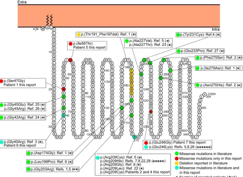
Figure 1 Membrane topology modeling ofGNAO1
Membrane topology was predicted using the Protter online tool29(P09471, GNAO_HUMAN). KnownGNAO1missense mutations are indicated in green, the
deletion is in yellow. Mutations found in this report and already described in the literature are indicated in light blue, novel mutations in red.
high temperature (patient 1), intention, and purpose-ful movements (patients 1 and 7). Attacks frequently presented in clusters, lasting minutes (patient 1), hours or weeks (patient 2), or months (patient 7) (videos 1, 2, and 5). Such exacerbations were pharma-coresistant to standard therapies. Patients 1, 2, and 7 also exhibited associated autonomic instability, sweat-ing, dehydration, and rise in CK, frequently requiring hospitalization and intensive care and, often, admin-istration of anesthetic agents (patients 2 and 7). Worsening of baseline dystonia with hyperkinetic movements and loss of fine motor skills were reported in patient 7 after each episode. One such exacerbation determined a 3-month admission to the intensive care unit and anesthetic agents. The emergency placement of a deep brain stimulator (DBS) device bilaterally into the globus pallidus interna was transformative
and resulted in almost complete remission of the pro-nounced hyperkinesia, although residual generalized dystonia persisted.
Six patients (1, 2, 4–7) exhibited epilepsy, with age of seizure onset ranging from 2 months to 10 years. While seizures were well controlled by medica-tion in 4 patients, patient 7 presented rare generalized tonic-clonic seizures and focal dyscognitive seizures and patient 6 had a history of EOEE followed by drug-resistant focal epilepsy with dyscognitive seiz-ures. Patient 3 never had seizures and died at age 4 years.
Additional clinical features were also reported in some patients. Patients 1 and 2 exhibited mild self-injurious behavior (lip biting, hair pulling). Patient 7 has a history of significant anxiety, requiring treat-ment with fluoxetine, sertraline, and risperidone.
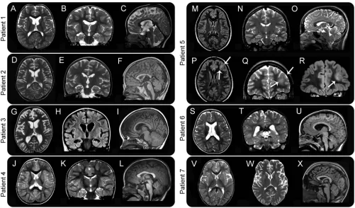
Figure 2 MRI characteristics of 7 patients withGNAO1mutations
In our cohort, diagnostic metabolic investigations were unremarkable, including CSF neurotrans-mitters, except for the reduction of CSF 5-methyltetrahydrofolate levels in patients 1 and 2. In both patients, calcium folinate administration was started but did not lead to discernible clinical improvement. Brain MRI (figure 2) was unremark-able in patients 4 and 7, while inconsistent changes were detected in the remaining patients (microceph-aly, macroceph(microceph-aly, progressive cerebral atrophy with dilated ventricles and subarachnoid spaces, caudate volume loss, and dysmorphic corpus callosum). In 1 patient (patient 5), a focal lesion involving the gray and white matter of the left frontal lobe was identified and confirmed as a diffuse astrocytoma (WHO grade 2) after surgical removal.
DISCUSSION Through its activation by GPCRs, Gao is involved as a modulator or transducer in several transmembrane signaling pathways.10,12,13
Gao also mediates the widespread presynaptic auto-inhibitory effect of many neurotransmitters (viaa2 adrenoreceptors, M2/M4 muscarinic, m/d opioid, GABAB, adenosine A1, or endocannabinoid CB1 receptors) through a reduction of sensitivity to membrane depolarization and a direct inhibitory effect on the vesicle fusion process.14Furthermore,
Gao indirectly activates G protein–coupled inwardly rectifying K1 channels that mediate neuronal excitability through a slower self-inhibitory postsynaptic potential.15
Gao-deficient mice (Gnao12/2) have occasional seizures, severe impairment of motor-control, hyper-algesia, and behavioral abnormalities with early post-natal lethality.16,17 Although heterozygous Gnao1
knockout mice do not manifest seizures, a gain-of-function knock-in mutant murine model (Gnao1 Gly184Ser/1) has severe seizures with markedly increased frequency of interictal epileptiform dis-charges and sudden premature death. Animal model data thus suggest that pathogenic monoallelic
GNAO1 mutations in humans may also result in a gain-of-function effect. Moreover, impaired protein localization and decreasedGNAO1-mediated inhibi-tion of calcium currents by norepinephrine compared to the wild type have been showed in in vitro func-tional expression systems.18 GNAO1 has also been
implicated in the etiology of brain tumors, such as ependymoma and glioblastoma multiforme.19,20
De novoGNAO1mutations were originally first reported in Ohtahara syndrome and EOEE in 4 children, associated with abnormal movements in 2.1Since this initial description, 30 additional
pa-tients have been reported with an emerging pheno-type characterized by neurodevelopmental delay with an early onset of a hyperkinetic movement
disorder, inconsistently associated with epilepsy. The epilepsy phenotype of the 7 patients we studied ranged from EOEE to mild drug-sensitive epilepsy, whose onset could precede or follow that of the movement disorder, which was certainly a promi-nent feature. The occurrence of stereotypies (pre-viously reported in 2 patients with EOEE)4,5 and
characteristic paroxysmal exacerbations, associated often with clear triggers, may both be considered as 2 further important discerning clinical features ofGNAO1encephalopathy.
As previously reported, standard investigations appear to be unyielding in patients with GNAO1 -related disease. Two of our patients had low levels of CSF 5-methyltetrahydrofolate, although this was not consistently seen in the cohort. Brain MRI (figure 2) showed a combination of minor features in most, including a thin corpus callosum with dilated ven-tricles in 4, as previously reported8as well as
hypo-plastic caudate nuclei in 3 and a diffuse astrocytoma (WHO grade II) in the oldest patient (patient 5). The latter finding raises concerns about the possible role of the Gly40Arg mutation in tumorigenesis, in view of the reported role ofGNAO1in promoting oncogenic transformation.19,21 A somatic GNAO1 mutation
(p.Arg243Hys) has been identified in breast carcino-mas where it promotes oncogenic transformation by rendering the Gasubunit constitutively activated and enhancing signaling pathways responsible for neo-plastic transformation.21AlthoughGNAO1was
iden-tified as part of the human plasma proteome, its high abundance was suggested to promote cancer cell via-bility via proapoptotic protein interference.20Larger
series and longer follow-up data will be fundamental to determine whether patients carrying specific
GNAO1 mutations are at higher risk of developing tumors.
TwentyGNAO1mutations, 19 missense and 1 dele-tion, have been previously reported (figure 1).1–8,22–28
The only reported deletion was described in a patient with a severe phenotype, including Ohtahara syn-drome, developmental delay, and severe intellectual disability, who died secondary to respiratory-tract complications.1The 7 patients we are reporting
har-bored 6 different mutations, one of which was evi-dent in 2 patients (figure 1). Two of the 6 mutations have been reported previously [p.(Arg209Cys) and p.(Gly40Arg)],3,5 while 4 are novel [p.(Ser47Gly),
c.72311G.A, p.(Ile56Thr), and p.(Glu246Gly)]. The c.72311G.A splicing mutation, likely result-ing in abnormal mRNA splicresult-ing, was found in patient 3, who died at 4 years of age and exhibited the most severe phenotype. The p.(Arg209Cys) mutation falls within the switch II domain, which is important for guanidine nucleotide-dependent regulation of downstream effectors and is highly
conserved across vertebrate species.5 The Arg209
amino acidic residue represents a mutation hotspot since, including our series, 10 patients carrying muta-tions affecting this residue have been reported. All these patients presented developmental delay and cho-rea or dystonia and 3 had seizures. The p.(Glu246Gly) mutation, although novel, affects an amino acid resi-due that represents a second mutation hotspot since 7 patients with mutations affecting this residue have been reported (including our own), all presenting with developmental delay and chorea, with only patient 7 developing late childhood–onset seizures. The Arg209 and Glu246 residues form a salt bridge that is important for the stabilization of the Ga-containing complexes, mainly in GTP-bound active state.5
Hence, the variants involving these residues should disrupt their interaction, resulting in destabilization of the Ga-containing complexes.5In our study, the
identification of 2 additional patients harboring mutations involving the Arg209 amino acid residue and 1 additional patient harboring a mutation involv-ing the Glu246 amino acid residue confirm that these residues areGNAO1mutation hotspots.
Tetrabenazine was the most effective drug in the baseline management of the severe involuntary move-ments in patients 1 and 7; its positive effect was pre-viously described in 6 patients8treated in association
with neuroleptics (risperidone and haloperidol). In addition, a recently reported patient harboring the c.626G.A (p.Arg209His) exhibited an initial signif-icant improvement with tetrabenazine and subse-quent response to trihexyphenidyl.28 Emergency
DBS, during a severe prolonged exacerbation, was life saving in patient 7. Three patients withGNAO1 mu-tations had previously been reported whose hyperki-netic exacerbations, usually preceded by illness, clearly improved after DBS insertion into the globus pallidus.7,27Our report provides further evidence that
DBS should be promptly considered in all the pa-tients with sustained intractable movement disorder due toGNAO1mutations.
Long-term outcome of early-onset GNAO1 -related disease remains yet to be determined. We would suggest that prognosis of these patients should remain guarded, as 3 previously reported patients1,8
and 1 in this series have died in childhood. Further-more, 2 reported patients have experienced definite motor regression in the early stages of disease.7,8
Identification of more patients will certainly aid delineating this newly identified disorder. Given the ever-growing disease spectrum, GNAO1 muta-tions should be considered in the differential diagno-sis for patients with unexplained paroxysmal/ nonparoxysmal early-onset hyperkinetic movement disorders, especially in the context of neurodevelop-mental delay with or without epilepsy.
AUTHOR CONTRIBUTIONS
Study concept and design: R. Guerrini and V. Leuzzi. Patient collection: F.R. Danti, S. Galosi, M. Montomoli, V. Leuzzi, M.A. Kurian, R. Guer-rini, N. Mahant, A. McTague, T. McShane, S.S. Mohammad, J. Ng, D. C. Russell, R. Samanta, U. Shah, and G. Vadlamani. Mutation screening and data analysis: F.R. Danti, M. Romani, E. Parrini, C. Bianchini, E.M. Valente, R. Guerrini, M.A. Kurian, A. McTague, K.J. Carss, and F.L. Raymond, NIHR Bioresource Rare Diseases Consortium. Drafting of the manuscript: F.R. Danti, M.A. Kurian, and R. Guerrini. Critical revi-sion of the manuscript for important intellectual content: M.A. Kurian, V. Leuzzi, E.M. Valente, and R. Guerrini. Obtained funding: F.L. Raymond, M.A. Kurian, and R. Guerrini.
STUDY FUNDING
This work was partly supported by the European Research Council Start-ing Grant 260888 (to E.M.V.), the EU seventh Framework Programme (FP7) under the project DESIRE grant N602531 (to R.G.), and by The National Institute for Health Research England (NIHR) for the NIHR BioResource–Rare Diseases project (grant RG65966) (to F.L.R.). M.A.K. is funded by a Wellcome Intermediate Clinical Fellowship (WT098524MA) and receives funding from Rosetrees Trust and Great Ormond Street Hospital Children’s Charity.
DISCLOSURE
Dr. Danti, Dr. Galosi, Dr. Romani, Dr. Montomoli, Dr. Carss, Dr. Raymond, Dr. Parrini, Ms. Bianchini, and Dr. McShane report no dis-closures. Dr. Dale has served on a scientific advisory board for Queens-land Children’s Medical Institute Research; has received speaker honoraria from Biogen Idec and Bristol-Myers Squibb; has served on the editorial boards ofMSARD,Neurology®Neuroimmunology &
Neuro-inflammation; and theEuropean Journal of Paediatric Neurology; receives publishing royalties from Biogen Idec (honoraria in 2008) and Bristol-Myers Squibb (in 2015); and has received research support from NHMRC and Multiple Sclerosis Research Australia. Dr. Mohammad has received travel funding from the Movement Disorders Society and has received research support from NHMRC. Dr. Shah and Dr. Mahant report no disclosures. Ms. Ng has received research support from MRC and Great Ormond Street Hospital Children’s Charity Rosetrees Trust. Ms. McTeague has received research support from MRC. Dr. Samanta and Dr. Vadlamani report no disclosures. Dr. Valente has received a speaker honorarium from Teva; has served on the editorial board of
Pediatric Research; and has received research support from the Italian Ministry of Health, European Community, the European Research Council, the Italian Ministry of University and Research, and Telethon Foundation Italy. Dr. Leuzzi reports no disclosures. Dr. Kurian has received speaker honoraria for 2 Recordati courses. Dr. Guerrini has received travel funding and honoraria for Advisory Board activities from Eisai Inc, Novartis, and Zogenix; has received travel funding from UCB; has served on the editorial boards ofEpilepsia,Progress in Epileptic Dis-orders,Neuropediatrics, the Journal of Child Neurology,Seizure, BMC Medical Genetics,Topics in Epilepsy, theJournal of Pediatric Epilepsy, Epileptic Disorders, theEuropean Neurological Journal,Neurology®, and
theJournal of Embryology & Developmental Biology; receives publishing royalties from Cambridge University Press, Lippincott Williams & Wilkins, John Libbey Eurotext, and Oxford University Press; and has received research support from the European Union, Tuscany Region Research Department, EC, Italian Ministry of Health and Tuscany Region, and the Pisa Foundation. Go to Neurology.org/ng for full dis-closure forms.
Received January 11, 2017. Accepted in final form February 13, 2017.
REFERENCES
1. Nakamura K, Kodera H, Akita T, et al. De Novo muta-tions inGNAO1, encoding a Galphao subunit of hetero-trimeric G proteins, cause epileptic encephalopathy. Am J Hum Genet 2013;93:496–505.
in synaptic transmission genes including DNM1 cause epileptic encephalopathies. Am J Hum Genet 2014;95:360–370.
3. Law CY, Chang STL, Cho SY, et al. Clinical whole-exome sequencing reveals a novel missense pathogenic variant of
GNAO1in a patient with infantile-onset epilepsy. Clin Chim Acta 2015;451:292–296.
4. Talvik I, Moller RS, Vaher M, et al. Clinical phenotype of de novo GNAO1 mutation: case report and review of literature. Child Neurol Open 2015;2:1–7.
5. Saitsu H, Fukai R, Ben-Zeev B, et al. Phenotypic spectrum ofGNAO1variants: epileptic encephalopathy to involun-tary movements with severe developmental delay. Eur J Hum Genet 2016;24:129–134.
6. Marcé-Grau A, Dalton J, López-Pisón J, et al.GNAO1
encephalopathy: further delineation of a severe neurodeve-lopmental syndrome affecting females. Orphanet J Rare Dis 2016;11:1–9.
7. Kulkarni N, Tang S, Bhardwaj R, Bernes S, Grebe TA. Progressive movement disorder in brothers carrying aGNAO1mutation responsive to deep brain stimulation. J Child Neurol 2016;31:211–214.
8. Ananth AL, Robichaux-Viehoever A, Kim YM, et al. Clinical course of six children with GNAO1 mutations causing a severe and distinctive movement disorder. Pediatr Neurol 2016;59:81–84.
9. Alkufri F, Shaag A, Abu-Libdeh B, Elpeleg O. Deleterious mutation in GPR88 is associated with chorea, speech delay, and learning disabilities. Neurol Genet 2016;2: e64. doi: 10.1212/NXG.0000000000000064.
10. Holz GG, Rane SG, Dunlap K. GTP-binding proteins mediate transmitter inhibition of voltage-dependent cal-cium channels. Nature 1986;319:670–672.
11. Yang H, Wang K. Genomic variant annotation and prior-itization with ANNOVAR and wANNOVAR. Nat Protoc 2015;10:1556–1566.
12. Rosenbaum DM, Rasmussen SG, Kobilka BK. The struc-ture and function of G-protein-coupled receptors. Nastruc-ture 2009;459:356–363.
13. Straiker AJ, Borden CR, Sullivan JM. G-protein alpha subunit isoforms couple differentially to receptors that mediate presynaptic inhibition at rat hippocampal synap-ses. J Neurosci 2002;22:2460–2468.
14. Hamid E, Church E, Wells CA, Zurawski Z, Hamm HE, Alford S. Modulation of neurotransmission by GPCRs is dependent upon the microarchitecture of the primed ves-icle complex. J Neurosci 2014;34:260–274.
15. Chung HJ, Qian X, Ehlers M, Jan YN, Jan LY. Neuronal activity regulates phosphorylation-dependent surface deliv-ery of G protein-activated inwardly rectifying potassium channels. Proc Natl Acad Sci USA 2009;106:629–634.
16. Valenzuela D, Han X, Mende U, et al. G alpha(o) is necessary for muscarinic regulation of Ca21channels in mouse heart. Proc Natl Acad Sci USA 1997;94:1727– 1732.
17. Jiang M, Gold MS, Boulay G, et al. Multiple neurological abnormalities in mice deficient in the G protein Go. Proc Natl Acad Sci USA 1998;95:3269–3274.
18. Kehrl JM, Sahaya K, Dalton HM, et al. Gain-of-function mutation in Gnao1: a murine model of epileptiform encephalopathy (EIEE17)? Mamm Genome 2014;25: 202–210.
19. Pérez-Ramírez M, Hernández-Jiménez AJ, Guerrero-Guerrero A, et al. Genomics and epigenetics: a study of ependymomas in pediatric patients. Clin Neurol Neurosurg 2016;144:53–58.
20. Zupancic K, Blejec A, Herman A, et al. Identification of plasma biomarker candidates in glioblastoma using an antibody-array-based proteomic approach. Radiol Oncol 2014;48:257–266.
21. Garcia-Marcos M, Ghosh P, Farquhar MG. Molecular basis of a novel oncogenic mutation in GNAO1. Oncogene 2011;30:2691–2696.
22. Menke LA, Engelen M, Alders M, Odekerken VJ, Baas F, Cobben JM. RecurrentGNAO1mutations associated with developmental delay and a movement disorder. J Child Neurol 2016; 31:1598–1601.
23. Li J, Cai T, Jiang Y, et al. Genes with de novo mutations are shared by four neuropsychiatric disorders discovered from NPdenovo database. Mol Psychiatry 2016;21:298. 24. Zhu X, Petrovski S, Xie P, et al. Whole-exome sequencing
in undiagnosed genetic diseases: interpreting 119 trios. Genet Med 2015;17:774–781.
25. Gawlinski P, Posmyk R, Gambin T, et al. PEHO syn-drome may represent phenotypic expansion at the severe end of the early-onset encephalopathies. Pediatr Neurol 2016;60:83–87.
26. Helbig KL, Farwell Hagman KD, Shinde DN, et al. Diag-nostic exome sequencing provides a molecular diagnosis for a significant proportion of patients with epilepsy. Genet Med 2016;18:898–905.
27. Yilmaz S, Turhan T, Ceylaner S, Gökben S, Tekgul H, Serdaroglu G. Excellent response to deep brain stimulation in a young girl withGNAO1-related progressive choreoa-thetosis. Childs Nerv Syst 2016;32:1567–1568. 28. Dhamija R, Mink JW, Shah BB, Goodkin HP.GNAO1
-associated movement disorder. Mov Disord Clin Pract 2016;3:615–617.
29. Omasits U, Ahrens CH, Müller S, Wollscheid B. Protter: interactive protein feature visualization and integration with experimental proteomic data. Bioinformatics 2014; 30:884–886.
DOI 10.1212/NXG.0000000000000143
2017;3;
Neurol Genet
Federica Rachele Danti, Serena Galosi, Marta Romani, et al.
outcome
encephalopathy: Broadening the phenotype and evaluating treatment and
GNAO1
This information is current as of March 21, 2017
reserved. Online ISSN: 2376-7839.
Published by Wolters Kluwer Health, Inc. on behalf of the American Academy of Neurology. All rights an open-access, online-only, continuous publication journal. Copyright Copyright © 2017 The Author(s).
is an official journal of the American Academy of Neurology. Published since April 2015, it is
Services
Updated Information &
http://ng.neurology.org/content/3/2/e143.full.html
including high resolution figures, can be found at:
Supplementary Material
http://ng.neurology.org/content/suppl/2017/03/21/3.2.e143.DC1
Supplementary material can be found at:
References
http://ng.neurology.org/content/3/2/e143.full.html##ref-list-1
This article cites 29 articles, 6 of which you can access for free at:
Citations
http://ng.neurology.org/content/3/2/e143.full.html##otherarticles
This article has been cited by 9 HighWire-hosted articles:
Subspecialty Collections
http://ng.neurology.org//cgi/collection/primary_brain_tumor
Primary brain tumor
http://ng.neurology.org//cgi/collection/all_pediatric
All Pediatric
http://ng.neurology.org//cgi/collection/all_movement_disorders
All Movement Disorders
http://ng.neurology.org//cgi/collection/all_genetics
All Genetics
http://ng.neurology.org//cgi/collection/all_epilepsy_seizures
All Epilepsy/Seizures
following collection(s):
This article, along with others on similar topics, appears in the
Permissions & Licensing
http://ng.neurology.org/misc/about.xhtml#permissions
its entirety can be found online at:
Information about reproducing this article in parts (figures,tables) or in
Reprints
http://ng.neurology.org/misc/addir.xhtml#reprintsus
Information about ordering reprints can be found online:
reserved. Online ISSN: 2376-7839.
Published by Wolters Kluwer Health, Inc. on behalf of the American Academy of Neurology. All rights an open-access, online-only, continuous publication journal. Copyright Copyright © 2017 The Author(s).
is an official journal of the American Academy of Neurology. Published since April 2015, it is