The outcome of stapedotomy in adult patients with clinical
otosclerosis in Erbil
Received: 14/5/2017 Accepted: 17/9/2017
Abstract
* Department of Otolaryngology, College of Medicine, Hawler Medical University, Erbil, Iraq. Introduction
Otosclerosis is defined as a continuous process of bone remodeling in which there is an alteration in bone metabolism of the ottic capsule in the form of bone resorption and re-deposition. Unlike other similar bone diseases, it does not occur outside of the temporal bone. The formation of centers of newly constructed bone, usually occurs in the area of the oval window and annular
ligament, leading to stapes fixation.1
Otosclerosis was first described by Vasalva in 1735 as ankylosis of the stapes to the
margins of the oval window.2 It is well
known that otosclerosis has clinical and histological forms. The clinical form of otosclerosis refers to the presence of symptoms like hearing loss and tinnitus. While the histological form the disease is present without symptoms. Histologically demonstrated that otosclerosis is about
ten times more common than clinical
otosclerosis.3 The overall incidence of
otosclerosis reveals the variability in distribution according to race, gender, geographic location, familial incidence, pregnancy, and age. The disease occurs more frequently in the Caucasian race
(white race) than in other races.4,5 It is less
common in Asians and rare in Africans. There has been an increasing incidence
of otosclerosis in Japan.6 Otosclerosis
process usually affects young adults and
people between 15 and 45 years of age.7
The incidence of otosclerosis described in the literature ranges between 0.3 and
2%. Souza et al.8 indicated that clinical
otosclerosis is present in 0.5% to 1.0% of
the population. In 2001, Declau et al.9
stated that clinical otosclerosis has a prevalence of 0.3% to 0.4% among the white ethnic population. A recent Jordanian
Background and objective: Otosclerosis is a primary disease of the temporal bone
that leads to stapes fixation. Hearing loss and tinnitus are the main symptoms. Treatment
includes surgery, medical treatment, and sound amplification therapy alone or in combination. This study aimed to evaluate the functional outcomes of patients with clinical
diagnosis of otosclerosis undergoing primary stapes surgery in Erbil city.
Methods: A retrospective descriptive study. A total of 32 patients with clinical otosclerosis
underwent unilateral stapedotomy in the specialized center between September 2011 and September 2013. These included 20 females and 12 males, aged 21 to 48 years, their mean age (±SD) was 31.9 (±10.91) years.
Results: The average preoperative and postoperative air conduction threshold was 51.13
and 23.91 dB, respectively. The mean preoperative and postoperative bone conduction
threshold was 21.53 and 16.21dB, respectively. The average preoperative and postoperative air-bone gap was 29.03 and 8.51 dB, respectively. All 32 ears (100%) had a residual air-bone gap <10 dB.
Conclusion: Stapes surgery showed significant functional hearing outcomes in this study.
The very significant reduction in the air-bone gap is a good indicator of the success of the surgery.
Keywords: Hearing loss; Otosclerosis; Stapes surgery.
always made by surgical exploration, which confirms the immobility of the stapes and then it is verified histopathologically. Clinical symptoms of otosclerosis include progressive hearing loss and tinnitus. In rare cases, dizziness may occur as well. Historically otosclerosis has been treated both medically and surgically. Of the factors that may inhibit the disease process, fluorides, cytokine inhibitors, and bisphosphonates, however, the medical intervention has not yet been shown to
prevent or slow the disease.20Amplification
with hearing aids or assistive devices has been indicated. Surgical correction of the conductive hearing loss is highly effective. One of the most important developments in the surgical treatment of conductive hearing loss caused by otosclerosis was the first stapedectomy, performed by John
J. Shea, Jr. in May 1956.21 Since the
surgery by Dr. John J. Shea, numerous techniques have been introduced in an effort to achieve optimal improvement in the hearing loss. Stapedotomy was later established as the gold standard procedure
because the limited opening of the vestibule was found to significantly reduce
the risk of inner ear damage. It was first performed by Professor Henri Andre
Martin.10 The main aim of stapes surgery
today remains elimination of the ABG or a significant reduction, to within 10 dB. Among large series of stapedotomies, reported air-bone gap (ABG) closure (closed to <10 dB) varied from 94%
(n=2368) to 75% (n=861).22,23 Patient
characteristics, surgical experience, and intraoperative findings may be considered potential prognostic factors affecting
postoperative audiometric results.9 This
study aimed to evaluate the effectiveness of stapedotomy in improving hearing in patients with conductive hearing loss due to otosclerosis.
study10 reported that the incidence of
clinical otosclerosis in the general population is about 2%. Majority of patients
with otosclerosis has the disease in both ears. Bilateral symptoms have been
reported in 70% to 85% of cases.8 Many
studies have indicated that the clinical form of otosclerosis is twice more common in
women than in men.11 However, when it
comes to the histological form of the disease, the ratio between women and
men is 1:1.4 Current research suggests
that heredity, genetic malformations, viral infection, trauma, endocrine disorders, and autoimmune diseases play a role in the etiology of otosclerosis. However, none of the hypotheses is accepted as a unique
etiopathogenetic theory.12 Most authors
agree that this disease is transmitted by autosomal dominant inheritance with varying degrees of penetration of the gene responsible for the development of the
disease.13,14 Recent studies indicate the
existence of nine different chromosomes
containing genes responsible for
the development of otosclerosis15.
Furthermore, genetic investigations have identified seven loci (OTSC15, OTSC7,
OTSC8), although none of the
corresponding genes have been found.16,17
It is well-known that pregnancy may trigger the onset of otosclerosis or worsen it. Measles Virus infection is another risk factor implicated in the development of
the disease.10 The involvement of stapes
footplate in patients with otosclerosis causes conductive or mixed hearing loss. Depending on the extent of the process, conductive hearing loss can range from 30
dB to 50 dB.18 However, sensorineural
hearing loss eventually occurs, and its cause has not yet been determined. One of the theories behind the sensorineural hearing loss in otosclerosis is the invasion
of the spiral ligament by the disease.19
In 1919, Wittmaack suggested that sensorineural hearing loss occurs as a consequence of toxic or inflammatory
material deposited within the cochlea.8
The definitive diagnosis of otosclerosis is
Methods
Patients
for conventional-frequency audiometry). First, pure tones at 0.25, 0.5, 1, 2, 3, 4, 6,
and 8 kHz were presented to one ear at
a time and thresholds for bone-conducted sound measured by placing a calibrated vibrator on the mastoid process while
presenting tones at 0.5, 1, 2, and 4 kHz.
The hearing threshold was identified using the modified Hughson-Westlake method as recommended by the International Standards Organization. These measures
were performed before surgery and 6–8 weeks after that. Tonal audiometry results obtained before and after surgery were compared. Bone conduction, air conduction and air-bone (AB gap) threshold at 500 Hz, 1000 Hz, 2000 Hz, and 4000 Hz were compared. Subsequently, we compared the average
hearing threshold at hearing frequencies
(PTA-pure tone average), bone
conduction, air conduction and air-bone
threshold before and after surgery. PTA was done for all four voice frequencies by adding the values expressed in dB at the aforementioned
frequencies. The values were then divided by four to obtain the value that can be used in further calculations as the relevant
one.
Statistical analysis
Data were analyzed using the statistical package for the social sciences (version 19). The paired sample t-test was used to compare means, before and after
the procedure. A P value of ≤0.05 was
considered as statistically significant. before and after surgical treatment of
otosclerosis using stapedotomy method at a specialized center in Erbil city, Kurdistan region. The study included patients who were surgically treated from September 2011 through September 2013. All patients were surgically treated by the same surgeon, using the same operative technique (stapedotomy). During that period, 32 patients underwent surgical treatment (20 females and 12 males) aged 21 to 48 years, their mean age (±SD) was 31.9 (±10.91) years.
Surgical procedure
The operation was performed with an
endaural procedure under general
anesthesia. The tympanomeatal flap was
elevated, and the bone from the posterior
scutum was removed with a curette or drill to expose the oval window and the stapes. The mobility of the stapes was checked by mobilizing the malleus handle by a needle. After separating the incudostapedial joint with a joint knife and cutting the stapedial tendon with scissors, the posterior crus of the stapes was divided with a scissor. The anterior crus of stapes was subsequently down-fractured with a microhook and removed. The prosthesis was sized by measuring the distance from the footplate to the long process of the incus. A fenestra of 0.6 mm in diameter was made at the junction of the posterior one-third and anterior two-thirds of the footplate by a micro-perforator (microdrill). Telfon piston prosthesis was used in all cases. The prosthesis was positioned after the adjustment of its length.
Audiometric assessment
The diagnostic algorithm included the following analysis procedures: anamnesis, clinical examination, tonal audiometry, tympanometry, stapedial reflex testing. All functional diagnostic procedures were conducted at the specialized Audiology
center applying the tonal Audiometer -
Interacoustics –AA-222 Audiotraveller and tympanometry. The hearing was measured in a soundproof booth with patients wearing calibrated headphones (TDH39
Results
Hearing results
The air conduction thresholds, bone conduction thresholds and air-bone gap (AB-gap) of all patients were evaluated in
500, 1000, 2000 and 4000 Hz frequencies. The preoperative and postoperative
hearing statuses of patients are
summarized in Table 1.
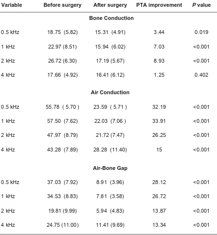
Table 1: Preoperative and postoperative audiometric results among 32 patients.
Pure Tone, Mean (SD), dB
Variable Before surgery After surgery PTA improvement P value
0.5 kHz 18.75 (5.82) 15.31 (4.91) 3.44 0.019
1 kHz 22.97 (8.51) 15.94 (6.02) 7.03 <0.001
2 kHz 26.72 (6.30) 17.19 (5.67) 8.93 <0.001
4 kHz 17.66 (4.92) 16.41 (6.12) 1.25 0.402
Air Conduction
0.5 kHz 55.78 ( 5.70 ) 23.59 ( 5.71 ) 32.19 <0.001
1 kHz 57.50 (7.62) 22.03 (7.06 ) 33.91 <0.001
2 kHz 47.97 (8.79) 21.72 (7.47) 26.25 <0.001
4 kHz 43.28 (7.89) 28.28 (11.40) 15 <0.001
Air-Bone Gap
0.5 kHz 37.03 (7.92) 8.91 (3.96) 28.12 <0.001
1 kHz 34.53 (8.83) 7.81 (3.58) 26.72 <0.001
2 kHz 19.81 (9.99) 5.94 (4.83) 13.87 <0.001
4 kHz 24.75 (11.00) 11.41 (9.69) 13.34 <0.001
Bone Conduction Threshold
The difference in bone conduction at 0.5 kHz, 1 kHz and 2 kHz before and after surgery was statistically significant
(P ≤0.001). The difference between bone
conduction at 4 kHz before and after surgery was not statistically significant
(P = 0.402).
Air Conduction Threshold
The differences in air conduction at all frequencies (0.5 kHz, 1 kHz, 2 kHz, 4 kHz) before and after surgery were highly
statistically significant (P≤0.001).
Air-Bone Gap (AB gap)
The differences in air-bone gaps at all frequencies (0.5 kHz, 1 kHz, 2 kHz, 4 kHz) before and after surgery were highly statistically significant
(P≤0.001).
Average
The mean air conduction thresholds of all patients were evaluated in 500, 1000, 2000 and 4000 Hz frequencies. The average preoperative bone conduction threshold was 21.53dB which was reduced to 16.21dB postoperatively. The difference in bone conduction thresholds was
statistically significant, P = 0.009 (before
X = 21.53; after X = 16.21). The difference
in air conduction PTA at all four frequencies before and after surgery was
highly statistically significant, P ≤0.001
(before X = 51.13; after X = 23.91). The difference in air-bone gap PTA at all four frequencies before and after surgery was highly statistically significant,
(P ≤0.001) (before X = 29.03; after
X = 8.51) (Table 2).
Table 2: The pure tone average of the bone conduction, air conduction and air-bone gap
before and after surgery.
PTA Bone conduction Air conduction AB-gap
Before surgery 21.53 dB 51.13 dB 29.03 dB
After surgery 16.21 dB 23.91 dB 8.51 dB
Figure 1: Case examples of Pure Tone Audiogram pre and post surgery.
Figure 1 shows three case examples of pure tone audiogram pre and
Case 1 Pre-Surgery Post- Surgery
post surgery.
Case 3 Pre-Surgery Post- Surgery
Various surgical techniques have been used to treat otosclerosis, but stapedotomy
still remains the method of choice.18 In our
study, all patients were surgically treated by small fenestra stapedotomy of the stapes footplate. The main goal of surgical techniques has been to improve patients’ hearing function and to eliminate the accompanying symptoms of the disease,
such as tinnitus and dizziness. Most
studies dealing with the outcomes of surgical treatment of otosclerosis depend on audiological testing before and after surgery, presuming that audiological measurements reflect the patient’s subjective experience regarding the treatment outcome.
Bone conduction
In the current study the average preoperative bone conduction value at
500 Hz was at the level of 18.75 dB preoperatively and 15.31dB
postoperatively, this was considered statistically significant. In addition, the change of average bone conduction value before and after surgery was also achieved a statistical significance at 1000 Hz and 2000Hz. However, at 4000Hz the mean values measured before and after surgery were 17.19 dB and 17.66 dB, respectively and thus showing no statistically significant difference at 4000 Hz. Thus in our study, there was a statistically significant difference in the mean of pre- and postoperative bone conduction values at frequencies 500, 1000 and 2000Hz but the difference was not statistically significant at 4000Hz. Similar to our data several studies have noted that in otosclerotic patients BC thresholds are better in the postoperative than in the preoperative period. However,
thedegree of BC improvement differs in
various studies. Awengen et al.24 noticed
an improvement in BC after stapedectomy in 500, 1,000 and 2,000 Hz but its deterioration in 4,000 Hz. Arnoldner
et al.25 showed some BC improvement in
conventional or laser-assisted surgery.
Aarnisalo et al26 showed an improvement
of BC about 4.5 dB in stapedectomy, and 3.1 dB is stapedotomy group. In a study by
Moscillo et al.,27 the BC improvement
was 4 and 7.1 dB in two different types of surgery, but the difference was not statistically significant. He also showed that this improvement might occur in a different frequency. On the other hand, various studies have stated that the surgery does not affect the bone conduction value. In
a study by Vincent et al.23 BC did not
change after stapes surgery. Similarly,
Quaranta et al.28 reported the changes of
average bone conduction value at 1000 Hz, 2000 Hz and 4000 Hz at the level of 0.2 dB. In 2005, Lazaro et al. reported a small change in the mean pre- and postoperative bone conduction values at 2000 Hz (32.73dB pre- and 30.78 dB
post-operatively.29 It is important to know
that BC threshold not only depends on the direct transmission of the vibration to the inner ear fluids through the skull, but it is also related to the relative movement of the footplate in the oval window due to the different inertia of the ossicular chain
and the otic capsule.30,31 The mechanical
process by which the energy of the sound waves entering the external canal and middle is utilized is known as
Carhart phenomenon.32 In patients with
otosclerosis, this energy is not utilized properly as there is a reduction of ossicular chain fluctuations caused by stapes fixation. This eventually will lead to difficulties in transmission of stimuli to the inner ear mostly at a frequency of
2000 Hz.32 Thus, the major drop in bone
conduction is observed at this frequency.
Air conduction
The average air conduction at 500 Hz before surgery was 55.78 dB. After surgery, the average air conduction was 23.59 dB. This result is highly statistically significant, indicating that otosclerosis surgery results in the improvement of conductive hearing loss. At 1000 Hz, the average preoperative air conduction was 57.50 dB, reaching 22.03 dB after surgery. This result is considered highly statistically
significant, confirming the successful outcome of the procedure at the frequency of 1000 Hz. The mean air conduction values at 2000 Hz were 47.97 dB and
21.72 dB before and after surgery,
respectively. This improvement is
considered highly statistically significant. The comparison of the mean pre- and postoperative air conduction values at 4000 Hz revealed a statistically significant difference. The mean values obtained before and after surgery were 43.28 dB and 28.28 dB, respectively. According to the above results, the functional hearing tests and their comparison demonstrate the effectiveness and success of stapedotomy
as a valuable method in treating otosclerosis. At all frequencies, the improvement in PTA value of air conduction before and after surgery was
statistically significant. The data in the present study are very similar to the results reported by Lazaro et al. in their study done
in 2005.29 Their data revealed the following:
the average preoperative air conduction at 500 was 60.2 dB before surgery and 36.8 dB after surgery. Preoperative and postoperative values at 1000 Hz were 56.6 dB 34.5 dB, respectively. The scores obtained at 2000 Hz were 52.7 dB before surgery and 36 dB after surgery, whereas mean preoperative and postoperative air conduction values at 4000 Hz were 54.3 dB and 45 dB, respectively.
Air-bone gap
The purpose of otosclerosis surgery and the greatest benefit resulting from it are the improvement of air conductivity and closure of the air-bone gap. The mean air-bone gap at 500 Hz before the surgery was 37.03 dB, whereas the value after surgery was 8.91 dB. This difference was highly statistically significant. The average air-bone gap at 1000 Hz before surgery was 34.53 dB while the postoperative value was 7.81 dB, which is considered highly statistically significant difference. At 2000Hz the values were 19.81dB before surgery and 5.94 dB after surgery, and the difference was highly statistically
significant. The average air-bone gap at 4000 Hz before the operation was at the level of 24.75 dB, whereas the value recorded after surgery was 11.41 dB, resulting in a statistically significant difference. Overall our study demonstrated
the following improvement of average air-bone gap values: at 500 Hz by 28.12 dB, at 1000 Hz by 26.72 dB, at 2000 Hz by 13.87 dB, and at 4000 Hz by 13.34 dB. Our data fully corresponds with the results
obtained by Vincent et al.23 Their study
demonstrated the following improvement of average air-bone gap values: at 500 Hz by 26.5 dB, at 1000 Hz by 25.4 dB, at 2000 Hz by 13.3dB, and at 4000 Hz by 8.2 dB. Very close results were also reported by the research of Belgin and Yilmaz
in 2004.33 The authors reported the
improvement of the air-bone gap of 30.8 dB at 500 Hz, whereas the scores at 1000 Hz and 2000 Hz were 25.5 dB and 14.3 dB, respectively. However, at 4000 Hz the increase of only 2.9 dB was demonstrated and this was not considered statistically significant. Contrary to the aforementioned study, our results at 4000 Hz showed a statistical significance, as the improvement of the average air-bone gap was 13.34 dB.
Average (all frequencies)
References
This study concludes that the significant
reduction in the air-bone gap, regardless of the change in bone conduction threshold, is a good indicator of the success of the stapedotomy surgery in patients with otosclerosis.
Conclusion
Competing interests
The authors declare that they have no competing interests.
before and after surgery was 20.52 dB. The goals of otosclerosis surgery are the closure of the air-bone gap and producing
the capability of hearing without
amplification. Success, defined as closure
of the air-bone gap to less than 10 dB.18
In our study, the closure of the air-bone gap to less than 10 dB was obtained in all of our patients as the average air-bone gap after surgery was 8.51 dB. In 2006, Vincent
et al.23 performed a prospective study over
a period of 14 years. The air-bone gap was
≤10 dB in 94.2% of cases. In 2013, Oeken
et al.34 published a study of 256 cases
of stapedotomy in which the postoperative
air-bone gap was ≤ 10 dB in 86%. In 2013,
Ataide et al.35 observed the same result in
75.8% of patients undergoing stapedotomy. In his study of otosclerosis, Sargent
et al.11 reported that in patients who had
undergone otosclerosis surgery the closure
of air-bone gap within a range up to 10 dB in 90% of patients. The research of
Rauka and Halik36 in 2005, who compared
the PTA– air-bone gap before and after surgery, a gap of 10 dB or less was observed in 85.19% of patients. Therefore, the audiometric results obtained in the present study are consistent with those in the literature. After all, it is argued that the relation between the average air-bone gap values before and after surgery cannot be considered a reliable indicator of the success of the surgical procedure, especially if there is a decrease in bone conduction during the postoperative
period8. In the aforementioned study, no
significant decrease in bone conduction was observed after surgery. Thus the average values for air-bone conduction were considered statistically significant. Unlike the above study, our research showed a significant reduction of the bone conduction after surgery except at 4000 Hz. Also as it is indicated in the earlier section, the reduction in bone conduction
thresholds after surgery was also reported
by Awengen24, Arnoldner et al.,25 Aarnisalo
et al.26 and Moscillo et al.27
1 . S t e f a n o v ić P . O t o r i n o l a r i n g o l o g i j a
maksilofacijalnom patologijom. Beograd:
Naučna knjiga; 1994.
2. Makarem AO, Hoang TA, Lo WW, Linthicum FH, Fayad JN. Cavitating otosclerosis: clinical, radiologic and histopathologic correlations. Otol Neurotol 2010; 31(3):381–4.
3. Menger DJ, Tange RA. The aetiology of otosclerosis: a review of the literature. Clin Otolaryngol Allied Sci 2003; 28(2):112–20.
4. Quinn BF, Ryan WM. Otosclerosis: grand rounds presentation, UTMB, Dept. of Otolaryngology; 2003.
5. Young G. Otosclerosis: grand rounds presentation. UTMB; 1996.
6. Ueda H, Miyazawa T, Asahi K, Yanagita N. Factors affecting hearing results after stapes surgery. J Laryngol Otol 1999; 113(5):417
–21.
7. Janošević LJ, Janošević S. Etiopatogeneza
otoskleroze. Beograd: Naučna knjiga; 1986. 8.
functional outcomes after stapes Surgery in patients with clinical otosclerosis in a teaching institution. Int Arch Otorhinolaryngol 2016; 20:39 –42.
9. Declau F, Van Spaendonck M, Timmermans JP, Prevalence of otosclerosis in an unselected series of temporal bones. Otol Neurotol 2001; 22(5):596–602.
10. Al- Husban H. Outcome of management of otosclerosis by stapedotomy compared to stapedectomy in a Jordanian population. Oman Med J 2013; 28(1):36–8.
11. Sargent E. Otosclerosis: A review for
audiologists. 2001. (Accessed on January 21, 2017, at http://www.audiologyonlin.com/Articles/ article_detail.asp?article_id=288).
12. Lolov S. Otosclerosis is a conformational disease. Med Hypotheses 2004; 62(1):121–3.
13. House HP, De la Cruz A, Friedman RA, Linthicum HF. Otosclerosis overview. Hearing
14. McGuirt WT, Fukushima K, Willems PJ, Van Camp G, Smith RJ. Linkage of a gene for otosclerosis to chromosome 15q25-q26 and identification of a candidate gene. Aggrecan. ARO abstracts; 1998:428.
15. Markou K, Goudakos J. An overview of the etiology of otosclerosis. Eur Arch Otorhinolaryngol 2009; 266:25–35.
16. Glasscock ME III, Storper IS, Haynes DS, Bohrer PS. Twenty-five years of experience with stapedectomy. Laryngoscope 1995; 105; (9 Pt 1):899–904.
17. Rondini-Gilli E, Bozorg Otosclerosis surgical techniques and results in 150 patients. Ann Otolaryngol Chir Cervicofac 2002; 119(4):227–33.
18. Dankuc D, Pejaković N, Komazec Z, Vlaški L.
Functional hearing results in patients with otosclerosis before and after stapedotomy. Med Pregl 2012; LXV(1-2):54–8.
19. Antoli-Candela F, McGill T, Peron D. Histopathological observations on the cochlear
changes in otosclerosis. Ann Otol Rhinol Laryngol 1977; 86(6 Pt 1):813–20.
20. Chole RA, McKenna M. Pathophysiology of Otosclerosis. Otology & Neurotology
prostheses over time. See comment in Pub Med Commons below Adv Otorhinolaryngol 2007; 65:174–8.
22. Kisilevsky VE, Dutt SN, Bailie NA, Halik JJ. Hearing results of 1145 stapedotomies evaluated
with Amsterdam hearing evaluation plots. J Laryngol Otol 2009; 123(7):730–6. 23. Vincent R, Sperling NM, Oates J, Jindal M.
Surgical findings and long-term hearing results in 3,050 stapedotomies for primary otosclerosis: a prospective study with the otology-neurotology database. Otol Neurotol 2006; 27(8)(suppl 2):S25 –47. 24. Awengen DF. Change of bone conduction
thresholds by total footplate stapedectomy in relation to age. Am J Otolaryngol 1993; 14:105– 10.
25. Arnoldner C, Schwab B, Lenarz T. Clinical results after stapedotomy: a comparison between the erbium: yttrium-aluminum- garnet laser and the conventional technique. Otol Neurotol 2006; 27:458–65.
26. Aarnisalo AA, Vasama JP, Hopsu E, Ramsay H. Long-term hearing results after stapes surgery: a 20-year follow-up. Otol Neurotol 2003; 24:567– 71.
27. Moscillo L, Imperiali M, Carra P, Catapano F,
Motta G. Bone conduction variation
poststapedotomy. Am J Otolaryngol 2006; 27:330 –3.
28. Quaranta N, Besozzi G, Fallacara AR, Quaranta A. Air and conduction change after
stapedotomy and partial stapedectomy for otosclerosis. Otolaryngol Head Neck Surg 2005; 133:116–20.
29. Pérez-Lázaro JJ, Urquiza R, Cabrera A, Guerrero C, Navarro E. Effectiveness assessment of otosclerosis surgery. Acta
Otolaryngol (Stockh) 2005; 125(9):935–45. 30. Schick F. Alterations of bone conducted
hearing in cases of modified middle ear mechanics. Eur Arch Otorhinolaryngol 1992; 249:268–72.
31. Tonndorf J. Bone conduction hearing. In: Keidel WD, NeV WD (eds) Handbook of sensory physiology. Berlin: Springer; 1994. P. 172–247.
32. House JW, Cunningham CDIII. Otosclerosis. In: Cummings CW (ed) Otolaryngology head and neck surgery, 4th ed. Mosby, Philadelphia;
2005.
33. Yilmaz ST, Belgin E. Otosclerosis disease and importance of long term follow-up after
stapedectomy. Otoscope 2004; 4:155–60. 34. Oeken J.
61(6):504–9. 35. Ataide AL, Bichinho GL, Patruni TM.
Otorhinolaryngol 2013; 79(3):325–35. 36. Raut V, Halik J. Argon laser assisted small