Comparative of Recombinant
Vespa affinis
Hyaluronidase Expressed
in Different Cloning Vectors and their Biological Properties
Piyapon Janpan
Faculty of Pharmaceutical Sciences, Khon Kaen University, Khon Kaen, Thailand
Yutthakan Saengkun Faculty of Pharmaceutical Sciences, Khon Kaen University, Khon Kaen, Thailand
Prapenpuksiri Rungsa Faculty of Pharmaceutical Sciences, Protein and Proteomics Research Center
for Commercial and Industrial Purposes Khon Kaen University, Khon Kaen, Thailand
Mongkol Vesaratchavest Research and Development Center
Betagro Public Company Limited Klong Nueng, Klong Luang, Pathumthani
Tewa Upathanpreecha Research and Development Center,
Betagro Public Company Limited, Klong Nueng, Klong Luang, Pathumthani
Patthana Tastub
Research and Development Center, Betagro Public Company Limited, Klong Nueng, Klong Luang, Pathumthani
Sakda Daduang∗
Faculty of Pharmaceutical Sciences
Protein and Proteomics Research Center for Commercial and Industrial Purposes Khon Kaen University, Khon Kaen, Thailand
Abstract:Cloning and expression of recombinantVespa affinishyaluronidase (rVesA2) were successfully expressed in
Escherichia colisystem. The VesA2 gene was cloned into pET-17b and pET-32a cloning vectors which had molecular weight 41.71 and 59.0 kDa, respectively. The recombinant plasmid of pET-17b was composed 1.08 kDa his-tag at the N-terminal. The 17.14 kDa of fusion tag; thioredoxin tag, histidine tag, and S-tag, was found in pET-32a. The verified expression conditions ofrVesA2 induced under the conditions of 0.1 mM IPTG at 37◦C for 4 hrs gave the highest quantity of protein expression. The colony PCR and sequencing analysis were used to verify therVesA2. The positive clones were detected the hyaluronidase activity by a zymographic gel. Recombinant proteins from both cloning vec-tors were insoluble. However, the recombinant from pET-32a showed higher solubility than that form pET-17b, after dissolving in 4 M urea solution. This result suggests that the fusion tag increase protein solubility.
Keywords:Hyaluronidase enzyme, hyaluronic acid, banded tiger wasp
Received:20 February 2018;Accepted:23 May 2018;Published:13 July 2018
I. INTRODUCTION
The banded tiger wasps (Vespa affinis) are insects in order Hymenoptera, and are found in Southeast Asia including Northeastern of Thailand [1]. They are dan-gerous to people because of their nest is, widespread
on trees or under eaves of houses. Their venom was used for defending themselves. Clinical manifestations when wasp bite is both local and systemic reactions such redness site bite area, pain, swelling, and headache [2]. The major proteins of venom gland include
phospholi-∗Correspondence concerning this article should be addressed to Sakda Daduang, Faculty of Pharmaceutical Sciences, Protein and Proteomics Research Center for Commercial and Industrial Purposes, Khon Kaen University, Khon Kaen, Thailand E-mail:[email protected]
c
pase, antigen 5, dipeptidyl peptidase and hyaluronidase [3]. Phospholipase A1 (PLA1) is one of the enzymes
in phospholipase family that hydrolyzes phospholipid at the 2-acyl-lysophospholipid position to fatty acid and lysophospholipid [4]. Antigen-5 is an allergenic protein which causes hypersensitivity in human. This protein can find in plants and animals including wasp venom [5, 6]. Dipeptidyl-peptidases (DPPs) are exopeptidase that cleaves the polypeptide at N-terminal of peptide bond [7]. The hyaluronidase plays a role in degrada-tion connective tissue of prey, and it facilitates spreading of another components into their body (spreading factor) [8]. In addition, that enzyme also is an allergenic fac-tor [9,10]. The hyaluronidases are a family of glycoside hydrolase (EC. 3.2.1.35) [11,12]. This enzyme cleaves hyaluronic acid to tetrasaccharides at beta-1,4-glycosidic bond [13,14]. In a previous study, hyaluronidase in ven-omous animals contains mixture biochemical and bio-physical properties depend on species of animal. For example, the hyaluronidase venom of V. tropica has a molecular weight ranging from 46-47 kDa, pI 8.91 and optimal pH 3-4 [15, 16, 17]. The aims of this study are cloning and expression of recombinant hyaluronidase from banded tiger wasp (V. affinis) venoms and compar-ison different expression vector (pET-17b and pET-32a) inE. colisystem.
II. MATERIAL AND METHODS
A. Total RNA Extraction and cDNA Synthesis
Total RNA was extracted from venom gland of V. affinis using Trizol⃝R reagent (Ambion and Life
tech-nologies, USA). The quality of total RNA was inves-tigated with absorbance at 260/280 nm by UV-Visible spectrophotometer and gel electrophoresis. After that, cDNA was synthesized from total RNA using Re-verseAid first strand cDNA synthesis kit (Thermo Sci-entific, USA) following manufacturer’s protocol.
B. Gene Cloning and Sequencing Analysis
The specific primers were designed from hyaluronidase gene sequence of another wasp [15]. In addition, the specific PCR primers was obtained KpnI andNotI restriction size position to ligate with cloning vector, pGEM-T easy vector (Invitrogen, USA) and ex-pression vector, pET-17b and pET-32a vector (Invitro-gen, USA) (Table1). After that, the hyaluronidase gene (VesA2 gene) was amplified with the cDNA as a tem-plate using polymerase chain reaction (PCR) followed by the conditions in Table2. The PCR product was de-tected by agarose gel electrophoresis and purified with GenepHlowTM Gel/PCR kit (Geneaid, Taiwan) follow-ing manufacturer’s protocol. TheVesA2gene was ligated with pGEM-T easy vector using T4 DNA ligase at 4◦C for 16-18 hrs and transformed into DH5α E. colistrain. The transformants (containing pGEM-VesA2 plasmid) was verified by colony PCR method. The positive clones were cultured in LB broth containing 100µg/ml ampi-cillin at 37◦C overnight. The recombinant plasmids (pGEM-VesA2) were isolated using the PrestoTM Mini Plasmid kit (Geneaid, Taiwan) according to manual’s protocol. The isolated plasmids were sequenced and analyzed using the basic local alignment search tool (BLAST), https://blast.ncbi.nlm.nih.gov/.
TABLE 1
SPECIFIC PCR PRIMERS FOR VESA2 GENE
Primers Product size (bp)
Forward 5-GGTACCGCACCATCATCATCATCATTCCGAGAGACCGAAAAAAG-3 993 Reverse 5-GCGGCCGCCACTAGTTAACGGCTTCTGTCA-3
TABLE 2
PCR CONDITION FOR VESA2 GENE
Temperature Time (min)
Pre-denaturation 94◦C 5:00
Denaturation 94◦C 0:30 25-35 cycles Annealing 50◦C 0:30
C. Gene Construction and Protein Expression
The VesA2 gene was sub-cloned from the pGEM-T easy vector into both pEpGEM-T-17b and pEpGEM-T-32a expres-sion vector. Briefly, the recombinant plasmids ( pGEM-VesA2), pET-17b and pET-32a vector were digested with restriction enzymesKpnI andNotI using double diges-tion method at 37◦C for 3 hrs. After digestion, the re-actions were analyzed by agarose gel and purified us-ing GenepHlowTMGel/PCR kit as described above. The
VesA2 gene was ligated with both 17b and pET-32a expression vectors at 4◦C for 16-18 hrs using T4 DNA ligase. The ligation products, pET-17b-VesA2
andpET-32a-VesA2construction, were transformed into BL21(DE3) expression E. coli host and cultured on LB agar plate containing 100µg/ml ampicillin at 37◦C overnight. The colonies on agar plate were cultured in 5 ml LB broth containing 100µg/ml ampicillin at 37◦C 14-16 hrs for the starter culture. After that, 100 µl of the starter culture was added in 10 ml of LB broth con-taining 100 µg/ml ampicillin and cultured at 37◦C un-til the cell density reached to OD600 nm between 0.4-0.6. The cultured cells were incubated at 37◦C for 4 hrs with 0.1mM IPTG. The cell pellet was kept by certifica-tion at 10,000×g for 3 minutes. The protein expression of hyaluronidase (recombinantVesA2protein) was ana-lyzed using SDS-PAGE gels.
D. Hyaluronidase Activity Assay
Hyaluronidase activity was confirmed by gel zymog-raphy method that was modified from Mio and Sterm’s reports [18]. The methods are similar to SDS-PAGE gels, but the hyaluronic acid substrate was obtained in separating gel. The 13% separating gel solution was pre-pared by adding 2,130µl of acrylamide solution (30%; 37.5:1), 1,250µl of 0.4 mg/ml of hyaluronic acid, 1,233 µl of 1.5 M Tris-HCl buffer pH 8.8, 333µl of water, 50 µl of 10% SDS solution, 33µl of 10% APS solution, and 2.1 µl of TEMED. After that, the 4% stacking gel was prepared following as: 265µl acrylamide solution (30%; 37.5:1), 500µl of 0.5 M Tris-HCl buffer pH 6.8, 1,215 µl of water, 20µl of 10% SDS solution, 12.5µl of 10% APS solution, and 6.25µl of TEMED. This method was used electric potential at 150 volts. The substrate gel was incubated with 3% Triton-X 100 in 50 mM Hepes for 1 hr to remove SDS. Then, the gel was rinsed 2 times with water. After that, the assay buffer (0.1M acetate buffer pH 4.0 in 0.15M NaCl) was added and incubated at 37◦C for 16 hrs. The gel was stained with Alcian blue solution for 1 hr and destained with 7% acetic acid until the ob-served lytic zone.
III. RESULT
A. Gene Cloning and Sequencing Analysis
Total RNA extraction from the venom gland of V. affiniswas extracted from venom gland (Figure1).
Fig. 1. Agarose gel analysis of (A) RNA extraction and (B)
cDNA synthesis from venom gland ofVespa affiniswere
am-plified using specific primer for hyaluronidase gene by RT-PCR technique. The product was about 1,000 bp in size. M: 100 bp DNA ladder, 1: hyaluronidase gene
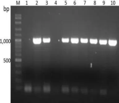
The RT-PCR product, about 1,000 bp in size, using gene-specific primers was obtained (Figure1). The PCR product band was purified and ligated with the pGEM-T easy vector. The recombinant plasmids (pGEM-VesA2) were transformed into DH5α E. colistrain and analyzed by colony PCR. The positive colonies clearly showed the products about 1000 bp in size (Figure2, Lane 2-3 and 5-10). Then, the purified recombinant plasmids ( pGEM-VesA2) were nucleotide sequenced to confirm the gene.
Fig. 2. Determination of recombinant gene (pGEM-VesA2) in
E. coliDH5α by colony PCR technique. Lane 2-3 and 5-10 shown product size about 1,000 bp. M: 100 bp DNA ladder,
The obtained nucleotide sequences correspond to wasp venom hyaluronidase records in the GenBank database. Then, the nucleotide sequences were deduced to 331 amino acid residues using BLASTX online
ac-cessible tools. The amino acid sequences composed 96 residues with above 90% similarity to those of wasps,
Vespa magnificaandVespula germanica(Table3).
TABLE 3
THE AMINO ACID SEQUENCE OF VESA2 WAS ANALYZED BY PROTEIN BLAST AFTER ALIGNMENT TO HYALURONIDASE FROM WASP VENOM
Description % Identities
Hyaluronidase, Vespa magnifica 96
Hyaluronidase, Vespula germanica 92
B. Protein Expression and Hyaluronidase Activity As-say
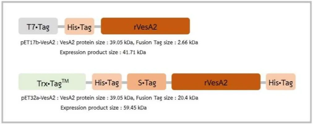
The solubility of the recombinant products originally from both expression vectors had been investigated. The basic structures of pET32a and pET17b were shown in Figure4. For the pET-17b plasmid, T7·tag and His·tag were at the N-terminus upstreamingly conjugated to the insertedVesA2(Figure3), whereas for pET-32a plasmid, the fusion tags were thioredoxin tag, His·tag, and S·tag at the N-terminus (Figure3).
The recombinant plasmids were transformed into BL21(DE3) expression host and induced by 0.1 mM IPTG. The recombinant VesA2 protein (39 kDa) from
pET17b-VesA2andpET32a-VesA2constructs were about 41 (Figure4A) and 59 kDa (Figure4B), respectively, in size after analysis by SDS-PAGE. The hyaluronidase ac-tivity was demonstrated on substrate gel (Figure4). Both recombinant VesA2 proteins showed clear bands in the size of 41 and 59 kDa, corresponding to those analyzed by SDS-PAGE.
Fig. 3. Schematic of rVesA2 protein with fusion protein from both expression vector. (A) Recombinant protein from pET17b
vector was fusion protein of T7·Tag, His·Tag and rVesA2 with product size about 41 kDa. (B) Recombinant protein from pET32a
vector was that of Trx·TagTM, His·Tag, S·Tag, rVesA2 and His·Tag with product size about 59 kDa
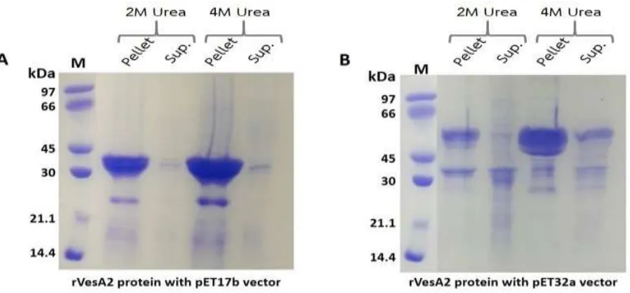
The induced cell pellets were determined their solu-bility. The pellet was freeze-thawed, sonicated and cen-trifuged at 10,000×g for 30 minutes. Then, the pellets were dissolved in 2 M and 4 M urea buffer. After cen-trifugation, the pellet and supernatant fractions of 2 M and 4 M urea were analyzed on SDS-PAGE (Figure5).
The insoluble rVesA2 proteins were found from both
Fig. 4. SDS-PAGE analysis of recombinant VesA2 protein expression. (A) The expression of rVesA2 inpET17b-VesA2 construc-tion (41 kDa) and its hyaluronidase activity. (B) The expression of rVesA2 in pET32a-VesA2 construcconstruc-tion (59.45 kDa) and its hyaluronidase activity. Products form both of vectors showed hyaluronidase activity in zymographic gel
The induced cell pellets were determined their solu-bility. The pellet was freeze-thawed, sonicated and cen-trifuged at 10,000×g for 30 minutes. Then, the pellets were dissolved in 2 M and 4 M urea buffer. After cen-trifugation, the pellet and supernatant fractions of 2 M and 4 M urea were analyzed on SDS-PAGE (Figure5).
The insoluble rVesA2 proteins were found from both
pET17b-VesA2 (A) and pET32a-VesA2 constructs (B). However, rVesA2 frompET32a-VesA2construct shown higher solubility in denaturing conditions (2 M and 4 M urea) than rVesA2 frompET17b-VesA2constructs.
IV. DISCUSSION AND CONCLUSION In this study, we described the cloning, expres-sion, and characterization of rVesA2 derived from the tiger wasp, V. affinis, venom. The theoretical molecu-lar weight and isoelectric points of mature VesA2 were about 39 kDa and 9.2, respectively, which corresponds to the experiments. Bacterial inclusion bodies are usu-ally expressed at the high levels of protein expression [19]. The VesA2 was overexpressed mainly in insolu-ble form. However, the insoluinsolu-ble form has no effects on the HAase activity according to the zymographic gel as-say [18]. The renaturing by 3% Triton-X detergent has a tendency to easily recover the activity. The refolding in gel, rVesA2 was able to degrade the substrate, hyaluronic acid, causing a lytic zone with electrophoretic mobility about 41 and 59 kDa in size after Alcian blue staining of the intact background polysaccharide, hyaluronic acid [18,20].
However, the solubility was different. High con-centration of urea (2 and 4 M urea) showed a tendency to synergize the solubility of fusion proteins expressed from the vectors with thioredoxin tag, a pET32a vector. Therefore, the addition of fusion Protein Tags, such as glutathione S-transferase (GST), thioredoxin (Trx), and Maltose binding protein (MBP) often improves the sol-ubility of recombinant expression products [21,22]. We clearly concluded that the tags just affected the solubility. The levels of fusion protein overexpression and HAase activity still showed similar results from both short-tag or long-tag vectors.
V. ACKNOWLEDGMENTS
This work was financially supported by Research and Researchers for Industries (RRI), The Thailand Re-search Fund (TRF) (Grant no. MSD59I0004) with the additional support from the Faculty of Pharmaceutical Sciences and the Khon Kaen University Research Fund, the fiscal year 2016 to 2017.
REFERENCES
[1] S. Sukprasert, P. Rungsa, N. Uawonggul, P. Incam-noi, S. Thammasirirak, J. Daduang, and S. Dad-uang, “Purification and structural characterisation of phospholipase A1 (Vespapase, Ves A 1) from Thai banded tiger wasp (Vespa affinis) venom,”
Toxicon, vol. 61, pp. 151–164, 2013. doi: https: //doi.org/10.1016/j.toxicon.2012.10.024
[2] N. Sookrung, S. Wong-din dam, A. Tungtrong-chitr, O. Reamtong, N. Indrawattana, Y. Sakolva-ree, N. Visitsunthorn, W. Manuyakorn, and
W. Chaicumpa, “Proteome and allergenome of Asian wasp, vespa affinis, venom and IgE reactiv-ity of the venom components,”Journal of Proteome Research, vol. 13, no. 3, pp. 1336–1344, 2014. doi:
https://doi.org/10.1021/pr4009139
[3] P. Rungsa, P. Incamnoi, S. Sukprasert, N. Ua-wonggul, S. Klaynongsruang, J. Daduang, R. Pa-tramanon, S. Roytrakul, and S. Daduang, “Com-parative proteomic analysis of two wasps venom, Vespa tropica and Vespa affinis,”Toxicon, vol. 119, pp. 159–167, 2016. doi: https://doi.org/10.1016/j. toxicon.2016.06.005
[4] G. S. Richmond and T. K. Smith, “Phospholipases A1,” International Journal of Molecular Sciences, vol. 12, no. 1, pp. 588–612, 2011. doi: https://doi. org/10.3390/ijms12010588
[5] A. Henriksen, T. P. King, O. Mirza, R. I. Mon-salve, K. Meno, H. Ipsen, J. N. Larsen, M. Gajhede, and M. D. Spangfort, “Major venom allergen of yellow jackets, Ves v 5: Structural characteriza-tion of a pathogenesis-related protein superfam-ily,” Proteins: Structure, Function, and Bioinfor-matics, vol. 45, no. 4, pp. 438–448, 2001. doi:
https://doi.org/10.1002/prot.1160
[6] P. Rungsa, S. Peigneur, S. Daduang, and J. Tyt-gat, “Purification and biochemical characterization of VesT1s, a novel phospholipase A1 isoform iso-lated from the venom of the greater banded wasp Vespa tropica,”Toxicon, vol. 148, pp. 74–84, 2018. doi: https://doi.org/10.1016/j.toxicon.2018.03.015
[7] T. K. Nemoto, T. Ono, and Y. Ohara-Nemoto, “Es-tablishment of potent and specific synthetic sub-strate for dipeptidyl-peptidase 7,” Analytical Bio-chemistry, vol. 548, pp. 78–81, 2018. doi: https: //doi.org/10.1016/j.ab.2018.02.008
[8] K. C. Bordon, M. G. Perino, J. R. Giglio, and E. C. Arantes, “Isolation, enzymatic characterization and antiedematogenic activity of the first reported rat-tlesnake hyaluronidase from Crotalus durissus ter-rificus venom,” Biochimie, vol. 94, no. 12, pp. 2740–2748, 2012. doi: https://doi.org/10.1016/j. biochi.2012.08.014
[10] D. Kolarich, R. Léonard, W. Hemmer, and F. Alt-mann, “The N-glycans of yellow jacket venom hyaluronidases and the protein sequence of its ma-jor isoform in Vespula vulgaris,” The FEBS Jour-nal, vol. 272, no. 20, pp. 5182–5190, 2005. doi:
https://doi.org/10.1111/j.1742-4658.2005.04841.x
[11] K. Girish and K. Kemparaju, “The magic glue hyaluronan and its eraser hyaluronidase: A bio-logical overview,” Life Sciences, vol. 80, no. 21, pp. 1921–1943, 2007. doi: https://doi.org/10.1016/ j.lfs.2007.02.037
[12] R. A. Hutagalung, S. Hermawan, I. Magdalena, Iskandar, and S. Mastrorillo, “Increasing growth and survival rate of land hermit crabs (Coenobita sp.) in artificial habitat through feeding habit,” In-ternational Journal of Applied and Physical Sci-ences, vol. 3, no. 3, pp. 55–59, 2017. doi: https: //doi.org/10.20469/ijaps.3.50001-3
[13] R. Stern and M. J. Jedrzejas, “Hyaluronidases: Their genomics, structures, and mechanisms of ac-tion,”Chemical Reviews, vol. 106, no. 3, pp. 818– 839, 2006. doi:https://doi.org/10.1021/cr050247k
[14] K. L. Chao, L. Muthukumar, and O. Herzberg, “Structure of human hyaluronidase-1, a hyaluro-nan hydrolyzing enzyme involved in tumor growth and angiogenesis,” Biochemistry, vol. 46, no. 23, pp. 6911–6920, 2007. doi: https://doi.org/10.1021/ bi700382g
[15] P. Rungsa, P. Incamnoi, S. Sukprasert, N. Ua-wonggul, S. Klaynongsruang, J. Daduang, R. Patramanon, S. Roytrakul, and S. Daduang, “Cloning, structural modelling and characteri-zation of VesT2s, a wasp venom hyaluronidase S(HAase) from Vespa tropica,” Journal of Ven-omous Animals and Toxins including Tropical Diseases, vol. 22, no. 1, pp. 28–39, 2016. doi:
https://doi.org/10.1186/s40409-016-0084-5
[16] P. Bunruk, D. Kantachote, and A. Sukhoom, “Iso-lation and selection of purple non-sulfur bacteria for phosphate removal in rearing water from shrimp cultivation,” International Journal of Applied and Physical Sciences, vol. 3, no. 2, pp. 73–80, 2017. doi: https://doi.org/10.20474/japs-3.2.5
[17] N. Mahmad, R. M. Taha, N. Rawi, and S. Mo-hajer, “The effects of picloram and 2, 4-dichlorophenoxyacetic acid on induction of red coloured callus from celosia plumosa, an attractive ornamental plant,” Journal of Applied and Physi-cal Sciences, vol. 1, no. 1, pp. 9–12, 2015. doi:
https://doi.org/10.20474/-japs1.1.2
[18] K. Mio and R. Stern, “Reverse hyaluronan sub-strate gel zymography procedure for the detection of hyaluronidase inhibitors,”Glycoconjugate Jour-nal, vol. 17, no. 11, pp. 761–766, 2000.
[19] S. M. Singh and A. K. Panda, “Solubilization and refolding of bacterial inclusion body proteins,”
Journal of Bioscience and Bioengineering, vol. 99, no. 4, pp. 303–310, 2005.
[20] M. W. Guntenhöner, M. A. Pogrel, and R. Stern, “A substrate-gel assay for hyaluronidase activity,”
Matrix, vol. 12, no. 5, pp. 388–396, 1992. doi:
https://doi.org/10.1016/s0934-8832(11)80035-1
[21] S. Maijaroen, P. Anwised, S. Klaynongsruang, S. Daduang, and A. Boonmee, “Comparison of recombinant α-hemoglobin from Crocodylus sia-mensis expressed in different cloning vectors and their biological properties,”Protein Expression and Purification, vol. 118, pp. 55–63, 2016. doi:https: //doi.org/10.1016/j.pep.2015.09.028
[22] Y. Li, “Carrier proteins for fusion expression of antimicrobial peptides in Escherichia coli,”