www.fm.viamedica.pl O R I G I N A L A R T I C L E
Address for correspondence: Dr Małgorzata Waszak, Department of Functional Anatomy, University of Physical Education, ul. Królowej Jadwigi 27/39, 61–871 Poznań, Poland, tel: +61 835 52 26, fax: +61 833 00 87, e-mail: [email protected]
Developmental interdependence between
selected somatic features and the weight
of internal organs in human male and female
foetuses
Małgorzata Waszak, Krystyna Cieślik
Department of Functional Anatomy, University of Physical Education in Poznań, Poland
[Received 24 October 2002; Accepted 10 December 2002]
The study material comprised 3889 foetuses of both sexes, aged 20–42 weeks and focused on developmental interdependence between the weight of the internal organs and their relation to selected somatic features during the prena-tal period. The study also attempted to distinguish potential sex-related differ-ences in the degree of statistical interdependence between the analysed vari-ables. As a result, the analysis of linear and canonical correlation coefficients for consecutive weeks of foetal life has been carried out. High correlation coeffi-cients have been obtained which indicate strong developmental interdepen-dence between those organs and the somatic features. However, no significant sex-related differences in the developmental interdependence of the analysed variables have been observed.
key words: developmental interdependence, correlation of morphological features, foetal sex dimorphism
INTRODUCTION
It seems interesting to determine the degree of interdependence of population describing measur-able morphological characteristics. The degree of in-terdependence of measurable morphological organ characteristics may provide a background for the analysis of biological relationships between those organs. Developmental interdependence between organs is related to the specificity of the relations between these organs and the selected somatic fea-tures of the foetus. This issue is, however, related to the question of whether body shape determines the shape of the internal organs or maybe the reverse is true, i.e. that it is the organs that determine the body shape. Or, possibly, the development of any of these is independent of one another?
Unfortu-nately, the results of available studies [2, 3, 6] are unconvincing and full of speculations and hypothe-sising, yet without providing definite answers.
delin-eate potential sex-related differences in the degree of statistical interdependence between the weight of the internal organs and the selected somatic fea-tures during the prenatal period.
MATERIAL AND METHODS
The study material comprised 3889 foetuses of both sexes (2203 males and 1686 females) aged 20–42 weeks. The foetuses with any indication of pathology were excluded from the study. Statistical interdependence between the weights of brain, heart, lungs, liver, spleen, kidneys, adrenals, thymus, as well as between the weights of these organs and selected somatic characteristics, such as total body length, crown-rump length, body weight and circum-ferences of the head, shoulders, chest and the ab-domen, were determined. Linear correlation coeffi-cients were calculated for male and female foetuses for consecutive weeks of foetal life and for the foe-tal age normalised data. In order to determine the sex-related correlations, an inter-sex comparison of the calculated correlation ratios was carried out by test-u statistical method:
2 2 1 2 2 1 Z + D Z D – Z Z u =
Afterwards, a more complex relationship, i.e. the interdependence between two groups of character-istics, i.e. somatic ones (independent variables — p) and the weight of internal organs (dependent vari-ables — q), carried out by means of canonical anal-ysis, was performed. As a result, if X = (X1, X2… Xp)
and Y = (Y1, Y2 … Yq) are column p and q vectors of
random variables (X — the set of somatic character-istics; Y — the set of weights of internal organs; p = 7; q = 8), then the covariance matrix of those p + q random variables will take the form of the following matrix:
covar q
p y x q22 p21 12 11 =
∑
∑
∑ ∑
,where
∑
21' =∑
12 row∑
12 = S ≤ min (p, q)As a result 7 pairs of canonical variables for each week and for each sex were obtained, as such was the number of variables in the smaller set of inde-pendent variables. The analysis comprised the first pair of canonical variables, which corresponded to the highest of the canonical roots, the latter being the squares of the canonical correlation coefficients.
The number of characteristic roots equals the mini-mum from the number of independent and depen-dent variables’ sets. The higher the value of the char-acteristic roots, the higher the correlation between the first and the second group of variables. As a re-sult, the first pair of canonical variables is the pair of variables characterised by the highest correlation between each other. Each of the characteristic roots had a characteristic vector assigned to it, the coeffi-cient of such a vector being referred to as a weight. The characteristic vector, when multiplied by the vector of independent variables, yields the first ca-nonical variable for these independent variables. Sim-ilarly, if the vector that is characteristic of dent variables is multiplied by the vector of depen-dent variables then the first canonical variable for the dependent variables is obtained.
The canonical variable for the primary X vari-ables is:
U = 1’X = 11X1 + 12X2 + … + 1pXp
The canonical variable for the primary Y vari-ables is:
V = m’Y = m1Y1 + m2Y2 + … + mqYq
The coefficients l = (l1, l2, …, lp)’ and m = (m1,
m2, …, mq)’, known as the weights of canonical
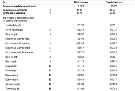
vari-ables, were calculated in such a way that the corre-lation between U and V variables is maximal. The value of the weights of the canonical variables indi-cates the share of specific somatic characteristics and weights of internal organs in the canonical correla-tion. The higher the absolute values of those coeffi-cients, the higher the impact of the X and Y sets variables on the degree of correlation of canonical variables. Moreover, the redundancy coefficients calculated for each of the analysed cases (each week, sex and the standardised data) for primary canonical variables — R2
YX [1] (Table 1) were calculated. Those
coefficients provide information about the share of primary variables in the new, canonical variable.
RESULTS
coefficients were also reported between the weights of the liver and the spleen — both of which are known to carry out their haematopoietic and immu-nological functions already in foetal life. The lowest, often insignificant correlation coefficients were found between the lungs and the spleen, the adrenals and the thymus. Interestingly, there is al-most a complete absence of correlation of the de-velopmental weight of the analysed organs with re-gard to their origin and localisation, e.g. the lungs and the thymus come from the endoderm and de-velop in the chest, yet no correlation between each other was detected. Considering, however, that each of these organs fulfils different functions during foe-tal life, it may be postulated that the existing/non-existing statistical correlations between these organs may be, at least partially, a consequence of their functional similarity/dissimilarity.
The analysis of developmental interdependence between the internal organs and foetal somatic features revealed that the highest correlation ra-tios are those of some internal organs, namely brain and liver, with the total body weight, and
this is understandable as the weight of those or-gans significantly determines the total body weight. High correlation ratios were also revealed between the brain weight and all the somatic fea-tures, in particular head and chest circumference and heart weight. Such results seem obvious once the localisation and the functions of those organs is taken into account.
The analysis of the results did not reveal any sig-nificant sex-related differences with regard to the correlations between the analysed characteristics and virtually all the correlations were found to be similar in both sexes. This similarity is exemplified by the fact that the weakest and the strongest correlations are found in both sexes for the same feature com-parisons and the respective correlation values are highly similar. However, the strongest sex dimor-phism was observed for the correlations between the head circumference and chest circumference, abdomen circumference, heart weight, and for the correlations between the total body length and heart weight, and between the total body weight and heart weight.
Table 1. Correlation and redundancy coefficients and the weights of canonical variables as calculated for the age norm-alised data
Sex Male foetuses Female foetuses
Canonical correlation coefficients 0.8930 0.9026
Redundancy coefficients x 47.46 49.55
for the set of variables y 47.78 50.98
The weights of canonical variables for specific characteristics
Total body length 1 0.1790 0.0551 Crown-rump length 2 –0.0248 0.0138
Body weight 3 0.8352 0.8426
Circumference of the head 4 0.0192 0.1358
Circumference of shoulders 5 –0.0166 0.0402
Circumference of the chest 6 0.0017 0.0129
Circumference of the abdomen 7 0.0772 0.0454
Brain weight 8 0.3930 0.3821
Heart weight 9 0.1123 0.2038
Lung weight 10 0.1113 0.1395
Liver weight 11 0.2743 0.2030
Spleen weight 12 0.0986 0.0590
Kidney weight 13 0.0988 0.1317
Adrenals weight 14 0.0109 –0.0050
analysed somatic characteristics, on the value of cor-responding canonical variables. In contrast, such reg-ularity could not be observed for the male foetuses. The correlation and redundancy coefficients and the weights of canonical variables for the age-norma-lised data are included in Table 3.
The analysis of the whole foetal period revealed higher redundancy coefficients in female foetuses, and in both sexes the organ weights had more ef-fect in the creation of the corresponding canonical variable than the somatic features. The weight val-ues of the canonical variables for the analysed fea-tures (Table 3) allowed us to find out which of the somatic features and internal organs weights had The canonical correlation coefficients for the first
pair of canonical variables for consecutive weeks of foetal life are presented in Table 2. These coefficients tend to decrease towards the end of the foetal peri-od, this trait being most pronounced in male foet-uses in week 42 and in female foetfoet-uses in week 40. These results may reveal some biological develop-mental regularity as the analysis of the linear corre-lation coefficients also revealed that during this pe-riod most coefficients were statistically insignificant. The analysis of the redundancy coefficients for the first set of canonical variables (Table 3) revealed that for female foetuses aged 27 to 42 weeks the set of primary dependent variables, i.e. the analysed internal organs’ weights, had a more potent effect, compared to primary independent variables, i.e. the
Table 3. Redundancy coefficients for dependent and in-dependent variables for consecutive weeks of foetal life in both sexes
The set of variables
Foetal Independent Dependent Independent Dependent
week (x) (y) (x) (y)
Male foetuses Female foetuses
20 72.12 55.51 64.72 54.61
21 60.68 54.05 82.92 66.21
22 54.65 39.95 57.08 68.92
23 52.01 46.80 59.89 45.93
24 49.85 46.72 53.11 42.30
25 40.08 44.60 53.80 56.27
26 49.08 52.81 55.33 49.85
27 47.30 57.69 53.40 54.70 28 56.58 60.36 45.83 55.12
29 54.06 46.46 56.99 57.89
30 62.57 65.13 43.00 54.23
31 49.82 59.53 51.96 53.68
32 57.57 61.18 56.73 61.74
33 62.36 60.08 70.99 70.87
34 59.62 58.97 62.68 63.18
35 50.01 57.57 55.73 59.17 36 55.33 51.17 63.37 65.99
37 39.64 45.96 59.33 60.13
38 55.73 49.07 62.53 63.22
39 47.12 52.02 49.09 61.85
40 48.68 45.03 43.93 44.66
41 60.32 55.45 53.63 55.52
42 38.47 38.47 58.04 55.28
Table 2. Canonical correlation coefficients for consecu-tive weeks of foetal life in both sexes
Foetal Male foetuses Female foetuses week
20 0.9461 0.9130
21 0.9472 0.9705
22 0.9267 0.9623
23 0.9300 0.9118 24 0.9065 0.9235
25 0.9103 0.9319
26 0.9156 0.9235
27 0.9379 0.9082
28 0.9298 0.9222
29 0.8966 0.9328
30 0.9436 0.9025
31 0.9277 0.9057 32 0.9381 0.9386
33 0.9313 0.9595
34 0.9483 0.9462
35 0.9356 0.9147
36 0.8984 0.9449
37 0.9021 0.9271
38 0.9082 0.9538
39 0.9228 0.9401 40 0.9149 0.8771
41 0.9236 0.9003
the most potent effect on the value of canonical correlations between these two groups of charac-teristics. Interestingly, the body weight was found to be most potently correlated with the internal organs group, both in male and in female foetus-es. In male foetuses the highest correlation between the weight of the internal organs and the somatic features was observed for brain, liver and heart weight, whereas in female foetuses these were brain, heart and liver weight (in order of impor-tance).
Because of the large sizes of the tables this arti-cle does not include the data on the canonical ables separately for each week and only the vari-ables for which those coefficients have the highest absolute values have been included (Table 4). Table 4 provides a confirmation of those observations, as in each foetal week (from week 20 to week 42) for both sexes it was total body weight (3) and brain weight (8) that were most critical for the correlation of the two groups of features.
The canonical correlation between the set of so-matic features and the set of the weights of internal organs has been graphically presented for the two first canonical variables and the degree of correla-tion for each foetus has been indicated (Fig. 1).
DISCUSSION
The analysis of the results from all the methods applied to determine the type and strength of the developmental interdependence between the inter-nal organs, as well as between the interinter-nal organs and selected somatic features, has revealed that the correlations are most pronounced between week 24 and week 38 of foetal life, whereas before week 24
Figure 1. Canonical correlation between the set of somatic features and the set of the weights of internal organs.
Table 4. Somatic features and the weights of the internal organs (1–15) that have the highest share in the canoni-cal correlation value for consecutive weeks of foetal life in both sexes
Foetal Male foetuses Female foetuses week Somatic Weights of the Somatic Weights of the
features internal organs features internal organs
20 3 8 1 8
21 3 8 3 11
22 3 8 3 8
23 3 8 3 8
24 3 8 3 8
25 3 8 3 13
26 3 8 3 8
27 3 8 3 13
28 3 8 3 8
29 3 11 3 8
30 3 11 3 8
31 3 9 3 9
32 3 8 3 8
33 3 8 3 8
34 3 8 3 8
35 3 13 3 8
36 3 8 3 8
37 3 10 3 10
38 3 8 3 8
39 3 8 3 8
40 1 8 3 9
41 3 8 3 8
ised by a limited morphological variability, which may be the consequence of intrauterine growth being dependent on the intrauterine environment which, in turn, precludes a more pronounced display of ge-netically determined variability.
The problem of sex-related differences in the developmental interdependence of measurable mor-phological features during the prenatal period has not been investigated so far, therefore the possibil-ity of any discussion is highly limited.
CONCLUSIONS
High correlation ratios between the organs in-dicate the strong interdependence of their devel-opment, particularly between week 24 and 38 of foetal life. This strong interdependence is strongly determined by their biochemical and physiological functions. The organs whose physiological func-tions are intertwined in the foetal life are charac-terised by the strongest developmental interdepen-dence.
A statistically significant dependence has been observed between the change in the organ weight and the somatic features, which confirms the cur-rent hypotheses on the influence of the internal or-gans on the growth and shape of some bodily parts of the foetus.
Strong developmental correlations have been observed in the foetuses of both sexes. No sex-relat-ed differences have been observsex-relat-ed with regard to developmental correlations between the analysed characteristics.
REFERENCES
1. Krzyśko M, Ratajczak W (1978) Analiza kanoniczna. Listy biometryczne. Pol Tow Biometr, pp. 65–67. 2. Malinowski A (1971) Problem rozwoju czaszki, mięśnia
skroniowego i mięśnia żwacza u płodów ludzkich. Przegl Antrop, 37: 19–36, 169–182.
3. Marecki B (1989) Development relations between the weight of internal organs and somatic features of foetus-es and new-borns. Z Morph Anthrop, 78, pp. 107–115. 4. Neligan G (1965) A community study of the
relation-ship between birth, weight and gestational age. Ges-tational age, size and maturity. London, pp. 28–32. 5. Wich J (1972) Z badań nad rozwojem płodowym
człowieka. Mat Prace Antrop, 83: 249–276.
6. Wolański N (1986) Rozwój biologiczny człowieka. Wyd. VI, PWN, Warszawa.
such correlations are much weaker and, in most cas-es, statistically insignificant. Similarly, after week 38, a gradual decrease in the correlation strength be-tween the analysed features can be observed. As a result, the weakest correlations have been observed at week 42 and only some of them were statistically significant. This phenomenon requires some attempt to be made to explain the implicated developmental mechanisms of the early and late period of intrau-terine growth. The embryonic period is known to be characterised predominantly by cell and tissue re-structuring and cell grouping in the process of or-gan development. During this period, when oror-gan development and somatic features formation occur along individual, genetically determined lines, and when most of the organs only begin to develop their functional capacity, the developmental interdepen-dence between the organs is weak, which is con-firmed by low correlation coefficients between those variables. The subsequent periods witness not only further development of the function and structure of the organs, but also functional adjustment of the organs to one another. In the late prenatal period the foetus undergoes partial functional reorganisa-tion before the expected environment change. The shift from intrauterine to extrauterine growth is pre-ceded by functional maturation of the lungs and energy storage in the form of carbohydrates and fats, to name a few. The foetal mechanisms, which con-trol the onset and maintain these processes, inhibit, or often block the functions of some organs to en-hance the function of others which play a more vital role in this period, e.g. liver glycogen storage de-pends chiefly on the degree of adrenals activity in the foetus. As a result, the mechanisms of function-al interdependence between some organs may be-come disrupted, as confirmed by weaker statistical correlations shown in our study.