Vol. 5, No. 4 (2017): 1151-1156 Research Article
Open Access
I
ISSSSNN::22332200--22224466
Evaluation of anti-inflammatory effect by using iron
nanoparticles prepared by
Juglans regia
water
extract
Hanen Abdulalsalam
1, Naksheen M. Ardalan
1and Sundus H. Ahmed
2,*
1
Department of Biology, College of Science for Women, University of Baghdad, Baghdad, Iraq.
2
Department of Biology, College of Science, University of Al- Mustansyriha.
* Corresponding author: Sundus H. Ahmed; e-mail: [email protected]
ABSTRACT
Synthesis of iron bio-nanoparticles was done by using Juglans regia water extract in a simple method using Ferric sulfate as un-oxidizing agent. Characterization of Iron nanoparticles was performed using UV, XRD, and FTIR. The diameter of iron nanoparticles was about 21 nm. In vitro anti-inflammatory activity was detected by using albumin denaturation assay, membrane stabilization, at different concentrations of bio-nanoparticles in comparison with standard drug. The results showed that heat induced haemolysis of erythrocyte was significantly inhibited at the concentration of 400 μg/ml, also the highest albumin denaturation inhibition appeared at 400 μg/ml.
Keywords:
bio-nanoparticles, UV, XRD, FTIR.1. INTRODUCTION
The family of Juglandaceae widely distributed in the world have a large number of species and one of its
genus was Juglans regia the medicinal part seeds, bark
and husk contains a large quantities of phytochemicals which are used in pharmaceutical industry [1]. The branches of nanotechnology science include organic chemistry, molecular biology, micro fabrication and science surface [2,3]. Many applications of nano technology products used in energy production, medicine and electronics (development of computer chips) etc. Synthesis of green iron nanoparticles is ecofriendly in comparison with chemical one [4, 5]. The green synthesis of iron nanoparticle used in the degradation of organic materials [6]. In our study green nanoparticle were synthesized and its antioxidant activity was studied. Anti-inflammatory of iron nanoparticles were characterized by physical methods
(FT-IR, XRD, and UV-VIS). Chronic Inflammation is the
initial response of the body, it is also achieved by the increased movement of plasma and leukocytes from the blood into the injured tissues. The inflammation
protocols today used for evaluation the activity of drugs and alternative drugs such as medicinal plant [7].
The peel of the Juglans fruit was collected
from northern Iraq. The peel of the fruits were dried in
the shade, and then grounded well into fine powder.
2. MATERIALS AND METHODS
2.1 Water extractFive grams of the peel fruit powder were add to 200 ml of distilled water; stirred well on hot plate till boiling for fifteen minutes, the solution was filtered and kept for 48 hours at a temperature of 4°C, the methods was described previously by [8].
2.2 Total Phenols:
The total phenolic compounds were detected by taking 150 μL of peel fruit crude extract (2 mg/ml D.W.) then reagent of Folin–Ciocalteu was added and mixed well for five minutes, then 2 ml of 20% sodium carbonate were added . The mixture was put in the dark for 60 minutes. Absorbance was measured at 650 nm. Total
phenols were quantified from calibration curve obtained by measuring the absorbance of known concentration of Gallic acid [9].
2.3 Detection of Flavonoids:
The totalflavonoids was measured according to [10].
Peel extract (0.1g) were added to 5 ml distilled water, then 5 ml ammonia solution was added, stirred well then mixed with 1 ml sulfuric acid. Yellow color refers flavonoids component.
2.4 Detection of Alkaloids
Peel extract 0.5 g was added to 3 ml of hexane mixed well then 5 ml of 1% HCl, was added heating the mixture till boiling, 1-3 drops of picric acid were added. Yellow- colored precipitate appeared indicated alkaloids component.
2.5 Detection of Terpenoids
Terpenoids content was determined as described by [10]. Peel extract powder (0.5 g) was mixed with 10 ml 90% methanol then 2 ml of chloroform and 3 ml of sulphuric acid were added and mixed well. Reddish brown color indicates the presence of terpenoids.
2.6 Detection of Tannins
Tannins were measured according to [10] by adding (0.5 g) of peel extract to 10ml distilled water then 2% of FeCl3. A blue-green color appeared indicated tannins.
2.7 Detection of Proteins
Protein content was measured by an assay as described by [10]. Violet color appearance suggest the presence of amino acids and proteins.
2.8 Denaturation inhibition of albumin
Human albumin (1%) was incubated at 37 Cᵒ for 20 minutes, heated at 51 ᵒC cooling. The turbidity was measured at 660 nm by UV Visible Spectrophotometer. The percentage of denaturation inhibition calculated by this equation = (Abs Control –Abs Sample) X 100/ Abs control.
2.9 Detection of membrane RBCs stabilization
Human blood cells centrifuged by 3000 rpm for (10) min, washed the precipitate with normal saline then resuspended to (10%) v/v in normal saline, incubated the suspension at 56ᵒC by water bath for 30 min, centrifuged at 3000 rpm for five minutes. Measured the absorbance at 560 nm.
The Percentage of haemolysis inhibition calculated:
% inhibition = (Abs control –Abs sample) X 100/ Abs control
2.10 Green Iron-Oxide Nanoparticles
Synthesis of green nanoparticles was done by method as reported by Salim S.A.T et al. (2016) [8] 0.05M FeSO4
was added to 800 ml of aqueous Jugulans peel extract.
The solution heated at 80ᵒC for one minute, the color of the mixture changed to black, then further mixing for
15 to 20 min produced the maximum yield of black iron particles. Iron particles separated by filtration using Whatmann cellulose nitrate membrane filter (0.45 μm) dried at 50˚C in an oven overnight.
2.11 UV-VIS Spectra Analysis
Small aliquot of sample diluting with distilled water by detecting absorbancy by UV- VIS spectrum between 300-500nm of the reaction medium by using shemadzu equipment.
2.12 X-Ray Diffraction (XRD)
In order to obtain the structural information of iron nanoparticle by using X-Ray Diffraction (XRD):
Experimental Condition: X-ray Tube : Cu(1.54060 A) Voltage : 40.0 kV Current : 30.0 mA Scan Range : 20.0000 <-> 100.0000 deg Step Size : 0.0200 deg Count Time : 0.12 sec Slit DS : 1.00 deg SS : 1.00 deg RS : 0.30 mm. The characteristic broad peak at 2θ of 45º indicates that the zero valent iron is predominantly present in the sample. The size of the particles was determined using the Scherrer equation [11].
CS= Kλ /β cos θ
Where CS is the crystallite size Constant (K) = 0.94
β is the full width at half maximum (FWHM)
Full width at half maximum in radius (β) = FWHM x π/180
λ = 1.5406 x 10⁻¹⁰, Cos θ = Bragg angle.
2.13 FTIR spectroscopy
The spectra of Fourier transform infrared generated by the radiation of electromagnetic absorption in the frequency range 500 to 4000 cm-1.
The absorption and intensity of different active functional group indicate geometry features of these groups. FTIR spectra were taken using Shimadzu model.
2.14 Statistical Analysis
Data were expressed as mean values ± SD by the statistical software package SPSS (version 16).
3. RESULTS AND DISCUSSION
Peel water extract consist of different groups of active components such as phenolic compounds, flavonoids,
Alkaloids, Terpenoids, Tannins, Proteins,
Table 1: Active components in Juglans regia.
Results Test
++++
Total phenol
+++
Alkaloids
++
Terpenoids
++
Tannin
+++
Protein
++
Carbohydrates
++
Steroids
++
Saponin
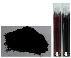
The changing of extract color from pale brown to black color (Figure 1) indicated Synthesized Iron Oxide
nanoparticles by the reduction of Fe+2 to Feᵒions, then
precipitate was washed with water and ethanol and kept for air dry. The UV Visible spectrum of nano iron particle is shown in Figure (2).
The absorption of peak at wavelengths of 375nm indicates the formation of nano iron particles. The pattern of XRD for synthesized as shown in Figure (3). The intensity and position counts of the peaks diffraction of the sample were match with the standard XRD data for bulk magnetite (JCPDS No. 88-0866). The peaks which appeared at 38, 45,65and 78 can be assigned to the peaks of the Fe3O4 and particle size about 21 calculated by Scherrer equation.
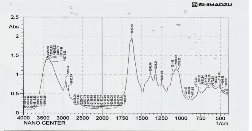
Figure (4) showed the spectrum of FTIR analysis, we found that the strong band absorption at 576 cm-1 assigned to Fe-O bond vibrations referred to the formation of Fe3O4 nanoparticles, it was reported that the wave number 570 and 600 cm-1 referred to Fe3O4 (14). The broad bands at 2000 cm- and 3934cm- are assigned to O-H bending vibrations of water, on iron oxide nanoparticles surface was due to the phenolic
compound in Jugulans extracts while the absorption at
1300-1950 cm-1 referred to C–C stretching of aromatic rings in the synthesis of Fe3O4 nanoparticles (15). The absorption of peak at wavelengths of 375nm indicates the formation of nano iron particles.
Figure 1: The changing of extract color from pale brown to black color indicated to Synthesized Iron Oxide nanoparticles: a) Powder b) nanoparticle colloidal.
Figure 3: XRD pattern of synthesized Iron nanoparticles.
Figure 4: FTIR spectrum of synthesized magnetite nanoparticles.
3.1 Denaturation of albumin Inhibition
Maximum inhibition of albumin was 75.8% observed at 400 μg/ml of nano particle compared with aspirin at the same concentration Table (2). Each value represents the mean ± SD. All values showed significant results when it compare with control p<0.01. This
Table 2: Results of albumin denaturation inhibition.
% inhibition of hemolysis Absorbance (nm) Concentration (μg/ml) Sample - 0.47±0.02 - Control 16.8 0.39± 0.04 100 1 37.8 0.29± 0.04 200 2 67.2 0.15± 0.02 300 3 75.8 0.09± 0.07 400 4 81.7 0.15±0.01 300 Aspirin
Experimental group were compared with control p<0.01, considered extremely significant; each valuerepresents the mean ± SD. N=3
Table 3: Inhibition of protein denaturation.
% inhibition of hemolysis Absorbance (nm) Concentration (μg/ml) Sample - 0.42±0.03 - Control 20 0.38±0.02 50 1 39 0.27±0.05 100 2 54 0. 20±0,01 200 3 78 0.15±0.03 300 4 85 0.13±0.07 300 Declofenac sodium
Experimental group were compared with control p<0.01, considered highly significant; Each value represents the mean ± SD. N=3.
3.2 Heat Induced Haemolysis
Table (3) showed the results activity of inhibiting haemolysis at different concentrations of iron nanoparticles. The effective concentration was 300μg/ml in comparing with standard drug Diclofenac sodium 300μg/ml resulted in good protection against damaging effect of heat solution.
4. CONCLUSION
In our study we have reported for the first time a simple, low cost and stable eco- friendly iron nanoparticles preparation, using water extract of
Juglans regia peels as stabilizing, reducing and capping agent. The iron nanoparticles have been characterized by FT-IR, XRD and UV- VIS spectroscopy. The iron nanoparticles has significant anti inflammatory activity and exhibited membrane stabilization of erythrocyte membrane in compare with standard drug (Aspirin and Diclofenac sodium). This finding suggests a novel source of membrane stabilizers and was capable of providing an alternative remedy for the management and treatment of inflammatory related disorders and diseases.
Acknowledgements
The authors are thankful to the Nanotechnology centre of Technological University for providing facilities to carry out the UV-VIS spectroscopy, FT-IR spectra and XRD pattern studies.
5. REFERENCES
1. Oliveira, A. Sousa, I. C. F. R. Ferreira, A. Bento, L. Estevinho, and J. A. Pereira, (2008) Total phenols, antioxidant potential and antimicrobial activity of walnut (Juglans regia L.) green husks Food Chem Toxicol. 46(7):2326-2331.
2. Goodsell D S 2004 Bionanotechnology Lessons from Nature John (Hoboken, NJ: Wiley)
3. Chandran, S.P., Chaudhary, M., Pasricha, R., Ahmad, A. and Sastry, M. (2006) Synthesis of Gold Nanotriangles and Silver Nanoparticles Using Aloe vera Plant Extract. Biotechnology Progress, 22, 577-583.
4. G. Hoag, J. Collins, J. Holcomb, J. Hoag, M. Nadagouda, R. Varma. Degradation of bromothylmol blue by ‘greener’ nano-scale zero-valent iron synthesized using tea polyphenols, J. Mater. Chem. 2009. 45: 8671–8677.
5. Nadagouda, M. N., A. B. Castle, R. C. Murdock, S. M. Hussain, and R. S. Varma. In vitro biocompatibility of nanoscale zerovalent iron particles (NZVI) synthesized using tea polyphenols, Green Chem. 12 (1): 114–122.
6. Shin, S. , H. Yoon, J. Jang, Polymer-encapsulated iron oxide nanoparticles as highly efficient Fenton catalysts, Catal. Commun. 2008. 10:178–182.
7. Gerard J Tortora, Sandra Reynolds, eds. Principles of Anatomy and Physiology. Harper Collins College Publishers, 1993, 7th edition: pp 695.
8. Salim S.A.T. and Sundus H. A. Evaluation of Anti Oxidant and Anti inflammatory Activity of Banana Peels, Advances in Life Science and Technology., 2016. 46: 10-15.
9. Wadood A, Mehreen G, Syed BJ, Muhammad N, Ajmal K, Rukhsana ,G and Asnad. Phytochemical Analysis of Medicinal Plants Occurring in Local Area of Mardan. Biochem Anal Biochem 2013. 2: 144.
10.Yadav RNS, Agarwala M (2011) Phytochemical analysis of some medicinal plants. Journal of Phytology 3: 2075-6240. 11.Prema P. (2011) Chemical mediated synthesis of silver
nanoparticles and its potential antibacterial application. Analysis and Modeling to Technol. Applications. 1(8): 151-166.
© 2017; AIZEON Publishers; All Rights Reserved
This is an Open Access article distributed under the terms of the Creative Commons Attribution License which permits unrestricted use, distribution, and reproduction in any medium, provided the original work is properly cited.
Ehrlich’s ascites carcinoma treated mice J. Ethnopharmacol. 129 (1): 131–134.
13.Jones, P and Vogt, T. (2001) Glycosyl transferases in secondary plant metabolism: tranquilizers and stimulant controllers Planta. 213 (2); 164–174.
14.Sadat, ME, Patel, R, Bud'ko, SL, et al. (2014) Dipole-interaction mediated hyperthermia heating mechanism of nanostructured Fe3O4 composites. Mater. Lett. 129: 57–60. 15.Venkateswarlu. S, B. Natesh Kumar, C.H. Prasad, P.
Venkateswarlu, N.V.V. Jyothi. (2014) Bio-inspired green synthesis of Fe3O4 spherical magnetic nanoparticles using Syzygium cuminiseed extract, Physica. 449: 67–71.
16.Oyedepo O.O and Femurewas A.J. (1995) Anti-protease and membrane stabilizing activities of extracts of Fagra santhoxiloides, Olax subscorpioides and Tetrapluera tetraptera. Int. J. Pharm. 33: 65-9.