Surgical Treatment of Mitral Valve Disease
ZUBAIR UL HASSAN, M.D.
Assistant Professor of Medicine, and Director, Cardiac Catheterization Laboratory, McGuire Veterans Administration Hospital, Richmond, Virginia
This paper discusses the indications for surgery in mitral valve disease by comparing the natural history of the disease, as it can best be determined from the literature, with the results of various surgical procedures. It may be stated at the outset, however, that there are no con-trolled studies comparing medical and surgical treatment nor are they likely to be carried out in the near future.
Mitral Stenosis
1. Detection. While the auscultatory find-ings of mitral stenosis are characteristic, many patients go undetected by routine clinical exam-ination; it is, therefore, important to be aware of the special syndromes with which they may present (Table 1 ).
Mitra! stenosis is much more common in women and should be looked for particularly in those who are young or middle-aged. The con-dition is predominantly a sequel to rheumatic heart disease which is prevalent among large families from lower socioeconomic groups. The availability of M-mode echocardiography in re-cent years has been of great help in the detec-tion of mitral stenosis, since this procedure has nearly 1 00% sensitivity and the specificity is al-most as good. With the two-dimensional echo-cardiogram the degree of stenosis can be mea-sured with great precision by mapping out the valve area. Echocardiography has thus elimi -nated the need for cardiac catheterization either for the detection or quantification of mitral ste-nosis, and cardiac catheterization is now
em-Correspondence and reprint requests to Dr. Zubair ul Hassan, McGuire VA Hospital, 1201 Broad Rock Road, Richmond, VA 23249.
ployed only preoperatively for the detection and assessment of associated lesions.
2. Natural history. Bland and .Jones in their classical 20-year study of 1 ,000 patients with acute rheumatic fever showed the extreme variability in the rate of progression of mitral ste-nosis in different patients. 1 Some patients devel-oped mitral stenosis within 5 years after the first attack of rheumatic fever while others were rela-tively well even 50 years later; however, a com-posite average can be deduced from this and other studies. In the preantibiotic era, mitral ste-nosis was clinically detectable approximately 1 0 years after the onset of acute rheumatic fe-ver, symptoms were minimal in the next 10 years unless pregnancy or complications oc-curred (for example, endocarditis or atrial fibril-lation), and in the next 20 years symptoms in-creased progressively with development of pulmonary hypertension, heart failure, and sys-temic embolism, leading to death, approxi-mately 40 years after the onset of rheumatic fe-ver, at an average age of 48 (Fig 1 ).1- 3
It is my impression that mitral stenosis today shows a slower rate of progression in the industrialized countries, mainly because the use of antibiotics has considerably improved the rate of recurrence of rheumatic fever and the treatment of endocarditis.
One of the most useful studies of the natu-ral history of mitnatu-ral stenosis is by Olesen and Baden who classified their patients according to the New York Heart Association (NYHA) classifi-cation and compared the non-surgical groups seen during the years 1933-1 950 with the ini-tial 1 65 consecutive patients operated on dur-ing the years 1950-1955 (Fig 2).4 Most studies of surgical treatment of mitral stenosis have
TABLE 1
Some Presenting Syndromes in Mitral Stenosis
EXERTIONAL DYSPNEA OR PULMONARY
EDEMA WITHOUT GENERALIZED CARDIOMEGALY
A TRIAL FIBRILLATION
SYSTEMIC EMBOLISM
(STROKE, MYOCARDIAL INFARCTION, HEMATURIA, ETC.)
HEMOPTYSIS WITHOUT PULMONARY INFECTION
RESTRICTIVE AND OBSTRUCTIVE LUNG DISEASE WITHOUT SMOKING HISTORY
used Olesen and Baden's non-surgical patients for comparison. In NYHA Class II patients with normal sinus rhythm the ten-year survival was 80%. At the other extreme over 50% of Class IV patients were dead within five years and all were dead within eight years.
Asymptomatic
Mild
Moderate
Severe
Total Disability
Death
Atrial
'.J
fibrulot,n Bacterial I endocarditisI
3. Choice of operations and results. The
possibility of correcting the mechanical problem of mitral stenosis surgically was first discussed around the turn of the 20th century. The first at-tempts at surgical correction were made in the
1 920s, but mitral valvotomy has come to be recognized as a generally acceptable method of treatment only since 1948. Today several dif-ferent surgical procedures are available for the repair of mitral valve disease, some of the more common being listed in Table 2.
The operative and postoperative morbidity and mortality rates are very low for mitral com-missurotomy but considerable when mitral valve
replacement with a prosthesis is necessary. It
is important, therefore, to determine pre-operatively whether the patient with mitral ste-nosis will require commissurotomy or valve re-placement. Table 3 gives some of the signs that determine this decision. Successful mitral
com-10 20 30 40
YEARS AFTER ACUTE ATTACK
4-6 cm2 1.5 -2.5 cm2 1.1-1.5 cm2 0.6-1.0 cm2
Rest LA N LA ... LA u
PA N PA
...
PA Uoruoco
Nco
Nco
t orfttExer LA
...
LAu
PA
...
PAu
co
...co
...
Fig 1-Natural progression of mitral stenosis. Clinical course, pathology and hemodynamic changes are correlated. Valve area is indicated below each diagram of valve. CO = cardiac output; Exer = exercise; LA = left atrial pressure; N = normal; PA = pulmonary arterial pressure;! = decreased; j = increased.
100 100
80 80
60 60
40 40
_.J
0 - - 0 MEDICALLY TREATED PATIENTS
<{
OPERATED PATIENTS
>
•
•
20>
20 a:::::::>
8.
Cl)
A.
0 0
lLJ 0 2 3 4 5 6 7 8 9 10 0 2 3 4 5 6 7 8 9 10 <..'.)
100 100
<{
I-2 LL.I
<.)
80 80
a:: lLJ
Q
60 60
40 40
20
o
C.
D.
O I 2 3 4 5 6 7 8 9 10 0
YEARS
0
2 3 4 5 6 7 8 910
Fig 2---Mitral Stenosis: Survival curves of surgery and medically treated patients. A. Normal sinus rhythm (NSR), NYHA Class II,
B. NSR, NYHA Class Ill; C. Atrial fibrillation and NYHA Class II or Ill; 0. NYHA Class IV. Vertical bars give 2 standard errors
above and below each point.
missurotomy is not possible if significant
regur-gitation accompanies stenosis or if the valve is
heavily scarred, markedly deformed, or
calci-fied and immobile. Attempts at valvotomy in
cal-cified valves result in fracture of the valve with
resultant insufficiency rather than cleavage of
the commissures. Lack of valve mobility is
evi-dent clinically by the absence of an opening
snap and the presence of a muffled first heart
sound; calcification can be detected by
fluoros-copy and echocardiography. Repeated
com-missurotomies also cause extensive scarring
and deformity which necessitate valve replace
-ment.
Olesen and Baden showed that 'closed'
TABLE 2
Common Operations Available for Mitral Valve Disease
• COMMISSUROTOMY
CLOSED OPEN
• VALVE REPAIR ANNULOPLASTY VAL VULOPLASTY
CARPENTIER RING
• VALVE REPLACEMENT
CAGE AND BALL VAL VE
TILTING DISC VAL VE
PORCINE TISSUE VAL VE
TABLE 3
Signs Indicating Probable Need for Valve Replacement in Mitral Stenosis
• INTENSITY OF S1 OR OPENING SNAP REDUCED
• SIGNIFICANT MITRAL INSUFFICIENCY • DENSE CALCIFICATION OF MITRAL VALVE • PRIOR MITRAL SURGERY
mitral commissurotomy (that is, commis-surotomy without cardiopulmonary bypass or 'open'-heart surgery) improved survival in all but minimally symptomatic patients in normal sinus rhythm. 4 Long-term survival was adversely
af-fected by the presence of preoperative pulmo-nary hypertension or postoperative mitral in-sufficiency. Ellis, Harken and Black reported essentially similar results in a much larger series of 1 ,000 surgical cases.5
In the United States, most institutions use open mitral commissurotomy (that is, with car-diopulmonary bypass) as the procedure of choice for mitral stenosis. The results of open commissurotomy are probably superior to the closed operation, although it is a more ex-pensive procedure. Improvements in surgical technique have markedly reduced the incidence of systemic embolization and mitral in-sufficiency, which were the principal dangers of mitral commissurotomy in earlier days. The mortality from mitral commissurotomy today is less than 1 % when the operation is done at an optimal time and most survivors experience im-provement in their symptoms. 6
When mitral stenosis can be treated with commissurotomy, the operation should be rec-ommended as soon as symptoms interfere with the normal enjoyment of life or at the earliest signs of complications. In a substantial minority of patients, the valve restenoses after several years with recurrence of symptoms. Additional surgery should be considered in these patients.
The other types of mitral valve surgery will be discussed with the treatment of mitral in
-sufficiency.
Mitral Regurgitation
The causes of mitral regurgitation are nu-merous; however, it is usually caused by con-genital heart defects in young children, rheu-matic heart disease or mitral valve prolapse in young adults, and coronary artery disease in older patients. The systolic apical murmur radi-ating into the axilla is usually easy to detect and
50 / HASSAN: SURGERY FOR MITRAL VALVE DISEASE
is almost diagnostic of mitral regurgitation, al-though occasional patients with aortic stenosis have their murmur referred to the mitral area. Rupture of chordae tendineae to the posterior mitral leaflet may produce a murmur loudest at the sternum, simulating aortic stenosis. After mitral valve replacement, significant mitral re-gurgitation may occur silently but should be sus-pected if left ventricular failure or hemolytic anemia develops unexpectedly.
Detailed studies of the natural history of mitral regurgitation according to severity of the lesion or staging by symptoms are not available.
Some impressions can be drawn from the pro-spective study of rheumatic fever by Bland and Jones.1 The prognosis of asymptomatic, mild, chronic mitral regurgitation without cardiome-galy is no different from that of the normal popu-lation; however, when significant symptoms are present with moderate-to-marked cardiome-galy, life expectancy is considerably shortened.
In Rapaport's experience approximately 20% of the patients with mitral regurgitation were dead within five years and 40% within ten years after their initial diagnosis. 3
Another consideration in mitral regurgita-tion regarding prognosis is the cause of the le-sion. Most patients with mitral valve prolapse (click-murmur, Barlow syndrome) have a nonprogressive lesion. Mitra! regurgitation sec-ondary to chronic left ventricular failure and di-latation (for example, cardiomyopathy, hyper-tension) usually does not respond well to surgi-cal treatment. In contrast with chronic mitral in-sufficiency, acute mitral insufficiency is very poorly tolerated and leads to rapid heart failure. Acute mitral regurgitation usually results from rupture of papillary muscles, chordae tendineae or valve leaflets due to infarction, infection or trauma, and occasionally spontaneously. In acute cases more attention should be paid to the severity of the hemodynamic lesion, as de-termined by physical signs and cardiac cathe-terization, than to the symptoms.
The combined lesion of mitral stenosis and regurgitation is usually due to rheumatic heart disease and is treated like mitral regurgita-tion because most patients require mitral valve replacement.
Mitral Valve Repair
at-tractive, particularly for younger patients. 7-9 It
avoids the problems of thromboembolism,
in-fection and hemolysis consequent upon the
in-troduction of a foreign body into the
blood-stream. The proponents of valve repair claim
that it is feasible in a majority of patients with
mi-tral regurgitation, but others have largely
avoided it. Several different techniques have
been described. Annuloplasty reduces the size
of the valve orifice by plication of the annulus
fusing the peripheral portions of the mitral
com-missures. Complete obliteration of mitral
in-sufficiency is frequently not possible by this
technique and postoperative dilatation of the
mitral annulus may necessitate valve
replace-ment at a later date. Valvuloplasty is a
spec-tacularly successful and curative operation for
many cases of ruptured chordae tendineae. The
redundant untethered portion of the posterior
leaflet is obliterated by pleating with sutures.
The Carpentier ring is a slightly irregular
biar-cuate stainless steel ring wrapped in a teflon
collar for sewing which has been recently
in-troduced as an aid to valvuloplasty. The ring
prevents further dilation of the valve, and the
sewing collar is used to anchor the sutures and
A
8
remold the deformed valves to approximate nor-mal anatomy. Risk of thromboembolism and endocarditis is not significantly increased with
the use of the Carpentier ring. At present, valve
repair should be strongly considered in younger patients and in those with mitral insufficiency secondary to rupture of the posterior leaflet chordae tendineae. With continuing improve-ments in technique, valve repair is likely to be-come possible in a larger proportion of patients.
The risk of valve repair is minimal, and marked
symptomatic improvement occurs after suc-cessful surgery, but it is not possible to decide preoperatively whether repair or valve replace-ment will be necessary in a specific patient. The indications for valve repair are therefore the same as for valve replacement as discussed be-low.
Mitral Valve Prostheses
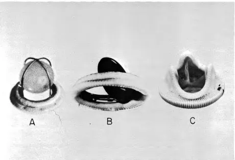
Three main types of prostheses are
cur-rently in use (Fig 3). Various models of the
Starr-Edward heart valves are examples of the cage-ball type of prosthesis. Blood flows turbulently around the occluding ball in contrast to the lami-nated flow in the normal valve. Long experience
c
Fig 3- Three commonly used mitral valve prostheses. A. Starr-Edward Model 6120; B. Bjork-Shiley; C. SGP Hancock Porcine
Model 342.
with these valves has shown them to be durable and reliable. The main problem is thromboem-bolism which is reduced but not eliminated by anticoagulants. Traumatic hemolysis may occur in models of the valve with cloth-covered struts. Another problem is the very loud closing click with some models. This sound can occasionally be heard by the unaided ear across the room from the patient. The incidence of infection of the prosthesis is probably similar to the other prosthetic valves.
Tilting disc valves, such as the Bjork-Shiley, have a more physiological blood flow and a low profile so that the valve does not oc-clude the left ventricular cavity. The major ad-vantage of this valve is that the sewing ring is not disproportionately larger than the internal lu-men and these valves are therefore associated with the lowest transvalvar gradients among all prosthetic heart valves. However, anti-coagulation is needed and the closing click can be quite noisy. This is the prosthetic valve of choice in patients with a small mitral valve an-nulus.
Glutaraldehyde-treated porcine hetero-grafts are becoming increasingly popular be-cause of low thrombogenicity even without anti-coagulation. These valves consist of porcine aortic valve tissue mounted on a man-made frame. Flow is central, very much as in the nor-mal valve, but the sewing ring is considerably larger than the internal lumen, and large trans-valvar gradients may be present post-operatively, particularly with the smaller-sized sewing rings. Long-term durability is another major concern. Nevertheless, this is the pros-thesis of choice when anticoagulation is con-traindicated.
Characteristics of the three valves dis-cussed above are compared in Table 4.
Results of valve replacement. Compara-tive studies of different types of valve pros-theses are not available at present. It appears,
however, that the operative mortality for the dif-ferent types of prostheses mentioned above is
roughly comparable, and the risk factors for op-erative mortality and long-term results are also similar. The most important risk factors for early operative mortality are advanced heart failure
and marked cardiomegaly, particularly left atrial
enlargement (Fig 4 ). 6· 10 The long-term results
are adversely affec!ed in patients with marked
left atrial enlargement or pulmonary hyper
-52 / HASSAN: SURGERY FOR MITRAL VALVE DISEASE
TABLE 4
Comparison of Three Most Commonly Used Prosthetic
Heart Valves. Pros and Cons Graded from One to
Three Plus. S-E
=
Starr-Edward; B-S=
Bjork-Shiley; P-H = Porcine Hancock.S-E B-S P-H
(/)
Q) Ol
ct!
c
DURABILITY +++ ++ +ct!
> LAMINATED FLOW + ++ +++
"O
<(
(/) THROMBOEMBOLISM +++ +++ +
Q)
HEMOLYSIS
Ol
ct! +++ ++ +
c
TRANSVALVAR ct!GRADIENT
> ++ + +++
"O
ct! NOISE +++ ++
-(/)
0 INFECTION + + +
tension. Most survivors of the operation show symptomatic improvement, but their survival curve does not parallel that of the general popu-lation (Fig 5). Young patients with severe mitral regurgitation show a remarkable reduction in
cardiac size following successful surgery, whether valve repair or valve replacement. Mi-tral valve replacement is recommended in all patients with significant symptoms, that is, NYHA Class Ill or IV. It is also indicated in acute severe mitral regurgitation and significant mitral regurgitation with progressive cardiomegaly or pulmonary hypertension, even when symptoms are less impressive (Table 5). It is advisable not to wait until advanced stages of heart failure, cardiac enlargement, or pulmonary vascular changes occur, but unfortunately patients are all too frequently in this condition when they first seek, or are referred for, valve surgery.
approxi-A
% MORTALITY
0
7
NYHA Class:
II
m
EARLY OPERATIVE MORTALITY
ISOLATED MITRAL VALVE REPLACEMENT MODEL 6120 STARR- EDWARD PROSTHESIS MAYO CLINIC 3/66 TO 1/72
MUIJU!lll
25
% MORTALITY2
N
LA Size: SMALLn
60
B
6
MODERATE
225
28
LARGE
57
Fig 4-Early operative mortality in isolated mitral valve replacement with Model 6120 Starr-Edward prosthesis, Mayo Clinic
3-66 to 1 -7 2; A = mortality according to preoperative NYHA classification; B = mortality according to preoperative left atrial (LA)
size.
...J
~
80
>
a::
:::>
en
~
60
>-
r-...J
CD
40-<t
CD
0
a::
a..
I
normal survival
EMBOkUS-FREE SURVIVAL
late systemic thrombo-embolism
1
2
3
4
5
TIME (years after mitral replacement)
Fig 5--Late survival and systemic thromboembolism in surgical survivors of isolated Starr-Edwards mitral valve replacement
with model 6120 prosthesis compared to normal life expectancy.
TABLE 5
Indications for Valve Replacement in Mitral Regurgitation
NYHA CLASS Ill OR IV
SIGNIFICANT ENLARGEMENT OF LEFT ATRIUM OR
VENTRICLE
PULMONARY HYPERTENSION
ACUTE SEVERE MITRAL REGURGITATION
mately 5%; thromboembolism is the most
seri-ous postoperative problem.
Surgical mortality is markedly increased in advanced heart disease (NYHA Class IV) and surgical treatment should therefore be consid-ered in all patients with mitral valve disease be-fore this stage is reached.
Figure 1 is taken in part from Proceedings of the Royal
Society of Medicine (60: 1 009-1 01 5, 1 96 7) and the
American Journal of Cardiology (35:221-227, 1975) by
permission.
Figure 2 is adapted from the Scandinavian Journal of
Thoracic and Cardiovascular Surgery (3:119-124, 1969)
by permission.
Figure 4 adapts data found in Barnhorst et al,
Ameri-can Journal of Cardiology (35:228-233, 1975).
Figure 5 is adapted from the American Journal of
Car-diology (35:228-233, 1975) by permission.
REFERENCES
1. BLAND EF, JONES TD: Rheumatic fever and rheumatic heart disease. A twenty-year report on 1 000 patients
54 / HASSAN: SURGERY FOR MITRAL VALVE DISEASE
followed since childhood. Circulation 4:836-843, 1951.
2. GOODWIN JF: The indications for surgery in acquired
heart disease. Proc Roy Soc Med 60: 1009-1015, 1967.
3. RAPAPORT E: Natural history of aortic and mitral valve
disease. Am J Cardiol35:221-227, 1975.
4. OLESEN KH, BADEN H: Mitral stenosis. Factors influenc-ing long-term survival rates after closed valvulotomy.
Scand J Thor Cardiovasc Surg3: 11 9-1 24, 1 969.
5. ELLIS LB, HARKEN DE, BLACK H: A clinical study of 1 000 consecutive cases of mitral stenosis two to nine years after mitral valvuloplasty. Circulation
19:803-820, 1959.
6. APPELBAUM A, KOUCHOUKOS NT, BLACKSTONE EH, ET
AL: Early risks of open heart surgery for mitral valve
dis-ease. Am J Cardiol37:201-209, 1976.
7. MANHAS DR, RITTENHOUSE EA, HESSEL ES, ET AL:
Re-constructive surgery for the treatment of mitral
in-competence: Early and late results in 91 patients. J
Thorac Cardiovasc Surg 62:781-787, 1971.
8. ELLIS FJ JR. FRYE RL, MCGOON DC: Results of
recon-structive operations for mitral insufficiency due to rup-tured chordae tendineae. Surgery 59:165-172, 1966.
9. CARPENTIER A, DELOCHE A, DAUPTAIN J, ET AL: A new reconstructive operation for correction of mitral and tri-cuspid insufficiency. J Thorac Cardiovasc Surg 61 : 1 - 1 3,
1971.
10. BARNHORST DA, OXMAN HA, CONNOLLY DC, ET AL:
Long-term follow-up of isolated replacement of the
aor-tic or mitral valve with the Starr-Edwards prosthesis.