Insulin Receptors
WILLIAM G. BLACKARD, M.D.
Director, Clinical Research Unit and Chairman, Division of Endocrinology and Metabolism, Department of Medicine, Medical College of Virginia, H ea/th Sciences Division of Virginia Commonwealth University, Richmond, Virginia
Much of the emphasis in the pathogenesis of diabetes mellitus has justifiably been placed on the endocrine gland, the pancreas. Extensive studies on the biosynthesis and release of insulin from the beta cell, bihormonal control of metabolism by insulin and glucagon, and more recently the role of soma-tostatin have attracted the attention of students of the subject; but considerable evidence exists to suggest at least some role of tissue resistance to insulin in the pathogenesis of this disorder. There have been many advocates for extra-pancreatic factors causing dia-betes. One of the first was Mirsky, who proposed that diabetes might be due to excessive amounts of hepatic insulinase, an enzyme which degrades insulin.' Val-lance-Owen suggested that a circulating insulin an-tagonist labeled synalbumin might be the cause of insulin resistance in diabetes.2 This factor was later shown to be an artifact. Others, such as Antoniades, proposed that insulin might circulate predominantly in a bound form in diabetic subjects and thus not exert full biologic activity.3
The most articulate spokesman for a role of insulin resistance in diabetes mellitus in recent years has been Gerald Reavan, and his group from Stan-ford University, who bases his theory on two obser-vations. The first is that a large number, if not the majority, of adult onset diabetics have increased cir-culating insulin concentrations rather than decreased concentrations as had been expected. This was first observed by Yalow and Berson shortly after the per-fection of the radioimmunoassay for insulin.4
Hyper-Correspondence and reprint requests to Dr. William G. Blackard, Clinical Research Center, Box 155, Medical College of Virginia, Richmond, Virginia 23298.
12
insulinism in diabetics has since been confirmed by many investigators. A second observation supporting the role of insulin resistance in diabetes is that of "glucose impedance" in diabetic patients. Glucose impedance was demonstrated by Reaven and col-leagues5 by infusing glucose and insulin at a constant rate in diabetic and non-diabetic subjects whose en-dogenous insulin release had been shut off by admin-istration of epinephrine and propranolol. New steady states for glucose and insulin were achieved in both groups, with comparable insulin concentrations in diabetics and non-diabetics, whereas the new steady state glucose concentration was considerably higher in diabetic subjects than in non-diabetic subjects.
These excellent studies indicated that for a given con-centration of insulin, the blood glucose-lowering ef-fect was less in diabetics than in non-diabetic sub-jects. More recently, Reaven and Olefsky have
suggested that insulin resistance in diabetic patients might be due to a decrease in the number of insulin receptors6 by showing a decrease in the number of insulin receptors on circulating monocytes in diabetic patients compared to those on monocytes of non-diabetic subjects. Additionally, treatment of their diabetic subjects with an oral hypoglycemic agent resulted in a return to normal of the number of insulin receptors on peripheral monocytes.7 A thor-ough understanding of these latter observations and their obvious, important implications for the patho-genesis of diabetes requires a certain knowledge of the insulin receptor and of recent advances in the field of receptor technology.
Properties of the insulin receptor are shown in Table I. As is the case with other polypeptide hor-mones, the receptor for insulin is located on the cell
TABLE I
Properties of Insulin Receptors
A) Located on cell membrane
B) Unevenly distributed and may occur in clumps C) Protein with mo! wt approx. 300,000-Tetramer
con-sisting of monomers of 75,000 mol wt each D) Continuous synthesis and degradation with relatively
slow turnover rate-2%/hr
E) Presence of insulin receptor on cell membrane requires protein synthesis and microfilament integrity
membrane. Evidence for intracellular distribution of
insulin receptors is very scant; they are not evenly
distributed over the cell surface but rather occur
ran-domly and in clumps at times.8 Present evidence
sug-gests that the insulin receptor is a protein with
mo-lecular weight of approximately 300,000. Studies
performed at the National Institutes of Health from
Dr. Jesse Roth's laboratory suggest that the insulin
receptor is a tetramer consisting of monomers of
75,000 dalton units each. Although the turnover rate
is low compared to many biologic processes (2% per
hour), continuous synthesis and degradation of the
insulin receptor occurs. Studies with inhibitors of
protein synthesis and microfilaments such as
pur-omycin and cytochalasin respectively indicate that
protein synthesis and microfilarhent integrity are
nec-essary for the presence of insulin receptors on the cell
membrane.9 Inhibitors of microtubular function
sur-prisingly had no effect on insulin receptors.9
Knowledge of three concepts involving the
in-sulin receptor (Table 2) is of critical importance in
interpreting studies in which receptor number and
affinity have been determined. The number of
recep-tors per cell varies with the parti9ular cell being
stud-ied. However, for the peripheral monocyte, which is
the most commonly studied cell in man because of its
accessibility, the numbers of receptors vary between
15,000 and 30,000 per cell. Clearly, only a fraction of
the receptor sites must be occupied for biological
activity, and the number of occupied sites required
for the different activities of insulin may vary. For
example, dose response data suggest that fewer sites
must be occupied to inhibit lipolysis than to stimulate
glucose oxidation. Thus, many of the insulin recep-tors on the cell surface will be spare or unused
recep-tors. Recent investigations have even shown that
some of these receptors rnay serve as a peripheral
reservoir for insulin,· releasing intact insulin under
appropriate circumstances.10
A second concept which is probably the most
13
important in understanding current receptor studies
is that insulin inhibits insulin receptor number.
Ex-periments by Gavin et al11 demonstrated that
pre-incubation of cultured lymphocytes with
phys-iological concentrations of insulin reduces the
number of insulin receptors on these cells. An
ob-vious corollary of this finding would be the presence
of decreased insulin receptors in states of
hyperinsu-linism such as obesity and some forms of diabetes.
Although not reported yet, reduced insulin receptors
would be anticipated in patients with islet cell
tu-mors. Thus, reduced receptor number might offer
some protection to the patient with an islet cell tu-mor.
The third important concept in understanding
insulin receptors is that of negative cooperativity.12
Simply stated, this concept refers to site interactions
on the cell surf::ice by which affinity of the receptor
for insulin is decreased as increasing numbers of
re-ceptors are occupied. This phenomenon might also
be considered a homeostatic mechanism which
pro-tects the individual from the effects of
hyperinsulin-1sm.
The insulin receptor perceives and either directly
or through a transducer substance influences the
ef-fector for a specific activity. Insulin binding is the
first step in biological activity of the hormone.
Al-though not all receptors are required for biological
activity, more receptors increase the likelihood of
binding for a given concentration of insulin. Binding
of insulin to its receptor is therefore determined by
insulin concentration, receptor number, and receptor
affinity. Radioimmunoassay techniques for the
mea-surement of insulin have been available for years;
now methods are available to measure insulin
recep-tor number and affinity.
As mentioned the most accessible cells for
mea-suring insulin receptors in vivo are peripheral
mon-TABLE 2
Important Concepts Involving Insulin Receptors
A) Spare receptors
I) Number of receptors per cell varies with cell type Peripheral monocytes have 15,000-30,000 receptors per cell
2) Only small percentage of receptors must be occupied for biologic activity
3) Spare receptors may serve as peripheral insulin reservoir
B) Feedback inhibition of insulin receptor number by insulin
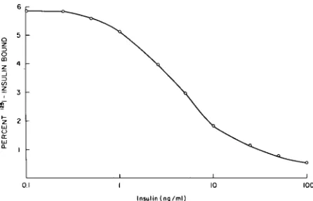
ocytes. These cells are obtained by Ficoll-Hypaque separation of the buffy coat of centrifuged blood. To determine receptor number and affinity, these cells are incubated with labeled insulin and increasing amounts of cold insulin, resulting in a binding curve (Fig I). Applying Scatchard analysis to this data results in a curvilinear plot (Fig 2B). Similar studies with the growth hormone receptors, or other hor-mones not showing negative cooperativity, produce a linear Scatchard plot (Fig 2A ). Although th,e favored interpretation of the curvilinear Scatchard plot for insulin receptors is negative cooperativity, the possi-bility that two types of insulin receptor sites (broken lines Fig 2) exist cannot be eliminated from present data. Employing Scatchard analysis, the number of receptors is calculated from the amount of bound insulin where the plot crosses the X axis. The slope of the plot reflects affinity, and new graphic analyses are
available to express affinity even from a curvilinear plot.13
Employing these techniques, insulin receptor number and affinity can be determined. The factors influencing affinity and receptor number are shown in Table 3. Some of these have already been discussed. One of the most important determinants of affinity is pH; its effect on insulin binding to receptors is shown in Figure 3. For both human monocytes and cultured lymphocytes, reducing pH from 7.4 to 6.8 results in greatly depressed insulin binding ahd may contribute to the insulin resistance observed in severe diabetic ketoacidosis.
0
z
:::,
0
<D
z :::;
:::,
v,
;; 3
!, 2
"'
u
"' "'
0.
0.1 10 100
Insulin {ng/ml)
Fig 1-1 nsulin binding to peripheral monocytes. 20 X I 0'
mon-onuclear cells ( 14% monocytes) were incubated in 0.5 ml buffer
containing 50-100 pg 125[-insulin and increasing amounts of
unla-beled insulin to give the final concentration indicated in the figure. After 3 hours incubation, 200 ul aliquots were centrifuged,
aspi-rated, and the sediment counted.
K
A
Ro B
LL
... CD
B
Ro B
Fig 2-Scatchard analysis of binding data. B/F = bound/free radioactive ligand. Horizontal axis (Ro) is amount of ligand bound to receptor in molar quantities. A. Scatchard plot for hormone not
exhibiting negative cooperativity; B. Plot is for hormone exhibiting
negative cooperativity or having two different receptor sites.
In-sulin receptor studies show curvilinear plot as in B.
The major factor so far uncovered altering in-sulin receptor number is inin-sulin acting in a type of feedback mechanism to inhibit insulin receptor num-ber. Thus, as previousiy pointed out, reduced insulin receptors are anticipated in obesity where insulin re-sistance and hyperinsulinism exist. Reduced insulin receptors have indeed been shown to occur in obese humans and animals. 1•-16 In addition, dieting and weight reduction result in normalization of the num-bers of insulin receptors.13 It is debatable whether
reduced receptor concentration is primary, resulting in insulin resistance and hyperinsulinism, or whether insulin resistance due to some other factor is primary, causing hyperinsulinism and, secondarily, reduced in-sulin receptors.
Returning to Reavan and Olefsky's observations in non-obese diabetic subjects,6 it is not clear whether the reduced insulin recej".ltor number is due to the hyperinsulinism exhibited by this group [fasting im· munoreactive insulin (IRI) 20 ± 2 versus 10 ± I in normals] or whether it might be primary and thus be important pathogenetically. Nevertheless the de-creased insulin receptors observed in the diabetic
sub-TABLE 3
Determinants of lnslllin Binding to Tissues
l. Receptor affinity
PH Temperaiure Ionic strength
Receptor occupancy (Negative cooperativity)
ll. Receptor number
35
;:::
z 25
w
~
w
"-<.'.)
z i5 Z 15
iii :a
::)
:;a; x <I
:a
- PERIPHERAL CELLS
o-o CULTURED CELLS (IM-91
6.8 7.2 7.6
pH OF INCUBATION MEDIUM
8.0 8.4
Fig 3-Effect of pH on insulin binding to receptors. Maximum binding refers to percent of 1251-insµlin bound to receptors in the absence of unlabeled insulin. For peripheral cells 20 X IQ', mon-onuclear cells (14% monocytes) were used; cultured lymphoblas-toid cells (IM-9) were used at 3.0 X 10' cells per ml concentration.
jects in Reaven and Olefsky's study would certainly
contribute to the insulin resistance observed. The
return of receptor number to normal with chronic
sulfonylurea treatment7 might in similar fashion be attributed to the reduced 'insulin concentrations in
well-controlled diabetics on chronic sulfonylurea therapy.
Although the role of reduced insulin receptors in
the pathogenesis of diabetes mellitus is equivocal, a
rare diabetic syndrome recently reported is clearly
related to decreased insulin receptors. 17 In several
patients with other evidence of immunologic disease
associated with severe insulin resistance, an antibody
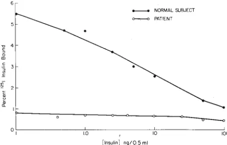
[Insulin] ng/0.5 ml
NORMAL SUBJECT PATIENT
Fig 4-1 nsulin binding to peripheral monocytes from normal sub-ject and patient with antibodies to the insulin receptors. Details as
for Fig I.
~ c a
0
CD
c ,i
30
E: 20
10
I.O
I M-9 CELLS PRE INCUBATED WITH HEPES NOR~L SERA (1•2) o----., PT. SERA (1•100)
10
[Insulin] ng/0.5 ml
100
Fig 5-Effect of serum preincubation on insulin binding to cul-tured lymphoblastoid cells (IM-9). IM-9 cells were preincubated with buffer, normal serum, or serum from insulin-resistant patient for 60 min, washed twice, and resuspended in 0.5 ml buffer at final concentration of 3 X 10' cells p~r ml. Binding curves were then obtained on these cells.
to the insulin receptor has been demonstrated. Since
this report we at the Medical College of Virginia have
had the opportunity to study two patients with this
syndrome. One of the patients requiring over 2,000
units of insulin daily had a strongly po~itive
antinu-clear antibody as the only other manifestation of
autoimmunity; the second patient had a
scleroderma-like illness and required 1200 units of insulin daily.
Insulin binding curves by peripheral monocytes from
one of these patients is shown in Figure 4. That a serum factor was responsible for the decreased
bind-ing was indicated by studies in which cultured
lym-phocytes (IM-9) were preincubated with the
pa-tient's sera ( I : I 00) and then used for binding studies (Fig 5). Scatchard analysis (Fig 6) revealed the
de-creased binding to be due to a reduction in numbers
of insulin receptors. Studies, not shown, in which
IM·9 CELLS PRE INOJBATED WITH
HEPES
1.0 12 14
NORMAL SERA 1,2 PT. SERA 1=100
ng Insulin Bound/3.0xl06 cel!
cultured lymphocytes were preincubated with IgG fraction of the patient's sera exhibited the same phe-nomenon, suggesting that the serum contained an antibody to the insulin receptor.
In summary, techniques are now available for measuring insulin receptors in vivo. So far, reduced
insulin receptors have been observed in obese persons
and in a selected group of adult onset diabetic
pa-tients. The pathogenetic significance of the latter ob-servation is uncertain and may possibly be a manifes-tation of the high insulin concentrations in these diabetics. However, a rare diabetic syndrome in which severe insulin resistance due to antibbdies to the insulin receptor has been reported and is now corroborated by our findings in two patients.
REFERENCES
I. MIRSKY IA: The metabolism of insulin. Diabetes 13:225-229, 1964.
2. VALLANCE-OWEN J, DENN ES E, CAMPBELL PN: Insulin antago-nism in plasma of diabetic patients and normal subjects. Lan-cet 2:336-338, 1958.
3. ANTONIADES HN: Studies on the state of insulin in blood: The state and transport of insulin in blood. Endocrinology 68: 7-16, 1961.
4. YALOW RS, BERSON SA: Immunoassay of endogenous plasma insulin in man.JC/in Invest 39: 1157-1175, 1960.
5. SHEN SW, REA VEN GM, FARQUHAR JW: Comparison of im-pedance to insulin-mediated glucose uptake in normal subjects
and in subjects with latent diabetes. J C/in Invest 49:2151-2160, 1970.
6. REA VEN GM, BERNSTEIN R, DAVIS B, ET AL: Nonketotic dia-betes mellitus: insulin deficiency or insulin resistance? Am J Med 60:80-88, 1976.
7. OLEFSKY JM, REA VEN GM: Effects of sulfonylurea therapy on insulin binding to mononuclear leukocytes of diabetic pa-tients. Am J Med 60:89-95, 1976.
8. JARETT L, SMITH RM: The random distribution, grouping and nonmigratory nature of insulin receptors. Diabetes 25:321, 1976.
9. VAN 0BBERGHEN ,E, DE MEYTS P, ROTH J: The cell surface distribution of peptide hormone receptors: possible role of microfilaments. Diabetes 25:321, 1976.
10. ZELEZNIK AJ, ROTH J: Plasma membrane receptors for pep-tide hormones in vivo role as reservoir for the circulating hormone. Program of the 58th Meeting, American Endocrine Society, 1976, p 27.
11. GAVIN JR, ROTH J, NEVILLE DM JR, ET AL: Insulin-dependent regulation of insulin receptor concentrations: a direct demon-stration in cell culture. Proc Nat Acad Sci USA 71:84-88, 1974.
12. DE MEYTS P, ROTH J, NEVILLE DM JR, ET AL: Insulin inter-actions with its receptors: experimental evidence for nega-tive cooperativity. Biochem Biophys Res Commun 55:154-161,
1973.
13. DE MEYTS P, RoTH J: Cooperativity in ligand binding: A new graphic analysis. Biochem Biophys Res Commun 66: 1118-1126, 1975.
14. BAR RS, GORDEN P, ROTH J, ET AL: Fluctuations in the affin-ity and concentration of insulin receptors on circulating mono-cytes of obese patients: effects of starvation, refeeding and dieting.JC/in Invest 58:1123-1135, 1976.
15. ROTH J, KAHN CR, LESNIAK MA, ET AL: Receptors for insulin, NSILA-s arid growth hormone: applications to disease states in man. Recent Prog Hor Res 31:95-139, 1975.
16. SOLL AH, KAHN CR, NEVILLE DM JR, ET AL: Insulin receptor deficiency in genetic and acquired obesity. J C/in Invest
56:769-780, 1975.
17. KAHN CR, FLIER JS, BAR RS, ET AL: The syndromes of insulin resistance and acanthosis nigricans. N Engl J Med