[1] Colonization and infection of the skin by S. aureus: Immune system evasion and the response to cationic antimicrobial... [2] Staphylococcus aureus in the community: Colonization... [3] Staphylococcus aureus infections: Epidemiology, pathophysiology, clinical... [4] Staphylococcus aureus nasal colonization: An Update on mechanisms, epidemiology, risk factors and subsequent infections. Front Microbiol. 2018; 9:2419.
[5] Methicillin resistance in Staphylococcus aureus. Pet-to-man travelling Staphylococci: Elsevier; 2018: 225-35. [6] Methicillin-resistant Staphylococcus aureus: An evolving... [7]
The molecular evolution of... [8] Prokaryotic taxonomy in the sequencing era-the polyphasic approach... [9] Stepwise decrease in daptomycin susceptibility in clinical Staphylococcus aureus isolates associated with an initial mutation.. [10] Mutations in HSP70-2 gene change the susceptibility to clinical mastitis in Chinese Holstein. Gene. 2015; 559(1):62-72. [11] Molecular identification and genotyping of MRSA isolates. FEMS Immunol Med Microbiol. 2009; 57(2):104-15. [12] Molecular cloning of two new heat shock genes related to the hsp70 genes in Staphylococcus aureus. J Bacteriol. 1994; 176(15):4779-83.
[13] Genotypic diversity of coagulase-negative... [14] Mechanisms of methicillin resistance in Staphylococcus aureus. Annu Rev Biochem. 2015; 84:577-601. [15] Focus: Infectious diseases: Vancomycin resistance in Staphylococcus aureus. Yale J Biol Med. 2017; 90(2):269-81. [16] Antibiotic resistance in Staphylococcus aureus. Current status... [17] Comparison of traditional and molecular methods of typing... [18] Spa typing and multilocus sequence typing show comparable performance in a macroepidemiologic study of Staphylococcus aureus in... [19] Multilocus sequence analysis (MLSA) in... [20] The role of universal stress proteins in Edwardsiella... [21] Genetic variation among Staphylococcus aureus strains from bovine milk and their... [22] Genetic variation in Staphylococcus aureus surface and immune evasion genes is lineage associated: Implications... [23] Phylogenetic relationships among Staphylococcus species and refinement of... [24] Comparison of complete rpoB gene sequence typing and multi-locus sequence typing for phylogenetic analysis of Staphylococcus aureus. J Gen ...
Molecular Characterization of Staphylococcus aureus
Isolated from Clinical Samples Based on 16srRNA, rpoB,
and hsp70 Genes by MLSA
A B S T R A C T
C I T A T I O N L I N K S A R T I C L E I N F O
Article Type Original Research
Authors
Elham Hoseiny Khorram Abadi1,
MSc,
Saeed Zaker Bostan Abad2*, PhD
How to cite this article Hoseiny Khorram Abadi E, Zaker Bostan Abad S. Molecular Characterization of Staphylococcus aureus Isolated from Clinical Samples Based on 16srRNA, rpoB, and hsp70 Genes by MLSA. Infection Epidemiology and Microbiology. 2019;5(3):27-38
1Department of biology, school
of Basic Sciences, Science and Research Branch, Islamic Azad University, Tehran, Iran
2Department of biotechnology,
School of Basic Sciences, Parand Branch, Islamic Azad University, Tehran, Iran
* Correspondence
Address: Department of Microbi-ology, Shiraz branch, Islamic Azad University, Shiraz, Iran
Fax: +98 21 88951392
Phone: +9821 4293 3150 E-mail: [email protected]
Article History
Received: July 27 ,2019 Accepted: September 27 ,2019 Published: October 12 ,2019
Aims: Staphylococcus aureus is a Gram-positive bacterium with the capability of causing a variety of nosocomial and community-acquired infections. Evaluating the genetic structure, polymorphism, genotyping, and phylogeny of S. aureus isolates could contribute to the prevention and treatment of infections caused by this microorganism.
Materials & Methods: In this study, the polymorphisms of 16S rRNA, rpoB, and hsp70 genes were investigated in a total of 50 S. aureus isolates using S. aureus NCTC 8325 as the reference strain. Polymerase chain reaction (PCR) was used for the detection and amplification of the studied genes. The amplicons were then sequenced using a Sanger sequencing method. Moreover, phylogeny of the isolates was studied using Neighbor-joining and Maximum Parsimony methods for 16S rRNA, rpoB, and hsp70 genes individually and in combination.
Findings:After Sanger sequencing, data obtained by Sequencher and Mesquite software programs revealed several polymorphisms of S. aureus isolates 16S rRNA, rpoB, and hsp70 genes, respectively. These polymorphisms included transversion, transition, insertion, and deletion. Among the studied strains, 10 cases showed no polymorphism. Multi-locus sequence analysis (MLSA) showed several genetic diversities in S. aureus isolates. Conclusion: It seems essential to rapidly and reliably identify the phylogenetic sources and characteristics of this microorganism and to have a better understanding of its molecular epidemiology in order for infection practical surveillance and control.
Keywords:Staphylococcus aureus, Polymorphism, Multi-locus sequence analysis (MLSA).
Introduction
Staphylococcus aureus is a commensal Gram-positive organism present on the skin and mucosal surface with the capability of surviving on dry surfaces due to its thick peptidoglycan layer [1]. Risk factors of S.
aureus infections include external devices, history of surgery, and extensive antibiotic use [2]. Patients at risk of S. aureus infections
include neonates, children with poor sanitation, women during menstruation, and patients with intravascular catheters. This microorganism could mainly lead to the bacteremia, endocarditis, osteomyelitis, pneumonia, skin and soft tissue infections [3].
Although S. aureus is generally considered as an opportunistic pathogen, some clones may be more capable of causing invasive disease due to the presence of certain virulence factors facilitating access to normally sterile sites [4].
Recently, S. aureus has exhibited great resistance against multiple antimicrobial agents, which is of great concern. Methicillin-resistant S. aureus (MRSA) strains; including hospital-acquired MRSA (HA-MRSA), community-acquired MRSA (CA-MRSA), and livestock-associated MRSA (LA-MRSA) strains; are resistant to all β-lactam antibiotics through acquiring mobile genetic elements called staphylococcal cassette chromosome mec (SCCmec) [5-7]. In addition
to acquiring antibacterial resistance, differences in staphylococcal pathogenicity, depending on different geographical regions and epigenetics, necessitate the investigation of the genomic structure, polymorphism, and phylogenetic relationships between different S. aureus clinical isolates.
Multi-locus sequence analysis (MLSA) is a powerful high-resolution method which provides data on genetic changes in housekeeping genes and could be served as a valid technique for the study of epidemiological relationships [8]. In fact,
MLSA is able to compare the primary DNA sequences of multiple conserved protein-coding loci in order to assess the diversity and relationship between different isolates and to determine the sources and evolutionary alterations of different taxa.
Objectives: This study aimed to investigate the genetic diversity of S. aureus clinical strains isolated from different body sites. In this study, a MLSA protocol was developed based on genes coding for β-subunit bacterial RNA polymerase (rpoB) and heat-shock protein 70 (hsp70) as well as 16S rRNA gene. These genes are essential for bacteria, and their polymorphism is highly important in determining bacterial genetic behaviors corresponding to different environmental factors [9-10].
Materials and Methods
employed for further analysis. Also, to confirm the quality of the extracted DNA, samples were analyzed by 1% agarose gel electrophoresis (Sigma, USA).
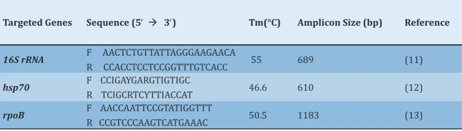
Polymerase Chain Reaction (PCR) and Sequencing: Amplification of 16SrRNA, rpoB, and hsp70 genes was performed in an automated thermal cycler (Bio-Rad, USA) in a total volume of 25 µL consisting of 2 µl of DNA template, 1 μL of the previously described primers (forward and reverse) (Table 1), 12.5 µL of Taq PCR Master Mix 2X (Fermentas, Lithuania), and 8.5 µL of DNase/RNAse free distilled water (Thermo Fisher Scientific). The PCR program included an initial denaturation step at 95°C for 5 min, followed by 35 cycles of denaturation at 95°C for 30 sec, annealing (annealing Tm for each primer is shown in Table 1) for 60 sec, extension at 72°C for 60 sec, and a final extension at 72°C for 5 min. Finally, amplicons in each reaction were analyzed on 1% agarose gel treated with safe stain (Thermo Fisher Scientific, USA) in 0.5X TBE after electrophoresis. Gels were visualized under the gel documentation system (Bio-Rad, UK).
The PCR positive products with the expected size were sequenced by Sanger method for five times (Forward and reverse). The sequences were then blasted against the 16S rRNA, rpoB, and hsp70 GenBank databases (NCBI).
Sequence data analyses: Chromatographs were annotated with the Sequencher DNA
analyzer program Ver.5.1 (Gene Codes Corpora tion). After finding contigs, they were blasted to find any change compared to the reference sequence of S. aureus NCTC 8325 standard strain.
Phylogeny of the isolates was analyzed using the maximum parsimony or neighbor-joining method using the Mega software Ver.6.0. Standard strain of S. aureus NCTC 8325 was used as the reference strain.
Findings
Genetic variations in the studied genes: All MLSA loci of 50 S. aureus isolates were successfully amplified. Sequencing analysis of the PCR products created sequences in both directions. The collections of sequences were transferred from the Sequencher to Mesquite software Ver.3.6 for alignment in order to find any type of polymorphism in the studied genes. According to the results, among the 50 studied S. aureus strains, 21, 33, and 23 strains showed genetic variations caused by mutations including insertion, transversion, transition, and deletion in 16S rRNA, rpoB, and hsp70 genes, respectively. Among the studied isolates, 10 cases showed no genetic variation compared to the reference strain (Tables 2, 3, and 4). Phylogenetic analysis of the strainsusing the Neighbor Joining method: Phylogenetic trees constructed based on 16s rRNA gene using the neighbor joining method showed that the Strain 14 was different from the
Table 1) Characteristics of primers used in this study
Targeted Genes Sequence (5′ à 3′) Tm(°C) Amplicon Size (bp) Reference
16S rRNA F AACTCTGTTATTAGGGAAGAACA
R CCACCTCCTCCGGTTTGTCACC 55 689 (11)
hsp70 F CCIGAYGARGTIGTIGC
R TCIGCRTCYTTIACCAT 46.6 610 (12)
rpoB F AACCAATTCCGTATIGGTTT
Table2) Polymorphisms in 16S rRNA gene of 50 S. aureus clinical strains with different sources compared to the reference strain
Strain No. Source Nucleotide Change (Base Number/ Type of Mutation) in hsp70 Gene
1 Urine AAC→AGC (273/ transition)
2 Urine TTT→TCT,TGT→TGTT(203, 297/transition/insertion)
3 Blood
-4 Blood
-5 Wound TTT,272,273,282/ transition, insertion)→TCT,CAC→CGC,AAT→CCC,TTC→TTT,AAT→GAAT,TAA→CAA (203,254,263,264,266,269 6 Blood ATA→AATA,TCA→TACA,CAA→ACAA (32,35,37/insertion)
7 Blood AAT→ATT (264, transversion)
8 Blood
-9 Urine
-10 Eye TTC→TTCC,CTA→CTTA (244, 260/ insertion)
11 Blood
-12 Wound AGA→AGCA (287/insertion)
13 Blood
-14 Blood TGT→TGTT (297/insertion)
15 Blood
-16 Wound
-17 Blood TTA→TTT (4/transversion)
18 Throat
-19 Urine
-20 Wound
-21 Blood
-22 Blood
-23 Blood
-24 Wound AAC→AGC (237/transition)
25 Blood
-26 Blood
-27 Blood ACT→ATT (179/transition)
28 Blood
-29 Blood
-30 Urine CTA→CAA,ACT→ATT,CTG→CAG,TTT→AAA (177, 179, 183, 266, 267, 268/ transversion, transition)
31 Blood
-32 Urine
-33 Blood
-34 Blood
-35 Blood AAT→ATTT,ATA→ACA,ACC→A-C (273, 275, 276, 287/ transition, insertion, deletion)
36 Blood
-37 Wound CAA→CGA,TCA→TCC,ATA→TTT (232,271,274,277/transversion) 38 Blood GTT→GTC (3/transition)
39 Blood
-40 Blood
-41 Urine
-42 Blood
-43 Wound 274, 277, 295, 296/ transversion, transition)ACA→ACT,ACG→AAC,TTT→TCT,TCA→TCT,AAT→ATT,TCA→TTC,GTG→GAT (6, 238, 267, 271,
44 Synovial
-45 Sputum CCG→CCAG,CAA→CTAA,AAT→AAA (19,37,92/insertion) 46 Blood GTG→GGG (295/tranversion)
47 Blood ACC→AGG (278, 279/transversion) 48 Blood ACC→ATC (278/transition)
Table3) Polymorphisms in rpoB gene of 50 S. aureus clinical strains with different sources compared to the reference strain
Strain No. Source Nucleotide Change (Base Number/ Type of Mutation) in rpoB Gene
1 Urine TGT→TAT, CTG→CCG (127, 331/transition)
2 Urine
-3 Blood
-4 Blood ATG→ATGG, CCA→CCCA, TTT→TTTT, AAT→ATT, TGT→TAT (5, 7, 14, 58, 619/transversion, transition, insertion)
5 Wound ATGCTG→→AGG, ATCCTGA, TAG→→ACC, CGATAA, TTA→→C-A, ATCTTT, CGA→→A-C (542,603,627,643,649,684,729,735,792,795,827,844,586,941) CAA, CAT→CCT, CGA→CCA, GTG→GGG, ACA→AAA, ATA→AAA, AGT→AAT, (transversion, transition, insertion, deletion)
6 Blood TCT→TCCT, CAA→CAGA (16,936/ insertion)
7 Blood CAC→CACC, TGC→TGCC, CAT→CATT, TAA→TAAA (476,555,570,582/ insertion)
8 Blood
-9 Urine ACC→ACCT (202/insertion)
10 Eye TCATCG→→TTG, CTGTTA, ATA→→AAA, CTCCGG, CAC→→CGC, AGACCC, CAT→→C-T, ACTAAA, ATC→→ATT, CCAACC, ACA→→CCC (287,323,331,342,345,388,399,404,421,477,533,53AAA, GTG→GGG, TCT→T-T, AAA→AA-, CGG→CGGG, 7,632,649,659,676/ transversion, transition, insertion, deletion)
11 Blood AATCAT→→-AT, CCACAA, AGC→-A, AGA→ACC, TAT→AGAT, TGG→TTT, AGT→TGGA, TCG→AAT, ATA→TCCC, TTC→A-A, ATG→TCC, CAC→CAG, ATC→CCC, CGA→ACC, TGT→CAA, AGA→GG- (1,8,271,272,273,511,534,5→AAA, AGT→AAT, CTT→CTTG, 35,596, 632,643,714,716,726,749,753,757,795,821,826,827,844,947,950/ transversion, transition, insertion, deletion)
12 Wound
-13 Blood ATG→ATGG,TGT→TAT,CAT→CCT,AGA→AAA,TGC→TTGC (5,6,7,619,700,714,720/ transversion, transition, insertion)
14 Blood
-15 Blood TGC→GGT,AAA→--A (399,401,789,790/ transversion, deletion)
16 Wound
-17 Blood ATT→ATAT,TTA→TAA(710,713/transversion, insertion)
18 Throat
-19 Urine TTC→TCC,TCA→TAA,TTG→TGG,CAT→CCT (411,417,435,488/ transversion, transition) 20 Wound CAA→ACAA,GCT→GGCT,CAA→AAA (78,720,739/ transversion, insertion)
21 Blood TGTtransition, insertion)→TGTT,CAT→CGC,TAC→TTC,TGT→TAT,TTT→TTTT,CTT→CTTG (138,293,382,619,720,726,733/ transversion, 22 Blood CCA→CCCA,TTC→TTTC,AAA→AAAA,GTG→GGTC (628,660,705,720/ insertion)
23 Blood
-24 Wound TGT→TAT (127/ transition)
25 Blood
-26 Blood AAT→TAT,TAC→TTC (1,382/ transversion)
27 Blood
-28 Blood GAC→GTC,TGT→TTT (296,303/ transversion)
29 Blood
-30 Urine ATT→ATTT,TGT→TAT,TTT→GGG,GCC→GAC,TCC→TGC (111,619,869,870,871,884,959/ transversion, transition, insertion)
31 Blood
-32 Urine TAC→CCC,ATA→CAC (990,991,999,1000/ transversion, transition)
33 Blood
34 Blood TGC→TGCC,--- →TTA,CTT→CTTT,CAA→CCAA (99,271,272,273,914,944/ insertion, deletion) 35 Blood TGT→TAT,TTG→TTTG (619,952/ transition, insertion)
36 Blood GTA→GAA,TTC→TTTC,CTG→CCG,GGT→GGGT,TGT→TAT (246,286,331,398,619/ transversion, transition, insertion) 37 Wound ATG→ATGG,AAT→A-T (5,23/ insertion, deletion)
38 Blood
-39 Blood ATG→ATGG,TTC→TTTC,TGT→TAT (5,14,619/ transition, insertion)
40 Blood AAT→TAT,GCC→G-C,AAT→A-T,CAA→CACA (1,8,23,76/transversion, insertion, deletion) 41 Urine TTC→TTT,TTC→TTA,ACA→AAA (663,767,988/ transversion, transition)
42 Blood
-43 Wound AAT→A-T,TAC→TTC,TCA→TCG (23,382,492/ transversion, transition, deletion) 44 Synovial TTC→TATC,CTG→CCG,TGT→TAT (275,331,619/transition, insertion) 45 Sputum TCG→TCCG,TGT→TAT (454,619/ transition, insertion)
46 Blood
-47 Blood TGT→TAT,TTC→TT- (619,663/ transition, deletion) 48 Blood CTG→CCG,TGT→TAT (330,619/ transition)
49 Blood
-Table4) Polymorphisms in hsp70 gene of 50 S. aureus clinical strains with different sources compared to the reference strain
Strain No. Source Nucleotide Change (Base Number/ Type of Mutation) in hsp70 Gene
1 Urine
-2 Urine GTA→GAA (476/ tranversion)
3 Blood
-4 Blood
-5 Wound
-6 Blood ATA→ACA (427/transition)
7 Blood
-8 Blood
-9 Urine
-10 Eye
-11 Blood
-12 Wound GCT→GCTT (433/ insertion)
13 Blood
-14 Blood AGAtransition, deletion)→AAA,AGT→AAA,TCC→TTC,CTC→TTT,GCT→GCTT (212,222,223,243,253,255,491/ transversion, 15 Blood GGT→GCT,CGT→CCT,CTT→GAT (12,19,35/ transversion, insertion)
16 Wound
-17 Blood
-18 Throat
-19 Urine GGT→GCT,GGC→G-C (11,489/ transversion, deletion)
20 Wound
-21 Blood
-22 Blood CCA→CCCA,CCT→C-T,TTT→-TT (156, 442, 492/insertion, deletion) 23 Blood TTT→TAT,GCT→GCTT (264, 491/ transversion, insertion)
24 Wound
-25 Blood
-26 Blood
-27 Blood
-28 Blood TCC→TCCC (14/ insertion)
29 Blood
-30 Urine
-31 Blood
-32 Urine
-33 Blood
-34 Blood
-35 Blood CAG→CGG (157/ transition)
36 Blood
-37 Wound GCT→GCTT (491/insertion)
38 Blood CTA→CGA,CTA→CAA (389, 443/ transversion) 39 Blood ATA→ACA,ATG→AGG (250, 278/ transversion)
40 Blood
-41 Urine AGAGCT→→GTT,AGAAGAA,CTC→→AAA, CTACTTC,GCA→→CAA,GCTGCCA,CCG→GCTTT,ATA→CGG,GAA→→AAA (218,245,260,269,281,307,321, 348,360,372,389 GAA, CTG→CTTC,AGT→AGGT,GAG →GGAAG, ,399,400,427/ transversion, transition, insertion)
42 Blood
-43 Wound CCT→CCCT (117/ insertion)
44 Synovial GCT→GGCT (462/ insertion)
45 Sputum
-46 Blood GCG→GCCG,GAA→GAAA,ACG→ACGG (47,281,342/ insertion) 47 Blood CTT→CTTT,TTA→TCA (491, 502/ transition, insertion) 48 Blood CGG→CTG,CTT→C-T,TTA→TAA (423, 434, 452/ deletion) 49 Blood CTC→CCTC,GCG→GGCG (56, 65/ insertion)
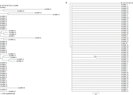
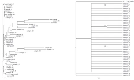
Strains 23, 49, and 42 (>99% difference) as well as the Strains 5 and 6 (>73%). Moreover, Strain 48 showed a sequence different from the Strains 6, 14, 15, 19, 22, 23, 42, and 49 (>99% difference). Finally, the Strain 50 was different in sequence from the Strains 1, 2, 3, 4, 5, 6, 11, 16, 20, 25, 30, 33, 37, 38, 39, 40, 46, 48, and 49 (Fig. 1A). Phylogenetic trees based on hsp70 gene showed that the Strains 5, 30, 43, and 49 were different from other strains in sequence (>99% difference). However, other strains were highly correlated, and the sequence similarity of the strains could be considered as an indicative of common ancestors (Fig. 2A). Based on the rpoB gene sequences, the Strain 11 showed a high correlation to the reference strain and Strains 2, 3, 5, 11, 24, 26, 37, 39, and 43, while it was different from other strains (>99% difference). Moreover, the Strain 15 was highly correlated with other strains, except for 19, 25, 29, 35, 41, 45, and 49 (>99% difference) (Fig. 3A). Phylogenetic trees constructed based on the concatenation of 16s rRNA, rpoB, and hsp70 genes sequences by maximum parsimony and neighbor-joining methods showed that the Strain 5 was different from the Strains 6, 15, 19, 30, 32, 42, 46, 47, 48, and 40 with a bootstrap value of 99%. Moreover, the Strain 11 showed a phylogenetic difference with the Strains 1, 2, 3, 4, 5, 6, 15, 19, 30, 32, 42, 46, 47, 48, and 49 with a bootstrap value of 99%. However, other strains were highly similar in sequence and showed a close relationship to one other. Notably, the Strain 24 only showed similar genetic sequence to the reference strain and Strain 7 and showed 99% difference with other strains (Fig. 4A). Phylogenetic analysis of the strains using the Maximum Parsimony method: According to the results of phylogenetic analysis based on 16s rRNA gene using maximum parsimony method, all S. aureus isolates showed similar genetic sequences
and were not phylogenetically different and were presented in a monophyletic manner. However, it must be noted that the Strains 13 and 39 showed a trivial difference with other isolates with a bootstrap value of 63% (Fig. 1B). Molecular analysis based on hsp70 gene indicated that the Strains 2, 5, 27, and 30 were different from other strains with a bootstrap value of 62%, while the Strains 1 and 24 were different from other strains with a bootstrap value of 61% (Fig. 2B). Finally, Phylogenetic analysis based on rpoB gene using the maximum parsimony method revealed the differentiation of the Strains 5 and 11 with 99% bootstrap value as well as the differentiation of the Strain 33 with 53% bootstrap value compared to the other strains (Fig. 3B). Phylogenetic analysis based on 16srRNA, rpoB, and hsp70 genes in concatenation indicated that the Strains 5 and 11, 1 and 24, and 37 and 43 were different from other strains with the respective bootstrap values of 99, 58, and 51% (Fig. 4B).
Discussion
S. aureus is one of the most common causes of both endemic and epidemic hospital-acquired infections with a high morbidity
and mortality rate [3]. The frequency of
multidrug-resistant staphylococci strains is reportedly increasing worldwide, including isolates that are resistant to methicillin, aminoglycosides, macrolides,
fluoroquinolones, lincosamides, or
combinations of these antibiotics. Therefore, the severe consequences of infections caused by S. aureus strains and the incidence of antibiotic resistance have increased the importance of prevention [14-16]. The study
Fig. 1. A) Neighbour-joining and B) Maximum Parsimony trees based on the 16S rRNA gene sequences of 50 S.
aureus clinical isolates
Fig. 2. A) Neighbour-joining and B) Maximum Parsimony trees based on the hsp70 gene sequences of 50 S.
Fig. 3. A) Neighbour-joining and B) Maximum Parsimony trees based on the rpoB gene sequences of 50 S.
aureus clinical isolates
Fig. 4. A) Neighbour-joining and B) Maximum Parsimony trees based on the concatenated sequences of the 16S
possible differences in their characteristics. Moreover, it could help us distinguish bacterial patterns regarding their hosts and sources. To date, several phenotypic and genotypic methods have been used for the typing of S. aureus strains, including biotyping, antibiotic susceptibility testing, pulsed-field gel electrophoresis (PFGE), multilocus sequence typing (MLST), and PCR-based techniques. Although PFGE has been introduced as an effective method for the typing of S. aureus strains, it is not cost-effective and requires technicality and effort. Therefore, in the current study, it was attempted to develop a MLSA method which is considered as an easy, useful, and affordable method [17-18]. Although the
16S rRNA gene sequencing has remained as the primary choice for the bacterial identification and different molecular targets, different factors such as genetic variation, horizontal gene transfer, and recombination may challenge this approach. Therefore, in this study, a three-locus MLSA scheme was used to reliably identify and characterize S. aureus isolates. In fact, using MLSA ensures that recombination at one locus could be compensated by the indications of relatedness between the strains provided by the others [19]. Therefore,
in the current study, the polymorphism and genetic diversity of S. aureus isolates was investigated based on 16S rRNA, hsp70, and rpoB genes which are important in bacterial cell cycle and protein synthesis, thereby affecting the clinical course of infection [20].
Polymorphism could disrupt cell processes including replication, transcription, and translation, which could be horizontally transferred to the next bacterial generations, leading to the emergence or appearance of a single or multiple bacterial characteristics. In fact, any change in genomic organization leads to the emergence of novel genotypes which could affect the rate of infection in any
given geographical region [21-22].
To the best of our knowledge, this was the first study using MLSA for S. aureus typing. However, similar studies have been conducted to determine the phylogenetic relationship between staphylococcal isolates. Lamers et al. (2012) used 16S rRNA, rpoB, and hsp60 genes to estimate the relationship between staphylococcal isolates using Bayesian partition modeling and maximum likelihood analysis. According to their phylogenetic estimates, they proposed a refined classification for Staphylococcus, in which species were classified into 15 cluster groups according to molecular data
[23]. Similar to the current study results, they
indicated that 16S rRNA, rpoB, and hsp60 genes were suitable for distinguishing S. aureus strains.
In another study by Seong et al. (2013), the complete rpoB and seven partial house-keeping genes sequences of 29 human and poultry isolated S. aureus strains were determined, and the phylogenetic analysis of these strains was conducted using the GenBank and EMBL databases. Their RS typing results showed the differentiation between the poultry and human isolated ST5 strains and the mutations related to the rifampin resistance in some human S. aureus strains [24], indicating that the study
of polymorphism and genetic variations in both antibacterial resistant and sensitive strains could be very useful for comparing the pathogenicity and genomic organization in these isolates. Similar to the current study, they showed that rpoB gene could be used to assess the genetic relationship between S. aureus isolates.
Conclusion
MLSA technique was developed for 50 S. aureus clinical isolates based on 16S rRNA, hsp70, and rpoB genes. The MLSA method was clearly capable of discriminating between the S. aureus genotypes. Overall, there was a high genetic diversity in the three studied MLSA loci among the 50 S. aureus clinical isolates compared to the reference strain of S. aureus NCTC 8325. The use of multi-locus sequence analysis and the study of polymorphisms in S. aureus clinical isolates are proposed for infection control and surveillance.
Acknowledgments: The authors would like to extend their sincere appreciation to all the staff in Biology Department of Islamic Azad University, Science and Research Branch, Tehran.
Ethical Permissions: The authors hereby declare all ethical standards have been respected in preparation of the submitted article.
Conflict of interests: The authors declare that they have no conflict of interest.
Author’s contribution: Hoseiny Khorram Abadi E. (First author), Sequence data analyses / Original researcher/ Discussion author (50%); Zaker Bostan Abad S. (Second author), Original researcher/ Methodologist /Discussion author (50%)
Fundings: This research was supported by Islamic Azad University, Science and Research Branch, Tehran.
References
1. Ryu S, Song P, Seo C, Cheong H, Park Y. Colonization and infection of the skin by S. aureus: Immune system evasion and the response to cationic antimicrobial peptides. Int J Mol Sci. 2014; 15(5):8753-72.
2. Miller M, Cook HA, Furuya EY, Bhat M, Lee M-H, Vavagiakis P, et al. Staphylococcus aureus in the community:
Colonization versus infection. PLoS One. 2009;4(8):e6708.
3. Tong SY, Davis JS, Eichenberger E, Holland TL, Fowler VG. Staphylococcus aureus infections: Epidemiology, pathophysiology, clinical manifestations, and management. Clin Microbiol Rev. 2015; 28(3):603-61. 4. Sakr A, Brégeon F, Mege J-L, Rolain J-M,
Blin O. Staphylococcus aureus nasal colonization: An Update on mechanisms, epidemiology, risk factors and subsequent infections. Front Microbiol. 2018; 9:2419. 5. Carretto E, Visiello R, Nardini P. Methicillin
resistance in Staphylococcus aureus. Pet-to-man travelling Staphylococci: Elsevier; 2018: 225-35.
6. Stryjewski ME, Corey GR. Methicillin-resistant Staphylococcus aureus: An evolving pathogen. Clin Infect Dis. 2014; 58(suppl_1):S10-9.
7. Deurenberg RH, Vink C, Kalenic S, Friedrich A, Bruggeman C, Stobberingh E. The molecular evolution of methicillin-resistant Staphylococcus aureus. Clin Microbiol Infect. 2007;13(3):222-35. 8. Kämpfer P, Glaeser SP. Prokaryotic
taxonomy in the sequencing era-the polyphasic approach revisited. Environ Microbiol. 2012; 14(2):291-317. 9. Bæk KT, Thøgersen L, Mogenssen RG,
Mellergaard M, Thomsen LE, Petersen A, et al. Stepwise decrease in daptomycin susceptibility in clinical Staphylococcus aureus isolates associated with an initial mutation in rpoB and a compensatory inactivation of the clpX gene. Antimicrob Agents Chemother. 2015;59(11):6983-91. 10. Huang P, Lu C, Li J, Xu J, Liu Z, Wang Q, et
al. Mutations in HSP70-2 gene change the susceptibility to clinical mastitis in Chinese Holstein. Gene. 2015; 559(1):62-72.
Microbiol. 2009; 57(2):104-15.
12. Ohta T, Saito K, Kuroda M, Honda K, Hirata H, Hayashi H. Molecular cloning of two new heat shock genes related to the hsp70 genes in Staphylococcus aureus. J Bacteriol. 1994; 176(15):4779-83.
13. Petti CA, Simmon KE, Miro JM, Hoen B, Marco F, Chu VH, et al. Genotypic diversity of coagulase-negative staphylococci causing endocarditis: A global perspective. J Clin Microbiol. 2008; 46(5):1780-4. 14. Peacock SJ, Paterson GK. Mechanisms of
methicillin resistance in Staphylococcus aureus. Annu Rev Biochem. 2015; 84:577-601.
15. McGuinness WA, Malachowa N, DeLeo FR. Focus: Infectious diseases: Vancomycin resistance in Staphylococcus aureus. Yale J Biol Med. 2017; 90(2):269-81.
16. Foster TJ. Antibiotic resistance in Staphylococcus aureus. Current status and future prospects. FEMS Microbiol Rev. 2017; 41(3):430-49. 17. Tenover FC, Arbeit R, Archer G, Biddle
J, Byrne S, Goering R, et al. Comparison of traditional and molecular methods of typing isolates of Staphylococcus aureus. J Clin Microbiol. 1994; 32(2):407-15. 18. O’Hara FP, Suaya JA, Ray GT, Baxter R, Brown
ML, Mera RM, et al. spa typing and multilocus sequence typing show comparable performance in a macroepidemiologic study of Staphylococcus aureus in the
United States. Microb Drug Resist. 2016; 22(1):88-96.
19. Glaeser SP, Kämpfer P. Multilocus sequence analysis (MLSA) in prokaryotic taxonomy. Syst Appl Microbiol. 2015; 38(4):237-45. 20. Akgul A. The role of universal stress
proteins in Edwardsiella ictaluri virulence: Mississippi State University; 2017. 21. Hata E, Katsuda K, Kobayashi H, Uchida
I, Tanaka K, Eguchi M. Genetic variation among Staphylococcus aureus strains from bovine milk and their relevance to methicillin-resistant isolates from humans. J Clin Microbiol. 2010; 48(6):2130-9. 22. McCarthy AJ, Lindsay JA. Genetic variation
in Staphylococcus aureus surface and immune evasion genes is lineage associated: Implications for vaccine design and host-pathogen interactions. BMC Microbiol. 2010; 10(1):173.
23. Lamers RP, Muthukrishnan G, Castoe TA, Tafur S, Cole AM, Parkinson CL. Phylogenetic relationships among Staphylococcus species and refinement of cluster groups based on multilocus data. BMC Evol Biol. 2012; 12(1):171.