319 *Corresponding author: Mahdi Aminian, PhD,
Depart-ment of Clinical Biochemistry, School of Medicine, Tehran University of Medical Sciences, Tehran, Iran.
Tel: +98 21 88953004 Fax: +98 21 64053385 E-mail: [email protected]
Rapid fluorometric quantification of bacterial cells using Redsafe nucleic
acid stain
Ehsan Khalili1, Vahid Hosseini1, Roya Solhi1, Mahdi Aminian1,2*
1Department of Clinical Biochemistry, School of Medicine, Tehran University of Medical Sciences,
Tehran, Iran
2Recombinant Vaccine Research Center, Tehran University of Medical Sciences, Tehran, Iran
Received: June 2015, Accepted: September 2015
ABSTRACT
Background and Objectives: Numerous procedures in biology and medicine require the counting of cells. Direct enumer-ation of Colony Forming Units (CFUs) is time-consuming and dreary accurate cell counting on plates with high numbers of CFUs is error prone. In this study we report a new indirect cell counting method that was developed based on the use of
Redsafe fluorometric assay. The usefulness of Redsafe, a nucleic acid stain, in liquid medium is based on the binding of the fluorescent dye to DNA.
Materials and Methods: Redsafe fluorometric assay was evaluated in comparison with MTT colorimetric assay as a colou -rimetric assay for enumeration of bacterial cells.
Results: Obtained results showed that fluorometric assay threshold for LB grown E. coli is 6×10⁴ CFU/ml. Redsafe fluo -rescent assay can be used as a rapid and inexpensive method for bacterial enumeration and quantification with increased
sensitivity.
Conclusion: The sensitivity of the Redsafe fluorometric assay for detection and enumeration of bacterial cells was 2-log-unit
more than that was observed for the MTT assay.
Keywords: Bacterial enumeration, Redsafe, MTT assay, fluorometric assay
ing on plates with high numbers of CFUs is error prone since it requires a high level of attention and so, often only parts of a plate are analyzed to esti-mate the whole plate count. Therefore, rapid methods
for quantification and enumeration of microorgan
-isms are important. Also, the number of rapid tests for the estimation of the microbiological quality of foods has increased in recent years. Many tests have been proposed, including methods based on antibody recognition, a wide range of biochemical and enzy-matic assays, phage probes, membrane filtration and impedance (5-7). Although, some of these methods have low accuracy for mixed bacterial populations, are expensive, and many are still too slow to provide useful result. Hence, detection and enumeration of microorganisms are very important, especially when
ORIGINAL
AR
TICLE
INTRODUCTION
count-investigating refrigerated food. Despite the high number of rapid methods that have been developed, the methods are generally unsatisfactory for the dairy industry as many of them are inaccurate or complex, and many are still too slow to provide useful results to the manufacturer. Furthermore, a number of the rapid methods available within the dairy industry are used to detect microorganisms rather than enumerate them. Enumeration, rather than detection, is a very important requirement when investigating food qual-ity.
Redsafe, a safe nucleic acid stain (8-10), is devel-oped to replace the Ethidium Bromide which is a carcinogenic fluorescent dye (11, 12). Data from the Ames-test supports that Redsafe is non carcinogenic and it can be disposed as any other non-carcinogenic
fluorescent dye (13). Until now, the Redsafe fluoro
-metric assay was not evaluated for the enumeration of E. coli in liquid medium.
Hence, in attempt to set up a novel and rapid meth-od for enumeration of E. coli, we aimed to assess the
ability of the Redsafe in order to establish a fluoro
-metric assay and also to compare it with traditional MTT assay which is based on dehydrogenase system of cells (14, 15). The present paper reports the
adap-tation of the Redsafe fluorometric assay for the esti
-mation of the microbiological quality and quantity in liquid medium.
MATERIALS AND METHODS
Bacterial growth and samples. E. coli origami strain was added to culture and was grown overnight in Luria-Bertani (LB) broth (1% bacto tryptone,
0.5% yeast extract, 1% NaCl) at 37 oC on a shaker
at 180 rpm. E. coli cell growth was determined by measuring the OD of the broth at 590 nm. Then, three samples consisting of untreated, washed, and boiled bacteria were prepared. The overnight grown
bacteria in LB broth was directly used as untreat
-ed bacteria. Then to wash bacteria, a 1.5 ml sample was removed from the overnight grown bacteria and placed into centrifuge tube and centrifuged at 5000g for 10 min at 4 °C. The pellet was washed with 1.5 ml of PBS for three times, resuspended in 1.5 ml of PBS and used as washed bacteria. Preparation of boiled bacterial sample was similar to washed bacteria and then followed by addition of a boiling step for 10 min in boiling water.
Determination of bacterial CFU. The number of colony forming unit (CFU) of E. coli cells was deter-mined by spread plating of log 10 serial dilutions of samples, consisting of untreated and washed bacte-ria, on LB-agar and incubated for approximately 20 hours at 37 oC. Numbers of CFU per milliliter were
calculated by multiplying the number of colonies by the dilution factor and the volume of the tested sample.
Redsafe fluorescent staining protocol. The fluo
-rometric system is a modification of previously pub
-lished by Kowalski (7), which was originally devised as a differential fluorescence system for detecting nicked circular DNA versus intact one (5). Three samples consisting of untreated, washed, and boiled bacteria were used in this regard. A total of 80µ of 1:3000 diluted Redsafe (iNtRON, South Korea) in
DW was added to 3 ml of diluted bacteria in a flu
-orimeter cuvette. The fluorescence of the resulting
solution was read just after mixing in spectrofluoro
-meter at an excitation wavelength of 270 nm and emission detection at 537 nm to measure the emis-sions from the Redsafe stained cells in suspension.
MTT assay. The MTT assay was performed as
described by Mosmann (16), with some modifica
-tions. Untreated and washed bacteria were used as bacterial samples in this assay. The bacterial serial dilutions (1:2) were made using PBS and two hun-dred microliters of each suspension was transferred to corresponded labeled tubes. Then, 20 µl of a stock solution of MTT (Sigma, UK) at 5 mg/ml in PBS was added to each tube and incubated for 20 min at 37 °C. The tubes were centrifuged at 10000×g for 1 min and the supernatants were discarded. Finally, the formazan crystals were solubilized using 100 μl of DMSO and pipetted into 96-well micro plates. Absorbance was measured with a microplate reader (Eon BioTek, USA) at wavelengths of 550 nm.
RESULTS
Fluorometric method for enumeration of E.
coli. To develop the Redsafe fluorometric assay for
enumeration of bacterial cells, serial diluted samples with defined CFU/ml were used. As illustrated in
Fig. 1, fluorometric assay lower threshold for untreat
http://ijm.tums.ac.ir
IRAN. J. MICROBIOL. Volume 7 Number 6 (December 2015) 319-323 321
http://ijm.tums.ac.ir
threshold was 7×10⁵ CFU/ml. In order to eliminate
possible quenching effectors of culture medium, fluorometric assay was performed with PBS washed bacteria. After washing samples with PBS, lower and higher threshold reached to 6×10⁴ and 1×10⁵ CFU/ml
respectively which was accompanied with increase in sensitivity by almost 7 fold. These data indicated that sensitivity of fluorometric assay is increased by washing of bacteria. Since the method described here is based on the ability of Redsafe to bind to DNA, binding of Redsafe to release bacterial DNA (boiled sample) was performed in comparison with DNA of intact bacterial cells. The results indicated that the fluorometric reading for intact bacteria and released DNA are the same (Fig. 1).
DISCUSSION
Enumeration and detection of bacterial cells is very important requirement for evaluating food quality. Several indirect methods have been yet proposed so far as rapid method for enumeration of cells (16-20). The use of Redsafe, a newer florescent dye, has been proposed for assessment of nucleic acids but it has
never been reported as a fluorometric dye for enumer
-ation of bacteria. This study, for the first time, presents a simple protocol for rapid enumeration of E. coli cells by the Redsafe fluorometric assay. The results showed that linear relationship between the emission of the Redsafe dye and the bacterial concentration was stron-ger when the concentration of E. coli cells ranged be-tween 8×10⁴ and 1×10⁵ CFU/ml. Notably, our results
showed that washing of bacterial cells can result in in-crease in sensitivity by almost 7 fold, probably due to elimination of quenching effectors in LB medium. We
also found no augmentation in the Redsafe fluoromet
-ric assay measurements following boiling of bacterial cells. Therefore, it can be deduced that Redsafe can conveniently access to DNA of intact bacterial cells and released bacterial DNA.
MTT reduction assay for enumeration of bacteri-al cells has been reported in previous studies (18, 19, 21). In consistence with other studies, this work showed that the linear relationship between the bac-terial concentration and the absorbance of the resul-tant formazan dye was stronger when the
concentra-Fig. 1. Redsafe fluorometric assay. The Redsafe fluoro -metric assay was performed on Untreated ( ), washed
( ) and boiled ( ) bacteria samples. Lower and
higher thresholds were 5x10⁵ CFU/ml and 7x10⁵ CFU/ml for untreated bacteria while for washed and boiled bacteria received to 6x10⁴ and 1x10⁵ CFU/ml.
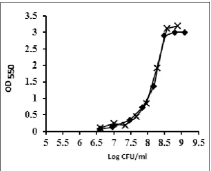
MTT assay for enumeration of E. coli.In order to perform the MTT assay, serial dilutions of
untreat-ed LB grown E. coli with defined CFU/ml were used.
Fig. 2 indicates that the lower threshold of this colo-rimetric assay is 5×10⁷ CFU/ml. The higher threshold
is about 5×10⁸ CFU/ml as the linearity of the assay is
lost at higher concentrations. In order to eliminate the possible effects of released reductases from dead cells the MTT assay was performed with washed bacterial cells as well. The results indicate that centrifugation has not effect on sensitivity of MTT assay (Fig. 2).
Fig. 2. MTT assay: The MTT assay was performed on
Un-treated bacteria ( ) and washed bacteria ( ). Lower and
tion of E. coli ranged between 5×10⁷ and 5×10⁸ CFU/
ml (18, 19, 21). These results showed that required amount of bacteria for Redsafe fluorometric assay is 2-log unit less than that needed for tradition MTT as-say. Since Redsafe binds to bacterial cell DNA, Red-safe fluorescent assay can be used to count all type of bacterial cells but MTT assay cannot be applied on bacterial cells without NAD(P)H-dependent oxidore-ductases and dehydrogenases (22). While MTT assay only count the living cells Redsafe fluorometric assay counts the dead and live cells which can be helpful in quality assessment of heated and refrigerated food. So, enumeration and detection of bacterial cells is very important requirement when assaying of food quality. In conclusion, the sensitivity of the Redsafe assay was 6 ×10⁴ CFU/ mL. This sensitivity limit complies
with the level of detection required to satisfy regu-lations in many countries. Results obtained from the
present study clearly indicates that the Redsafe fluo
-rescent assay can be used as a rapid and inexpensive method for bacterial quantification and enumeration. This method has the additional advantage that it can be performed in a 96-well microplate format for as-sessment of large numbers of samples. The sensitivity and the adaptability of the Redsafe fluorometric assay make it an efficient high throughput screening tool for epidemiological and diagnostic investigations, and rapid microbial analysis in food and drink industries.
ACKNOWLEDGEMENT
This research was supported financially by Tehran University of Medical Sciences (Grant 14007).
REFERENCES
1. Watson S, Novitsky T, Quinby HL, Valois F. Determi -nation of bacterial number and biomass in the marine environment. Appl Environ Microbio 1977; 33:940-946.
2. Zimmermann R, Iturriaga R, Becker-Birck J. Simul-taneous determination of the total number of aquatic bacteria and the number thereof involved in respira-tion. Appl Environ Microbiol 1978; 36:926-35.
3. Rosche WA, Foster PL. Determining mutation rates in
bacterial populations. Methods 2000; 20:4-17.
4. Pirnay J-P, De Vos D, Duinslaeger L, Reper P, Vanden -velde C, Cornelis P, et al. Quantitation of Pseudomonas
aeruginosa in wound biopsy samples: from bacterial culture to rapid ‘real-time’polymerase chain reaction.
Crit Care 2000; 4:255-261.
5. Kowalski D. A procedure for the quantitation of re-laxed closed circular DNA in the presence of
superhe-lical DNA: an improved fluorometric assay for nick -ing-closing enzyme. Anal Biochem 1979 ;93:346-354. 6. Vasavada PC. Rapid methods and automation in dairy
microbiology. J Dairy Sc 1993; 76:3101-3113.
7. Manzano S, Antonio Ordonez J, de la Hoz L, Fernan -dez M. A rapid method for the estimation of the micro-biological quality of refrigerated raw milk based on the aminopeptidase activity of Gram-negative bacteria. Int Dairy J 2005; 15:79-84.
8. Machida RJ, Knowlton N. PCR primers for metazoan nuclear 18S and 28S ribosomal DNA sequences. PloS one. 2012; 7:e46180.
9. Nady N, Krichevsky L, Zhong N, Duan S, Tempel W,
Amaya MF, et al. Histone recognition by human malig-nant brain tumor domains. J Mol Biol 2012; 423:702-718.
10. Ibeagha-Awemu EM, Ibeagha AE, Messier S, Zhao X. Proteomics, genomics, and pathway analyses of
Escherichia coli and Staphylococcus aureus infected milk whey reveal molecular pathways and networks in-volved in mastitis. J Proteome Res 2010; 9:4604-4619. 11. Schaeffer WI, Melamede R. Fluorometric
quantita-tion of broth-cultured mycoplasmas by using alkaline ethidium bromide. J Clin Microbio 1993; 31:1303-1307.
12. Lepecq JB, Paoletti C. A fluorescent complex between
ethidium bromide and nucleic acids: Physical—Chem-ical characterization. J Mol Biol 1967; 27:87-106. 13. Ames BN, McCann J, Yamasaki E. Methods for
de-tecting carcinogens and mutagens with the Salmonella/ mammalian-microsome mutagenicity test. Mutat Res
1975; 31:347-364.
14. Mosmann T. Rapid colorimetric assay for cellular growth and survival: Application to proliferation and cytotoxicity assays. J Immunol Methods 1983; 65:55-63.
15. Denizot F, Lang R. Rapid colorimetric assay for cell growth and survival: modifications to the tetrazolium
dye procedure giving improved sensitivity and reliabil-ity. J Immunol Methods 1986; 89:271-277.
16. Mosmann T. Rapid colorimetric assay for cellular growth and survival: application to proliferation and cytotoxicity assays. J Immunol Methods 1983; 65:55-63.
17. Wang H, Cheng H, Wang F, Wei D, Wang X. An improved 3-(4,5-dimethylthiazol-2-yl)-2,5-diphenyl tetrazolium bromide (MTT) reduction assay for evaluating the vi-ability of Escherichia coli cells. J Immunol Methods
2010; 82:330-333.
-http://ijm.tums.ac.ir
IRAN. J. MICROBIOL. Volume 7 Number 6 (December 2015) 319-323 323 http://ijm.tums.ac.ir
riers N. A colorimetric assay for the enumeration of
Candida albicans in biological samples after amplifica -tion in a selective medium. J Microbiol Methods 1993; 18:151-61.
19. Mshana RN, Tadesse G, Abate G, Miorner H. Use of 3-(4,5-dimethylthiazol-2-yl)-2,5-diphenyl tetrazolium bromide for rapid detection of rifampin-resistant My-cobacterium tuberculosis. J Clin Microbiol 1998; 36:1214-1219.
20. Skehan P, Storeng R, Scudiero D, Monks A, McMahon J, Vistica D, et al. New colorimetric cytotoxicity assay
for anticancer-drug screening. J Natl Cancer Inst 1990; 82:1107-1112.
21. De Logu A, Uda P, Pellerano ML, Pusceddu MC, Saddi B, Schivo ML. Comparison of two rapid colorimetric
methods for determining resistance of Mycobacterium tuberculosis to rifampin, isoniazid, and streptomycin in liquid medium. Eur J Clin Microbiol Infect Dis 2001; 20:33-9.