Pacemaker Concepts and Terminology*
BAROUH V BERKOVITS, E.E., Ing.
Associate in Surgery, Harvard Medical School, Boston, Mass., Associate in Electrophysiology, Miami University, Miami, Fla., Senior Research Scientist, Cedars-Sif1£li Medical Research Institute, Los Angeles, Calif., Consultant in Surgery, Peter Bent Brigham Hospital, Boston, Mass., Cardiovascular Research Manager, American Optical Corporation, Framingham, Mass.
Since the introduction of implantable cardiac pacing systems there have been marked advances in concept and design of the equipment available. Whereas intra-cardiac pacing is usually identified with A-V con-duction disturbances, recently gained knowledge of cardiac physiology has shown that electrical stimula-tion of the heart can be beneficial in the treatment of many arrhythmias. It is important that the phy-sician familiarize himself with the different modalities of pacing. The terminology used to describe the different concepts must be clear and precise so the physician can select the proper pacemaker concept for his patient without ambiguity
Pacemakers can be divided into two categories. parasystolic and nonparasystolic. While parasystolic-stimulation is independent of intrinsic activity; non-parasystolic-stimulation is controlled by the intrinsic activity The nonparasystolic pacemakers can be either inhibited, in which intrinsic signals suppress stimula-tion, or triggered, in which intrinsic signals induce stimulation. According to the site of stimulation the pacemakers can be atrial, ventricular, or A-V sequen-tial. Pacemaker types in present use are
1 Parasystolic (Continuous) a. Fixed or set rate atrial pacer b. Fixed or set rate ventricular pacer c. Fixed or set rate A-V sequential pacer 2. Triggered (Synchronous)
a. P-wave triggered ventricular pacer b. QRS triggered ventricular pacer 3. Inhibited (Demand)
a. P-wave inhibited atrial pacer b. QRS inhibited ventricular pacer c. QRS inhibited A-V sequential (Bifocal) The parasystolic pacemakers ( continuous fixed rate) may be used to stimulate the atria, ventricles, or stimulate both the atria and ventricles with a preset sequential delay In the presence of natural beats the modality of stimulation is not affected and it competes with the intrinsic activity The stimuli falling in the absolute refractory period will induce no response,
*
Presented at the American College of Cardiology and the Medical College of Virginia Cardiac Pacing Sympo-sium, April 16 and 17, 1971, Williamsburg, Virginia.MCV QUARTERLY 7(4): 131-135, 1971
but those falling in the vulnerable interval may induce repetitive response or even fibrillation.
Figure 1 illustrates the differences between the various modalities of nonparasystolic pacing.
Characteristics of P-wave triggered ventricular pac-ing are ( 1) The P waves trigger the pacemaker, which in turn stimulates the ventricles synchronously after a preset delay of 120 milliseconds. (2) This pacer usually has a refractory period of 500 milli-seconds (measured from the beginning of the P wave) which prevents the pacemaker from following atrial tachycardia or fibrillation. ( 3) When natural P waves do not appear for a preset interval ( 1 .04 sec.) the P-wave triggered pacemaker escapes. This escape mechanism protects against asystole when there are no P waves present. Thus, the pacemaker works either in synchronous or escape modality
Characteristics of QRS triggered ventricular pacing are· ( 1) The QRS complexes trigger the pacemaker,
which in turn stimulates the ventricles during its absolute refractory period. In the latest models, this synchronized stimulation is delivered immediately
after the detection of the ventricular endocardiac signal. (2) The pacemaker has a refractory period of 400 to 500 milliseconds. This built-in refractory
period is designed to protect the unit from running at fast rates. A long refractory time may prevent the
recognition of premature beats and thus causes escape stimulation competing with these premature beats. Competition with premature beats is even more hazardous than with the normal beats. ( 3) When natural QRS complexes do not appear for a preset interval ( 840 milliseconds), pacemaker escapes occur to protect against asystole.
Contrary to the triggered pacemakers, the demand pacemakers work only in escape mode. ( 1) In the presence of a faster natural rhythm, the demand pacemaker is dormant and no stimuli are delivered to the heart. (2) Stimuli are delivered only if the natural beat fails to occur for a preselected escape interval. Consequently, the heart is stimulated only
when needed ( on demand), and competitive rhythms or stimulation during the vulnerable phase are thus avoided.
Figure 2 shows how the ventricular demand or
DEMAND
A TRIO-SYNCHRONIZED
VENTRICULO-SYNCHRONIZED
VENTRICULO-SYNCHRONIZED
---Escape Interval
Fig 1-Differences between the various modalities of non-parasystolic pacing.
P-WAVE TRIGGERED
ATRIAL-CONTINUOUS
A-V SEQUENTIAL
BIFOCAL DEMAND
.
.
•.
A. T,tANlll(IIIPANI POT[NTIALS IN AUTOMATIC Fll[lt INITIATINt [SCAPE
IU,TI.
EL!CTLltlC CHAlllOE: ON TH[ TIIIIING CANCITOR OF TH[ O(ltAND PAC(IIAIC[R INITIATIN. tsc,,, IU,TI.
Fig 2-Actual workings of the ventricular demand or
in-hibited pacemaker
MAGNETIC
REED SWITCH
DEMAND PACEMAKER
VENTRICULAR DEMAND STIMULATOR
ORS DETECTOR
VENTRICLE
BIFOCAL DEMAND PACEMAKER
MAGNETIC
REED SWITCH
VENTRICULAR DEMAND STIMULATOR
ORS DETECTOR
ATRIAL DEMAND STIMULATOR
VENTRICLE
ATRIUM
Fig 4- Basic construction of the Bifocal demand pacemak er compared to the conventional demand unit.
F
kirLLLLRL
rr
rr
r
rr
ff
i
AV
AV
A
AV
AV
A
Fig 5 -The Bifocal demand pacemaker facilitating the natural depolarization sequence without competing with spont
BIFOCAL DEMAND PAC EM AKER
WORKING IN ATRIAL MODE,OCCASSIONAL PVC SC COMPENSATORY
PAUSE IN ATRIA
Fig 6-Bifocal pacemaker placed in a patient with sinus bradycardia and premature ventricular contraction.
B FOCAL DEMAND PACEMAKER
Fig 7-Bifocal pacemaker placed in a patient with first degree A-V block.
B FOCAL DE MAND PACEMAKER
L.~WL,
·
A
A
ill
~
· ·
I
)
1
;
l '
.
'
ON- MAGNET- OFF
TESTING RESPONSE OF VENTRICULAR STIMULATION
Fig 8-Testing of a pacemaker with a magnet; unit is
con-verted to a fixed-rate mode.
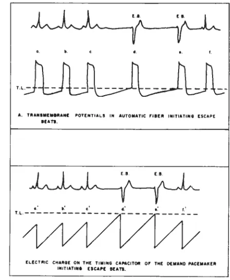
inhibited pacemaker actually works, imitating the natural escape rhythm of an automatic fiber The upper part of the tracing demonstrates how the
auto-matic fiber works and how the threshold potential is reached during phase four depolarization so that these fibers can fire by themselves and produce
escape beats. The bottom portion of the figure
dem-onstrates how the pacemaker is controlled by a capa-citor that restores its charge with each detected beat. In short, each time a depolarization signal is de-tected the pacemaker is reset and the timing cycle
is started again. When the capacitor's charge reaches the critical level because of the prolonged interval,
it permits escape and a stimulus is delivered to the ventricle. The pacemaker actually delivers its
stimu-lation in the same fashion as an automatic fiber Figure 3 demonstrates schematically the different modalities of pacing. As shown, Bifocal pacing is
similar to A-V sequential pacing, except that it is on demand and it is inhibited and reset by endocardial ventricular depolarizations. The Bifocal demand pace-maker adapts its modality of stimulation to the pa-tient's need. It combines the advantages of atrial, A-V
sequential, and demand stimulation. It may remain dormant, it may stimulate only the atria, or it may
stimulate both the atria and the ventricles with a preset
sequential A-V interval. The Bifocal demand pace-maker does not compete with the spontaneous
ven-tricular activity and it has no significant refractory
time.
Figure 4 illustrates the basic construction of the Bifocal demand pacemaker and compares it to the conventional demand unit. In conventional demand pacing the ventricular signal detected by a QRS de-tector will control the timing circuit of the ventricular
demand stimulator A magnetic switch is incorpo-rated in these pacers for evaluation of pacemaker function by preventing inhibition and thus converting
the unit to a fixed-rate mode. When testing the pace-maker, the rate produced by the magnetic switch is independent of the patient's physiological condition
and should be used to determine the condition of the batteries. It is important that during each outpatient
visit the demand pacemaker be checked by applying a magnet and this rate recorded for control. Con-ceptually, the Bifocal pacemaker is comparable to the conventional demand pacemaker except that
atrial stimulation controlled by the same QRS de-tector has been added.
Two functions of the Bifocal pacemaker are dem-onstrated in Figs. 5, 6, and 7 Figure 6 shows a
Bifocal pacemaker in a patient with sinus bradycardia
and premature ventricular contractions. It can be observed that the atrial pacemaker compensates for
the premature ventricular beats. Figure 7 shows a
Bifocal pacemaker in a patient with first-degree A-V
similar to the patient's own conducted A-V interval. Therefore, the different degrees of fusion and changes in morphology can be observed.
Figure 8 depicts the testing of a Bifocal pacemaker with a magnet, similar to the ventricular demand
pacemaker. The measurement of the interval during
a magnet-induced fixed-rate mode should be recorded
in order to follow the pacemaker function and de-termine battery condition. A change greater than 10 percent indicates battery failure.
During the past 18 months we have implanted 60
Bifocal pacers with encouraging results. In the early stages, the basic indication for implantation of Bifocal
pacers was to improve cardiac output. Recently,
Bi-focal pacemakers have also been used for patients with sick sinus syndrome-atrial
brady-tachyrhyth-mias. In a number of patients, we have found that
2 to 3 months after implantation, drugs could be progressively discontinued, and that the atrial
stimu-lation not only protected against the bradycardia but
also suppressed the episodes of tachycardia.
Pacing therapy has undergone marked evolution during its relatively brief existence. Further develop-ments in concept, clinical applicability and technical
areas are forthcoming. The physician has the
obliga-tion to understand the various pacing modalities and
to select the most suitable concept of pacing for each particular disorder (Table I)
Author's note. "BIFOCAL" is a trademark of the
American Optical Corporation for the QRS inhibited
A-V sequential pacemaker
TABLE I
THREE DIFFERENT MODALITIES OF A-V PACING
P-wave Triggered A-V Sequential Bifocal Demand (continuous
For Normal Atrial Activity For Atrial Bradycardia For Atrial Bradycardia
With A-V Block With A-V Block With or Without A-V Block
Monitors P-waves No monitoring Monitors QRS Complexes
P-waves Control ventricular Continuous Atrial and QRS Complexes Control Both
Stimulation No Atrial Ventricular Stimulation Atrial and Ventricular
Stimulation Available Stimulation
Stimulation is Delivered to Stimulation is Delivered Both Stimulation May Be:
to the Atria and Ventricles a Totally Absent
the Ventricles Continuously
Continuously b Delivered only to the Atria