M. ROBERT COOPER Department of Medicine, Bowman-Gray School of Medicine of Wake Forest University, Winston-Salem, North Carolina 27103
In 19iO a Chicago physician, Dr. J. B. Herrick, called attention to a peculiarly formed red cell which he observed in the blood smear of an anemic West India .Negro student (Necheles, Allen and Finkel, 1969). Linus Pauling, a physical chemist, demon-strated in 1949 that the protein portion of the hemo-globin molecule found in patients having sickle cell disease differed from that found in normal adults. In-gram ( 1957) further defined the molecular defect in that sickled hemoglobin differed from normal adult hemoglobin in one amino-acid residue per half mole-cule. The specific molecular identification of the defect in sickle cell anemia was that of valine replacing the normally occurring glutamic acid in the number 6 posi-tion of the beta chain. This finding provided inspiraposi-tion and technical knowledge whereby over 100 different hemoglobinopathies have now been described. The clinical explorations which have resulted from studies of mutant hemoglobin have allowed definition of a variety of syndromes whose manifestations require explanation in terms of the effects of molecular distor-tion on physiqlogical processes. The complex patterns of inheritance which lead to amino-acid substitution in either the alpha or beta chain dictate a variety of structural alterations which may modify hemoglobin affinity for oxygen, afford protection against fal-ciparum malaria, and provoke changes in micro-circulation which influence renal and splenic function. Whether abnormal hemoglobins induce these changes is determined at least in part by the site of replacement, changes on external surfaces exerting little effect, and the co-existence of abnormalities elsewhere in the molecule. The discovery of various hemoglobinopathies has demonstrated that specific molecular defects may express themselves in various pathophysiological man-ifestations. For instance, sickle cell anemia, a single molecular defect of the beta chain in which valine is substituted for the number 6 amino-acid glutamine, presents a wide spectrum of clinical manifestations. A marked dissociation between the degree of anemia and
*
Presented at the 23rd Annual Stoneburner Lecture Series, February 19, 1970, at the Medical College of Virginia, Richmond.64
the occurrence of other complications may be found. More detailed pathophysiological studies of patients with hemoglobinopathies should be performed to gain a better understanding of the expression of molecular disease.
Classification of Hemoglobinopathies
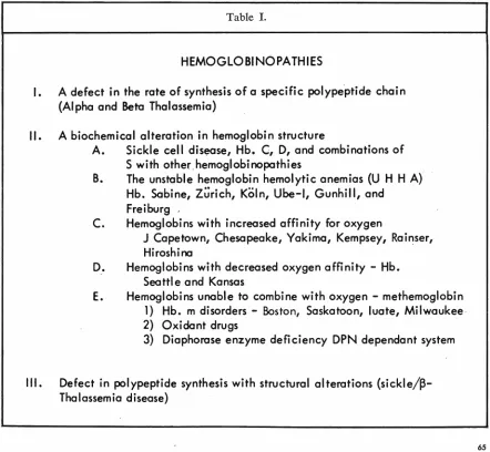
A better understanding of the hemoglobinopathies can be obtained if they are broken down into various categories. Table I shows these different types of hemoglobinopathies. The first is a defect in the rate of synthesis of a specific polypeptide chain. The most frequently considered clinical disease in this category is that of thalassemia. In this group of hemoglobin-opathies, there is either a defect in the synthesis of one or more alpha chains or one or more beta chains. These molecular defects result in specific clinical syndromes. Beta thalassemia minor may range in severity from completely asymptomatic elevations of one of the minor hemoglobin components to a syn-drome consisting of splenomegaly, intermittent jaun-dice, and mild anemia. The peripheral blood in patients with thalassemia minor shows hypochromia, microcytosis, anisocytosis, and poikilocytosis. Target cells ate quite common and the red cells show a decreased osmotic fragility. Beta thalassemia major is a more severe disease and is characterized by hypo-chromic anemia, splenomegaly, and normoblastemia. These patients are severely affected and have signs of severe anemia and phenotypically show expanded facial bones and increased pigmentation. Alpha thal-assemia is usually incompatible with life and results in hydrops fetalis.
The second major categorization of the hemoglobin-opathies are those hemoglobins in which a biochemical alteration in hemoglobin structure has occurred. The first significant subdivision within this category con-sists of patients with sickle cell disease, hemoglobin C, hemoglobin D, and combinations of hemoglobin S with other hemoglobinopathies such as hemoglobin SC, SD, and AS.
The next sub-category includes the unstable hemo-globin hemolytic anemias (UHHA). The unstable hemoglobin hemolytic anemias are characterized by the
M. ROBERT COOPER
presence of hemolytic anemia-· from infancy. Heinz bodies are usually seen in the red cells of patients with this type of anemia. Splenomegaly is usually pr:esent
and when splenectomy is performed the peripheral blood shows a marked increase in the number of red
cells containing Heinz bodies. Dacie (Dacie et al, 1964) reported studies on five patients with mild
anemia which he characterized as hereditary Heinz body anemia. A distinctive characteristic of this
group of patients with hemolytic anemia was the
pres-ence of Heinz bodies in· a blood suspension stained
with methyl violet. When hemolysates prepared from
washed red cells of all five patients were heated to 50
C for one hour, easily visible precipitates devel-oped. Since •this time, numerous hemoglobinopathies
characterized by hemolytic anemia and thermal
labil-ity of the hemolystate have been demonstrated. On
paper or starch gel electrophoresis at an alkaline pH,
the abnormal hemoglobin band is seen as a minor
component moving more slowly than hemoglobin A. It is extremely difficult to demonstrate these
hemo-globinopathies on conventional paper or Agar gel electrophoresis. Carrell and Lehmann ( 1969) have
reviewed in detail the unstable hemoglobin hemolytic
anemias. It is felt that the globin of the unstable
hemo-globins precipitates because it is no longer stabilized by firm heme to globin bonding. Hemoglobin Zurich
is an example of the unstable hemoglobin hemolytic
anemias in which there is a replacement of the non-heme linked histidine beta 63 by an arginine
amio-acid. This hemoglobinopathy is characterized by an increased susceptibility of the individual to hemolysis
as a result of taking sulfonamides. Other examples of
the unstable hemoglobin hemolytic anemias consist of
Ube-I disease. This was described in 1963 in a
fifteen-year-old Japanese girl. This has subsequently been
Table
I.
HEMOGLOBINOPATHIES
I.
A defect in the rote of synthes
i
s of a specific polypeptide chain
(Alpha and Beta Thalassemia)
II.
A biochemical alteration
i
n hemoglobin st
r
ucture
A.
Sickle cell disease, Hb. C
,
D, and combinations of
S with other hemoglobinopathies
B
.
The unstable hemoglob
i
n hemolytic anemias (U H H A)
Hb. Sabine, Zurich, Ko
l
n
,
Ube-I, Gunhill, and
Freiburg
.
C.
Hemoglobins with increased affinity for oxygen
J
Capetown, Chesapeake
,
Yak
i
ma, Kempsey, Rainser,
H
i
roshi no
D.
Hemoglob
i
ns wi
t
h decr
ea
sed oxygen
a
ffinity - Hb.
Seatt
l
e and Kan
s
as
E.
Hemoglobins unab
l
e
t
o co
mbi
ne
w
ith oxygen - methemoglobin
1) Hb. m d
i
sorder
s -
Bo
st
on
,
Saskatoon, luate, Milwaukee
2) Oxidant drugs
3) Diaphorose enzyme de
fi
c
i
ency DPN dependant system
Ill
.
Defect in polypeptide synthesis w
i
th
s
tru
c
tural alterations
(sickle/~Thalassemia disease)
shown to be similar to hemoglobin Koln which is char-acterized by blocking of the 93 amino-acid cysteine in the beta chain.
Schneider (Schneider et al, 1969) described hemoglobin Sabin disease in which there is a substitution of the beta chain at the 91 amino-acid of a lucine amino-acid by a praline residue at helical position F7 of the beta polypeptide chain. Other hemoglobinopathies in this group are those of hemoglobin Sydney (Raik, Hunter and Lindsay, 1967; Carrell et al, 1967) in which variant a substitution of valine for alanine at the beta 67 position occurs. Santa Ana (Opfell, Lorkin and Lehmann, 1968), Gun Hill (Bradley, Wohl and Rieder, 1967), Torino (Beretta et al, 1968), Hammersmith (Dacie et al, 1967), Genova (Sansone, Carrell and Lehmann, 1967), and numerous other hemoglobinopathies have been described in this category. On a molecular basis, the instability of hemo-globin molecules is thought to result from either a de-fect in the heme-binding site, a change in the overall globin confirmation, or a defect at the site of subunit interaction where the alpha and beta chains react to-gether (Carrell and Lehmann, 1969). It has been pointed out that the concentration cf amino-acid sub-stitutions of the unstable hemoglobins in the heme globin binding site is striking. Of the 11 positions cited by Perutz ( 1965) as being most closely related to the heme group, seven are found to be replaced in the un-stable hemoglobins. A molecular change which would alter the stability of the heme globin binding sites would result in instability of the hemoglobin molecule. Substitutions of amino-acids lining the heme pocket or crevice by others that are too large, alter the distribu
-tion of electric charges, or permit forma-tion of abnor-mal cross links within the molecule, decrease its stability with resultant precipitation of hemoglobin within the red cell and associated hemolytic anemia. Another group of changes consists of the substitution of praline for lucine residues. This changes the overall globin conformation with loss of stability of the mole-cule. Another aspect leading to stability of the globin heme molecule is the site of subunit interaction. It appears that it is necessary that heme and globin be bound together for normal solubility and function of hemoglobin.
The instability of hemoglobin H and Barts which are tetramers of normal beta and gamma chains re-spectively, has been previously demonstrated. Substi-tutions in the area of subunit contact promote dissoci-ation to monomers which are inherently unstable.
The third sub-grouping within this category of al-terations in hemoglobin structure consists of those hemoglobins associated with increased affinity for oxygen. Charache (Charache, Weatherall and Clegg, 1966) investigated hemoglobin Chesapeake, an alpha chain variant, which was found to have an increased oxygen affinity. This hemoglobin is characterized by a
substitution at the 92 residue of the alpha chain of lucine for arginine. The amino-acid substitutions in hemoglobins with increased oxygen affinity have been found in two areas. One is the area of contact be
-tween the alpha 1 and the beta 2 subunits in the tetramer; the other is near the C-terminal portion of the beta chain. Hemoglobin Rainier (Stamatoyan-nopoulos, Yoshida and Adamson, 1968) is the substi-tution of histidine for invariant residue 23 tyrosine in the beta hemoglobin polypeptide chain. Hemoglobin Chesapeake, Yakima, Cape Town, and Kempsey all have changes in the region between the F and H helical segments (Jones et al, 1967). These areas provide the end chain contact between the polar groups of the alpha and beta chains and are in
-volved in alpha-beta chain interaction. Substitution in these areas seems to alter the normal respiratory movements of the subunits such that oxygen .affinity
is increased. It should also be noted that four of these hemoglobinopathies have remarkably decreased n values with nearly normal effects.
The fourth sub-grouping within this major cate-gory is that of hemoglobins associated with decreased oxygen affinity characterized by hemoglobin Kansas (Reissmann, Ruth and Wohours, 1961) in which there is a substition of asparagine at the 102 residue of the beta chain by threonine. It is also a result of a substitution in the area of alpha 1, beta 2 contact.
The heme and globin components of the polypeptide are linked together by the iron of the heme binding to a specific histidine residue. This occurs at position number 87 in the alpha chain and number 92 in the beta chain. In addition, the histidine residue at num
-ber 58 in the alpha chain and number 63 in the beta chain are bound to the iron of the heme by either an oxygen molecule in oxyhemoglobin or a water molecule in desoxyhemoglobin (Necheles, Allen and Finkel, 1969).
Four of the five hemoglobin M's are produced by substitutions of the invariant histidine residues on either side of the iron atom in the heme area (Conley and Char ache, 1969). The presence of oxygen tends to change the iron to the ferric form producing metbemoglobin, a compound that cannot combine reversibily with oxygen. The substitution of tyrosine for histidine renders ferric iron so stable that the usual intracellular enzymes cannot reduce it to the functional ferrous (oxygen binding) state.
M. ROBERT COOPER
an increased affinity of the remaining ferrous atoms for oxygen.
Thus, amino acid substitutions and heme-globin interactions are important in the molecular behavior of hemoglobin. In addition, cell constituents influence the oxygen affinity of hemoglobin. Benesch (Benesch and Benesch, 1967) has shown that the affinity of hemoglobin may be increased or decreased by its inter-action with organic phosphates. Increased intra-corpuscular levels of 2-3-Diphosphoglycerate (2, 3-DPG) combine reversibly with deoxyhemoglobin, shifting the oxygen dissociation curve to the right. Charache (Charache et al, 1970) has shown that the concentration of 2-3-DPG is increased in hemo-globin SS erythrocytes as compared to hemoglobin AA red cells. This may partially explain the decreased oxy-gen affinity of hemoglobin SS. In addition, increased levels of 2-3-DPG have been found in low oxygen tension states and may be an important compensatory mechanism in hypoxemia.
The third major category of the hemoglobinopathies
are those in which there is a defect in polypeptide synthesis with a structural alteration combined with an inability of the individual to synthesize certain of the alpha or beta polypeptides. An example of this is sickled-beta thalassemic disease.
Recently we described an example of methemo-globin anemia associated with a Coombs positive hemolytic anemia. This patient (G.R.) was found to have increased levels of methemoglobin in his red cells during a severe hemolytic episode. However, the majority of his methemoglobin was extracellular con-sisting of hemoglobin which had been converted to methemoglobin. The patient showed multisystem de -generation and died in hepatic and renal failure. Methemoglobin has a characteristic spectral absorption curve which can be employed in its identification. Fig 1 shows a spectroscopic analysis of this pa
-tient's plasma. The methemoglobin is treated with cyanide to form cyano-methemoglobin and the change in light absorption at 632 m/L is measured. This method is quite specific except for the M hemoglobins
I - Plasma 1: 30 dilution with 0.1 M P04, pH 6.4
2-Plasma + 25,l 596 KCN (pH t )
3- Plasma+ additional 25-l KCN ( pHt)
2
700mµ 660 630 620 600 580 560
Fig I-Spectroscopic analysis of plasma from patient G. R. with hemolytic anemia and free methemoglobin.
which have anomalous spectroscopic properties. A simple screening method for this hemoglobin can be performed by obtaining three tubes of hemolysate or plasma. One tube is shaken with room air and if it does not turn red with adequate oxygenation, this is an indication of an abnormal hemoglobin. Potassium cyanide should be added to the second tube and if it turns red this indicates the presence of methemoglobin. Sulfhemoglobin produces a dark band at 618 mµ. which is difficult to distinguish from that of methemo-globin in the hand spectroscope. However, it can be recognized by the fact that it is not removed by the addition of cyanide. Hydrogen peroxide (3 percent) causes both of these bands to disappear. Addition of potassium cyanide and hydrogen peroxide to h emoly-sates is a simple screening procedure. A more specific identification can be obtained with a spectroscopic examination as demonstrated in Fig 1.
Methemoglobin reduction in normal cells may be accomplished by four known systems. These are ascorbic acid, glutathione, DPNH diaphorase, and TPNH diaphorase. Glutathione is maintained in the reduced state by glutathione reductase, an enzyme chiefly active with TPNH but also capable of using DPNH as a substrate. The DPNH diaphorase is by far the most active reducing methemoglobin.
Another new hemoglobinopathy which has enabled us to gain insight into both the molecular and pathophysiology of sickle cell disease is that of hemo-globin Memphis/S disease. This was initially discov -ered by Lorraine and Alfred Kraus at the University of Tennessee (Kraus et al, 1966; Kraus et al, 1967). This hemoglobinopathy is characterized by a normal and mutant alpha chain with an alpha .23 glutamine substi-tution for glutamic acid at residue 23 of the alpha chain. They have identified two hemoglobin S mole-cules-one consisting of
a/
/
{3
.5,
and hemoglobin Memphis/S consisting ofa
,M
/
/3
."
in two subjects. These patients, homozygous for hemoglobin S and heterozygous for hemoglobin Memphis, have very mild sickle cell disease suggesting that the serious conse-quences of some hemoglobinopathies may be amelior-ated by structural changes at other chain positions. Following the initial investigations by Kraus, we found a third patient with homozygous hemoglobin S hetero -zygous for hemoglobin Memphis who exhibited a sim-ilar profound dissociation between the degree of his anemia and the presence of serious complications. Spe-cial attention has been directed toward elucidating the effect of this hemoglobin on blood viscosity in an effort to account for the benignity of the process (Cooper et al, 1969).Our interest in this area was stimulated by (J.P.) a 53-year-old Negro male who has had episodic ab-dominal and musculoskeletal pain since childhood and has been repeatedly hospitalized for treatment of anemia (Fig 2). Despite shortness of breath and
Fig 2-Patient J. P., 53-year-old Negro man with hemo
-globin Memphis/S disease.
need for frequent blood transfusions, he worked as a farm laborer until four years ago when pulmonary insufficiency forced his retirement. He is 5' 1" tall, weighs 105 lbs., and his blood pressure ranges from
130-190 systolic and 85-100 diastolic. Funduscopic examination reveals slightly distended retinal veins. Slit lamp studies have shown no comma-shaped or curlicue segments of conjunctiva! vessels and no sludging. He exhibits signs of mild pulmonary emphy -sema and a Grade IV ejection systolic murmur was heard at the apex. The liver was smooth and non-tender and was felt 5 cm below the right costal margin. The spleen was nonpalpable. Double distal interphalangeal palmar creases were present in each middle finger and on the left fifth finger.
Admission laboratory data shows a urine specific gravity of 1.010; hemoglobin 9.5 gm/100 mg.; hematocrit 27 vol percent; RBC 3.1 million/cu ml; MCV 100 µ.3; MCHC 33 vol percent; reticulocytes
M. ROBERT COOPER
an unincubated sample; with incubation, marked re-sistance to hemolysis was exhibited at lower concen-trations of sodium chloride (Fig 3).
Hemoglobin electrophoresis revealed no hemoglobin
A with hemoglobin either in the S or F forms (Fig 4).
Alkali denaturization showed fetal hemoglobin to be
6 percent. Bone marrow aspiration yielded a marrow
which was 100 percent cellular showing marked
ery-throid hyperplasia and many sickled red blood cells
(Fig 5). Peripheral blood smears contained many
sickled cells and nucleated erythrocytes (Fig 6).
Poikilocytosis was prominent and Howell Jolly bodies
were present. Liver function studies disclosed no
evi-dence of abnormality. An oral cholecystogram
demon-strated many small gallstones and a chest film showed
mild left ventricular hypertrophy. Radiographic bone survey revealed diffuse osteoporotic and osteosclerotic
changes as well as concavity of the vertebral bodies
consistent with sickle cell anemia. No bone infarcts
were seen and at no time could the spleen be visualized
by X-ray. Serum bilirubin was 2.6 mg/100 ml of which
2.0 mg/100 ml was conjugated. The lactate
dehydro-genase was 1980 units and the direct and indirect
Coombs tests were negative.
Routine hematological data were derived by stand -ard techniques. Sickling was ascertained using Sher-man's test and minimal concentration gelling of
hemoglobin solution was determined according to the
method of Singer. Numerous isotopic studies
consist-ing of 51Cr labeled erythrocytes, 51Cr labeled platelets,
and heat-treated 51Cr labeled erythrocytes were
em-ployed. Hemoglobin electrophoresis was done on starch
gel and cellulose acetate using a discontinuous buffer
at pH 8.6. Individual erythrocytes were stained for
fetal hemoglobin which was then quantitated by alkali
denaturization using Betke's method. After
electro-phoresis, hemoglobin was characterized by chain
sepa-ration, tryptic digestion with peptide mapping, and
amino acid analysis of aberrant polypeptides according
to Kraus (Kraus, Miyaji and luchi, 1966). Whole blood
viscosity was determined with the falling ball
vis-cometer. Oxygenated and deoxygenated blood
viscosi-ties were also determined by using a modified
Brookfield viscometer. Renal function studies were
determined and the effect of hyperosmolar solution on
the patient's hemoglobin was also studied.
Family studies were performed and the results
showed a co-dominant inheritance pattern. The
Sher-man test (Table II) demonstrated 4.5 percent sickled
cells in arterial blood and 21.5 percent in venous
blood. Venous blood from homozygous SS patients
contains 30-60 percent sickled forms and 5-20
per-cent sickled forms in the arterial blood. A minimal
gelling concentration of 27.2 gm/100 ml was
ob-tained for S hemoglobin and 35.6 gm/I 00 ml for hemoglobin Memphis/S (Table III). The patient's
deoxygenated blood viscosity was determined by the
UNINCUBATED OSMOTIC FRAGILITY HEMOLYSIS BEGINS: 0.36% NaCl
HEMOLYSIS COMPLETED: 0. OB% NaCl
NORMAL UNINCUBATED FRAGILITY HEMOLYSIS BEGINS: 0.44% NaCl HEMOLYSIS COMPLETED: 0.28% NaCl 100
90
80
70
60
30
20
10
x
PROPOSIT
~
INCUBATED OSMOTIC FRAGILITY
0.10 0.20 0.30 0.40 0.50 0.60 0.70 0.80 0.90 % NaCl
Fig 3-Tncubated osmotic fragility of J. P.'s RBC.
'
•
NORMAL
CONTROL j
PATJ,ENT )
Fig 4- H. J. Electrophoresis with cellulose acetate strips
pH 8.6.
Fig 5- Bone marrow aspiration showing hemoglobin Memphis/S sickled erythrocyte.
Table II.
MODIFIED SHERMAN TEST
% SICKLED CELLS (200 CELLS COUNTED)
o:_Acx_A I
fJ
SJJ
s o:.Ao:.AljJAJJS
ct.A o:.M IfJ s
fJ s
VENOUS 30 - 60 <1% 21.5
ARTERIAL 5 -20 0 4.5
Table III.
THE LOWEST GELLING POINT CONCENTRATION OF HEMOGLOBINS CONTAINING HEMOLYSATES
\gm% S)
a Aa A I
/3
SfJ s aAaM lfJ SJJ s (XAa_A/fJAfJS BOWMAN GRAY 27. 0 gm/IOO ml 35.4 gm/IOO mlS± .54 S±2. 18
MEMPHIS• 19-24 gm/100 ml 26.8 gm/100 ml
CHICAGO" 21.B gm/100 ml 13.2 gm/100 ml
S±l.4 S±l .4
.
Krous - Trans. Ass. Amer. Physicians, 1967 u Singer, K. & Singer, L. Blood, 1953falling ball method (Fig 7). The result showed that the viscosity was 3. 7 centipoise units in contrast to 2.77 centipoise units for hemoglobin AA blood and
4.9 centipoise units for hemoglobin SS blood. For
oxygenated blood, comparable values were 3.2, 2.73 and 3.71 centipoise units.
When the Brookfield viscometer was used, no dif
-ference in viscosity of oxygenated and deoxygenated
hemoglobin AA blood was found, while patients with
sickle cell trait displayed an increase of 1 ± 0.46 cen-tipoise units in deoxygenated blood. Blood viscosity
increased approximately 27 centipoise units from the oxygenated to the deoxygenated state at all shear rates
for homozygous SS and SC disease (Fig 8).
Al-though the propositus is homozygous for hemoglobin
SS, his blood viscosity increased by only 1.1 ± 0.1
centipoise units with deoxygenation. The difference in
viscosity between oxygenated and deoxygenated blood from hemoglobin AA subjects was not significant. For sickle cell trait, the difference at all shear rates was
> 0.1 and < 0.5 significance level. The difference for
hemoglobin Memphis/S was >0.01 and <0.001. The
difference in viscosity of deoxygenated blood between
hemoglobin Memphis/S and sickle cell anemia was
signicant at >0.001 percent level (Fig 9). How-ever, the difference between hemoglobin Memphis/S and AS disease blood both with oxygenation and de-oxygenation was not significant.
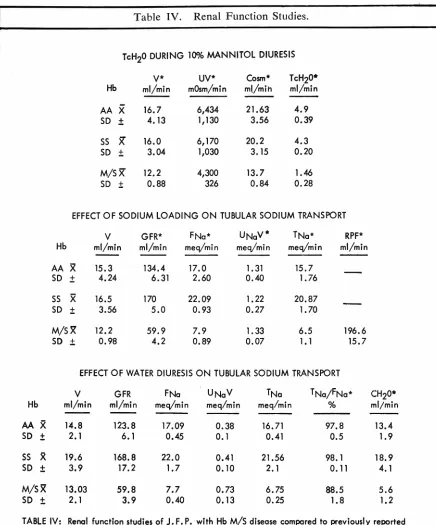
Renal function studies were performed during
Man-nitol diuresis, sodium loading, and water diuresis (Ta-ble IV). During Mannitol infusion Cosm TcH,o, UV,
and urine flow were decreased in comparison to
nor-mal subjects and individuals with hemoglobin SS. With sodium loading, glomerular filtration and renal plasma flow were abnormally low and the filtration fraction
was high reflecting a proportionately greater decrease
of the latter. Because of diminished glomerular
filtra-tion, filtered sodium was decreased but UN,V was not
suppressed indicating a decreased tubular
re-absorp-tion of sodium. Filtered sodium was decreased
sec-ondary to suppressed filtration during water loading
but UN, V was greater than in normal hemoglobin SS
subjects. TN• was considerably depressed. Tc"'° was
decreased, indicative of restriction of tubular diluting
activity.
Hemoglobin gelling point of the propositus required
a much higher concentration of hemolysate than that
which was necessary with hemoglobin SS. The
quan-tity of hemoglobin S necessary for gel formation is
decreased by the presence of hemoglobin A and C.
Because our patient demonstrated a high gelling con
-centration in contradistinction to subjects with sickle
cell anemia or sickle cell trait, the gelling point must
be altered as a result of the structural change in the
hemoglobin molecule resulting from the alpha 23
glu-tamine substitution.
M. ROBERT COOPER
Brookfield viscometer appear more accurate and
graphic than rheologic activity defined by the falling ball viscometer (Wells, Denton and Merrill, 1961). Blood is a non-Newtonian fluid (the relationship
be-tween shear stress and shear rate in such a fluid is non-linear), so the uniform shear rate during
measurement provided by the Brookfield device
per-mits more precise determinations. With the falling ball, shear rate varies continuously from 0 at the cap-illary tube and within the fluid not in contact with or
near the ball to some maximal value at the surface
of the ball or at the wall of the tube. In addition, the
shear rate under these conditions is dependent on the
dimensions of the instruments necessitating correction
for all but the lowest degree of non-Newtonian b
e-havior. Viscosity is also temperature dependent. Our
studies were performed at 37 C.
With the Brookfield instrument we found the
vis-cosity in hemoglobin Memphis/S disease to be similar
to that found in sickle cell trait disease but
signifi-cantly lower than in normal subjects when the hemato -crit was adjusted to 42-49 vols percent. The difference
found in viscosity of oxygenated and deoxygenated
blood in these two conditions was significant but the
Fig 6-Peripheral blood from patient J. P. with
hemo-globin Memphis/S disease.
differences between sickle cell trait and hemoglobin
S/Memphis were not. Thus, the alpha chain variant
decreases the viscosity of hemoglobin S to the range
found in sickle cell trait.
Sickle cell disease may result in a variety of renal
lesions: impairment in urine concentrating ability, medullary necrosis, hematuria, and the nephrotic syn -drome. Frequent examinations of the urine from our patient failed to demonstrate erythrocytes, the typical
features of medullary necrosis were not delineated by
intravenous pyelography, and proteinuria was less than 100 mg/day. The renal concentrating defect in sickle cell disease is characterized by a failure of TcH'2o
to increase progressively with hypertonic saline
load-ing with retention of the capacity to attain maximal levels in response to Mannitol infusion. Our patient
was unable to increase the Tctt2o during Mannitol
di-uresis, probably reflecting chronic renal disease. This
was additionally manifested by depressed glomerular
filtration and marked diminution in tubular sodium re-absorption in response to sodium loading and water diuresis. Diluting capacity response following oral or parenteral water loading was also impaired, a feature characteristic of chronic renal disease. While it could
5.00
4.00
"'
·~
·~ 3.00
~
~
I.> .!;:
~ ·~ 2.00 I.>
~
1.00
0
-~
...
~
...
VISCOSITY in centipoisies _falling Boll Technique_ Hct's. adjusted to - 33vol %
-D
02 E
0
x
y
G E
N
A T E
D
HANSEN
(AA)
-D E
0
x
02 yG E N
A
T
E D
PENN (Memphis/SJ
-D
E 0
02
x
y GE
N A T
E D
FLUE TT
(SS)
Fig 7- Viscosity of Hb. AA, Memphis/S, and SS-Falling
Table IV. Renal Function
Studie
s.
TcH20 DURING 100k MANNITOL DIURESIS
v•
UV* Cosm* TcH20* Hb ml/min mOsm/min m!/mih ml/minAA
X
16.7 6,434 21.634.9
SD
±
4.13 1,130 3.56 0.39SS
x
16.0 6,170 20.2 4.3 SD±
3.04 1,030 3. 15 0.20M/SX 12.2 4,300 13.7 1.46 SD
±
0.88 326 0.84 0.28EFFECT OF SODIUM LOADING ON TUBULAR SODIUM TRANSPORT
v
GFR* FNa* UNaV* TNa* RPF*Hb ml/min ml/min meq/min meq/min meq/min ml/min
AA
X
15.3 134.4 17.0 1. 31 15.7SD
±
4.24
6.31 2.60 0.40 1.76SS
x
16.5 170 22.09 1.22 20.87SD
±
3.56 5.0 0.93 0.27 1.70M/S~ 12.2 59.9 7.9 1. 33 6.5 196.6
SD
±
0.98 4.2 0.89 0.07 1. l 15.7EFFECT OF WATER DIURESIS ON TUBULAR SODIUM TRANSPORT
v
GFR FNa UNaV TNa TNa/FNa* CH20*Hb ml/min ml/min meq/min meq/min meq/min % ml/min
AA
x
14.8 123.8 17.09 0.38 16.71 97.8 13.4SD
±
2.1 6.10.45
0.1 0.41 0.5 1. 9SS 5( 19.6 168.8 22.0 0.41 21.56 98. l 18.9
SD
±
3.9 17.2 1.7 0.10 2.1 0.11 4.1M/S~ 13.03 59.8 7.7 0.73 6.75 88.5 5.6
SD
±
2.1 3.9 0.40 0.13 0.25 1.8 1. 2TABLE IV: Renal function studies of J. F. P. with Hb M/S disease compared to previously reported studies of patients with Hb. AA and SS Hatch et. al. J.C. I.Vol. 46, No. 3, pp. 336-345, 1967.
•Abbreviations - V= urine flow; UV= solute excretion; COsm= osmolar clearance; TcH20= free water
M. ROBERT COOPER
be argued that these changes could result from
Mem-phis hemoglobinopathy or that hemoglobin SS patients
of comparable age might present similar findings, the
weight of clinical evidence seems to favor chronic
renal disease. Renal biopsy failed to reveal the vascu-lar congestion and the accumulation of sickled cells usually found in glomerular capillaries. Instead, glo-merular hypercellularity with endothelial proliferation
and arteriolar nephrosclerosis with arteriolar thicken-ing were found. Thus, it is not possible to assume that
the alpha chain variation modifies the renal concen-trating defect of sickle cell disease, although these studies are suggestive and typical clinical features of sickle cell disease have not been observed. Younger and older subjects with hemoglobin Memphis/S (with and without hemoglobin S) without chronic renal dis-ease will have to be examined to clarify this point.
The pathological changes of sickle cell disease are
thought to be a result of a formation of sickled cells
under conditions of deoxygenation with increased vis-cosity, vascular stasis, and occlusion of the micro-circulation. Charache and Conley ( 1964)
demon-strated that the viscosity of deoxygenated blood varied directly with the percentage of sickle cells and ex-ponentially with the hematocrit, concluding that the
clinical manifestations of sickle cell anemia could be
attributed to intravascular sickling. To account for
the occasional patient who has a more benign course even with marked sickling and increased viscosity, protective extracorpuscular factors were postulated. Recent studies by Murayama (1964; 1966) and
Perutz (1965; Perutz and Lehmann, 1968) of ef-fects of amino acid substitution on the functional activity of hemoglobin may afford explanation of these
clinical variations. Murayama proposed that the sub-stitution of a glutamic acid residue at the number 6 position by a valyl residue results in an intramolecular hydrophobic bond with the N-terminal valine; a cyclic
structure on the beta chain is formed which can lock
into the alpha chain. Muirhead and Perutz (1963) demonstrated that the beta chains of reduced
hemo-globin are 7 Angstrom units further apart than those
of the oxygenated form. Thus, the reduced beta chains of hemoglobin/S would fit into the alpha chains which are not affected by decreased oxygen tension, forming a rigid tactoid. Murayama postulated that the binding
sites produced by cyclization at the N-terminal part of the beta chain must fit precisely with the binding
sites on the alpha chain. When the binding sites on the beta chain move closer together on oxygenation, the key of the beta chain no longer fits the lock of the alpha chain. Murayama demonstrated by polarized
light, magnetic orientation, and electron microscopy that microtubules of hemoglobin/S were linearly ar-ranged with resulting stacking up of molecules. The previous patients reported with hemoglobin Memphis/S disease have been found to have sickled red cells, but
our patient has been shown to have normal viscosity
with deoxygenation. This molecular defect on the al-pha chain must modify the lock so that less rigid tac-toids form.
Perutz (Perutz, Kendrew and Watson, 1965) h~s
suggested that the presence of histidine, glutamic acid, or aspartic acid is necessary for the formation of cor-ners or non-helical regions on globin molecules, and has found that one of these amino-acids is a constituent of every corner and occurs in an adjacent area of
every non-helical region. Perutz's model of
hemoglo-27
25
23
21
19
Ci:: 17 ~ ~ 15 <;;
Cl
~ 13 ::;;
11
9
7
3
6 12
HEMATOCRITS ADJUSTED TO 33 VOL."!.
DEOXYGENATION' 30 MIN. 95"1. N2_5'J'.C02 OXYGENATION' 15 MIN. 100"1.02 35•c
- DEOXYGENATEDaAcxA/thi 5
. . . OXYGENATED cxAcxA//JS/JS
... DEOXYGENATEDcxAcxA/jJS,aC
· - -OXYGENATED CXACXA/jlsjlc
- DEOXYGENATED ex AaA1jJAjJA
_""'l~~@! ... oxYGENATED aAcxAtpAjJA
30
... DEOXYGENATEDcxAaA/jl AjJS
.111.--A OXYGENATED cxAaA /fJAjJS
60 RPM
Fig 8-Viscosity Measured with the Brookfield
Visco-meter.
15
13
~" 0:
<:i
..._9
"-...
iO 7Cl
<.>
"'
:;; 5
3
6 12
STATISTICAL ANALYSIS
aAaM/pSpS_ OXYGENATED VB.
DEOXYGENATED >-01 < .001
cxAaA1jJApS-OXYGENATED vs.
DEOXYGENATED>-1 <.05
DI FF -tv1scos ITY
a.Aa.A/jJApS vs.a.Aa.M/jJApS
NORMAL VISCOSITY >.S <· 3
BLOOD
(HEMATOCRIT 42-49 HEMATOCRIT 33 vol.%
vol.%) ... OXYGENATED cxAcxM //Jsjls
30
e--e DEOXYGENATEDa.Aa.M,,pSpS
..:.. .... OXYGENATED cxAaA /fJS jJA
... DEOXYGENATEOaAcxA/pSJ)A
60 RPM
Fig 9-Brookfield Viscometer.
bin describes the 23rd residue of the alpha chain in-teracting with Number 21 histidine, stabilizing the cor-ner between the alpha and beta helical regions. The loss of stability at this position with glutamine substitution for glutamic acid must result in the change in tertiary conformation. Thus, the instability of the alpha Mem-phis and beta chain interaction appears to decrease rigidity of the "lock and key" mechanism-resulting in normal viscosity, increased gelling concentration and decreased sickling in the deoxygenated state. Finding whether or not the alpha chain defect can completely modify sickle cell disease must await the discovery of homozygous hemoglobin Memphis/S disease. The heterozygous alpha chain defect by itself is not sy mp-tomatically expressed.
Additional studies have been performed measuring the effect of hyperosmolar solutions on the viscosity
of various hemoglobinopathies. Perillie and Epstein
( 1963) reported the effect of hypertonic solutions on the various hemoglobinopathies. They reported that sickling occurred within a few seconds in all patients with sickle cell anemia and sickle cell variants when their blood was mixed with hypertonic saline. An in -crease in the sickling in the blood of patients with sickle cell anemia (SS disease) was first observed in osmolality of 600 mosm/kg. As the concentration of salt solution increased, the percentage of sickle cells reached a maximum at 600 to 1,000 mosm/kg. It did not usually increase with further increase in extracel -lular tonicity. The same phenomena was observed in patients with sickle cell trait, but the sickle cells were less numerous than in subjects with sickle cell anemia. They proposed that in patients with sickle cell anemia, sickling may take place in the capillaries of the renal medulla but that this is partly related to an-other property of this tissue-its hypertonicity. Thus,
sickling would increase the viscosity of blood entering
the medulla and, therefore, the resistance to blood flow through the medullary capillaries would be in-creased. They postulated that by decreasing intermo-lecular distances within the red cell, intracellular dehy-dration induced by hypertonicity might be expected to enhance the additional tendency to sickling pro-duced by hypoxia or acidity. Ham (Ham et al, 1968) reported on a series of studies investigating the effect of various solutions on the viscosity of red cells. His group found that red cells fixed in glutaraldehyde or formaldehyde were more viscous than normal cells and the flow properties were more Newtonian in behavior, thus being less dependent on shear rate. These authors showed that hyperosmolar solutions increased the rigidity of the red cells with an increase in viscosity. They reported that the in-crease in viscosity, decreased filtration, and resistance to packing and morphological changes were identical for oxygenated blood for dogs, normal humans, sickle cell trait blood, and sickle cell anemia erythrocytes.
They found no increase in sickling as the osmolar s o-lutions were increased. When red cells from sickle cell trait and homozygous SS disease were suspended in 600 mosm sodium chloride and then reduced, the vis-cosity was maximal or became unmeasurable because the cells behaved like a gel. They postulated that the hypertonicity of sodium resulted in increased viscosity of all cells but did not cause sickling of oxygenated blood. This increase in viscosity could result in de-creased flow in the vasa recti with rising concentrations of the sodium chloride. The decreased rate of flow would lead to lowered p02 and pH and these in turn could result in sickling in both homozygous and heter-ozygous hemoglobin S patients. Hatch (Hatch, Cul-bertson and Diggs, 1967) stated that although vascular damage undoubtedly plays an increasingly important role in sickle cell nephropathy with advancing age, it does not explain adequately the renal concentrating defect that is present almost from birth. He proposed that indirect evidence suggests a possible increase ra-ther than a decrease in medullary blood flow because of an inability of patients with sickle cell disease dur-ing hypertonic saline diuresis to exceed the maximum clearance of water as obtained during Mannitol diu-resis. Thus an increase in medullary blood flow would produce a loss in renal concentrating ability through a decrease in medullary solute concentration.
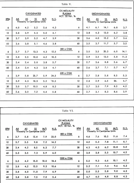
We measured the viscosity of the various hemoglo-binopathies at different osmolalities with a Brookfield viscometer (Table V). The viscosity was measured at 6, 12, 30, and 60 rpm on blood suspended in plasma, in 300 mosm sodium chloride solution, 600 mosm so-lution, 800 mosm solution, and 1,000 mosm solution concentrations (Table VI). Oxygenated cells suspended in plasma with an hematocrit of 33 vols percent
showed no significant difference between the viscosity
of hemoglobin AA and AS. However, there was a
sig-nificant increase in the viscosity of hemoglobin AA, AS, and HS blood at the 0.01 level when compared to Memphis/S and SS hemoglobin. Oxygenated blood at 300 mosm concentration showed a significant in-crease of hereditary spherocytosis erythrocytes and hemoglobin SS blood as compared to Memphis/S and hemoglobin AS and AA. At 600 mosm, hemoglobin SS and hereditary spherocytosis blood showed a s
ig-nificant increase at the 0.01 level in viscosity as com -pared to hemoglobin AA, AS, and Memphis/S. With 800 mosm solutions, hemoglobin SS and hereditary
spherocytosis (HS) showed a significant increase in
6
12
30
60
6
12
30
60
6
12
30
60
6
12
30
60
6
12
30
60
M. ROBERT COOPER
OXYGENATED
AA AS SS ~ H.S.
Viscosity in Centipoise Units
4.0 4.2 5.2 5.6 4.3
3.8 3.9 5.3 5.0 4. 1
3.7 3.9 5.2 4.7 3.9
3.6 3.8 5. 1 4.6 3.8
2.7 2.7 13.3 4.3 12.5
2.8 2.5 10.0 4.2 10.3
2.4 2.4 5.8 3.8 5.7
2.4 2.4 4.3 3.8 4.1
3.9 5.8 25.7 5.9 24.5
3.9 4.3 18.3 5.2 '15.5
3.0 3. 7 10.5 4.8 8.2
2.6 3.0 7.2 4.4 5.8
OXYGENATED
AA AS SS ~
Viscosity in Centipoise Units
3.7 5.8 12.4 7.9 23.0
3.7 5.5 9.8 7.2 14.2
3.4 4.2 8.5 6.3 7.7
3.3 3.5 7.3 5.9 6.0
3.5 4.2 15.0 10.0 18.4
3.8 4.2 12.5 9.3 10.6
3.8 4.0 11.0 7.9 6.9
3.8 3.8 7.5 7.0 5.4
Table V.
OS MOLALITY PLASMA HCT 33 Vol.%
300 m OSM
600 m OSM
Table VI.
OS MOLALITY PLASMA
HCT. 33 Vol. %
800 m OSM
1000 m OSM
RPM
6
12
30
60
6
12
30
60
6
12
30
60
RPM
6
12
30
60
6
12
30
60
DEOXYGENATED
AA AS ~
!:M1
H.S.Viscosity in Centipoise Units
4. 1 6. 1 14.1 6.8 3.7
3.8 4.3 13.0 6.2 3.8
3.6 4.0 12.2 5.7 3.6
3.5 3.7 11.7 5.7 3.5
3.0 3.5 20.3 6.0 14.1
2.9 3.5 13.0 5.8 11. 3
2.7 3.6 8.8 5.6 6.7
2.6 3.7 7. 1 5.7 4.7
2.7 2.8 5.8 8.5 5.3
2.8 2.9 6.5 84. 4.7
2.7 3.0 7.9 8.5 4.2
2.7 3. 1 8.3 8.6 3.9
DEOXYGENATED
AA AS SS M/S
Viscosity in Centipoise Units
4.6 7.6 10.5 11.6 7.4
4.5 5.3 7.8 11. 1 6.1
4.3 4.3 6.2 10.8 5.0
3.8 3.8 5.5 10.7 4.5
5.5 9.3 6.6 10.1 4.2
5.2 7. l 7.3 9.6 4.3
5.0 5.5 5.8 9.0 4.3
4.7 4.0 4.8 8.8 4.3
In the oxygenated blood at varying osmolalities from 300 to 1000 mosm solutions, hemoglobin SS and HS
blood behaved in a similar manner. Hemoglobin AA and AS did not show increasing viscosity with
increas-ing osmolar concentrations, as previously reported by
Hamm and his group. Hemoglobin Memphis/S did show an increasing viscosity with increasing osmolali-ties. Hemoglobin SS and red cells from patients with hereditary spherocytosis showed a similar pattern which was of a much greater magnitude than that of Memphis/S. The same type of studies were performed with deoxygenated blood. The hemoglobins listed in
order of ascending viscosity suspended in plasma were hereditary spherocytic red cells, hemoglobin AA,
he-moglobin AS, Memphis/S, and SS disease. At 300 mosm solution under deoxygenated conditions
hemo-globin AA and AS showed significantly less increased viscosity as compared to Memphis/S or as compared to HS or SS blood. At 300 mosm solutions,
hemoglo-bin HS and SS behaved similarly and showed a
sig-nificantly higher viscosity than the other
hemoglobi-nopathies. Hemoglobin Memphis/S showed increasing viscosity with increasing osmolar solutions and at 600, 800, and 1000 mosm concentrations was significantly
higher than any of the other hemoglobinopathies. Sherman tests were performed on the various
hemo-globin solutions at varying osmolar concentrations
and showed that there was no increase in the number
of sickled cells in hemoglobin AS or SS disease at in-creasing osmolar concentrations, similar to the results obtainel by Ham and his group at Case Western
Reserve. Hemoglobin Memphis/S showed an increased number of sickled cells in hyperosmolar solutions, but
this was a constant number throughout all of the os
-molar solutions. The consistent finding was that the red cells were spherical and crenated with numerous
spicules.
These data suggest that there may be an association between hereditary spherocytosis and sickle cell
dis-ease. A group of investigators from Japan (Matsuda
et al, 1969) reported a case of nephrotic syndrome in
a ten-year-old Japanese boy with hereditary sphero-cytosis. They pointed out that spherocytosis and sickle cell anemia have some features in common such as
ab-normal shape, short erythrocyte survival, persistent
icterus, and occurrence of crises. In addition, there
may be some association between hereditary sphero-cytosis and sickle cell anemia in reference to a marked
increase in viscosity with hyperosmolar solutions in
oxygenated blood. It has been pointed out by Jensen
( 1969) that the repeated sickling and unsickling of a cell may cause repetitive fragmentation with
propor-tionally greater membrane than volume Joss which eventually will result in the production of a spherocyte. This situation would be most likely to happen during a
sickle cell crisis in which the cells would be subjected
to maximal damage. This may also explain the varying
76
HEMATOCRITS ADJUSTED TO 33 VOL.96 DEOXYGENATION 30 MIN. 9596 N2-596 C02
~~.;gENATION' 15 MIN. 10096 02
-Dextron 40 0.3396
24 -Oextron 40 0.1096
28
~·D
e
xtro
n
40
196~ --Dextron 40 O. 6696
:§ 20
"'
.!Q"'
·9"&
"'
~
~
"
·
"'
~16
12
8
4
Oeoxygenoted - - - < > - - - .-·a A aA;fJSfJS
(without Oextron)
0 ':-:-:~---'----'---1
6RPM 12RPM 30RPM 60RPM DEXTRAN 40-HbSS
Fig IO-Viscosity measured with Brookfield Viscometer.
Hb. SS-Oxygenated versus deoxygenated RBC.
28
24
HEMATOCRITS ADJUSTED TO 33 VOL.% DEOXYGENATION' 30 MIN. 95% N2-5% co2
~~;~ENATION' 15 MIN.100% 02
~-+---e..-296 Dextron 40
0 =--=-~-L-,,-
__
i _ _ _ _ _ J _6 RPM 12RPM 30RPM 60RPM
DEXTRAN 40-HbSS
Fig I I-Viscosity of Hb. SS oxygenated versus
M. ROBERT COOPER
results reported in patients with hemoglobin SS when
subjected to hyperosmolar solutions.
Some investigators have suggested the use of low
molecular weight dextran in the therapy of patients
with sickle cell anemia. We have measured in an in
vitro situation the effect of various dextran 40 and
dextran 70 concentrations on the viscosity of hemo-globin SS disease (Fig 10, 11, 12). Our findings sug-gest that dextran actually increases the viscosity of
hemoglobin SS disease. Oski (Oski et al, 1965)
re-ported that low molecular weight dextran was of no benefit in the therapy of sickle cell crisis.
Eisenberg ( 1969) reported that low molecular
weight dextran had no specific effect on blood
vis-cosity. However, it did prevent erythrocyte rouleau formation and aggregation. It is also known that the
infusion of low molecular weight dextran is associated
with acute renal failure in at least fourteen patients.
Mailloux (Mailloux et al, 1967) produced acute
anuria in experimental animals with low molecular weight dextran. They postulated that a reduction in filtration pressure combined with a marked increase
in urinary viscosity generated by the dextran may lead
to tubular stasis and subsequent tubular blockade.
Laszlo (Laszlo, Obenour and Saltzman, 1969)
studied the effect of hyperbaric oxygenation on eryth-rocyte sickling during sickle cell crisis. His group at
Durham studied five patients during crises. They
found that the percentage of circulating sickle cells
did decrease during hyperbaric oxygenation. However,
there was no improvement of the sickle cell crisis and there was no tendency toward improved renal
concen-trating ability during the hyperbaric oxygenation. A
28
HEMATOCRITS ADJUSTED TO 33 VOU6 OEOXYGENATION ,30 MIN. 9596 Nz-596 COz
OXYGENATION' 15 MIN. 10096 Oz 35°C
~ ~D%
~ - - -- - - - --·3.0%
" 20
-~
~ 16 ~.3%
~ ~ ~==:=!~~~~~r:--1.2%
12... ~~~~~~;enoted without Dextran 70-75
~
-
~
8 /3.0%~
i~~~~~~i~
'
j
'
~.2.0% -1.2%4 ,~'0.6%
\b.3%
'Oxygenated without Dextron 70-75
o'----'---~--~
6RPM 12RPM 30RPM 60RPM
DEX TRAN 70-75 Hb SS
Fig 12-Viscosity of Hb. SS-oxygenated and
deoxygen-ated RBC using Dextran 70-75.
possible explanation for the ineffectiveness· of
hyper-baric oxygenation on sickle cell crisis in this study
may be that with hyperoxygenation in hyperosmolar solutions, the viscosity of sickle cell hemoglobin is ac-tually increased.
Further studies are needed to elucidate the
patho-physiology of the various hemoglobinopathies. Physi-cians involved in the care of pa:ients with sickle cell anemia as well as the other hemoglobinopathies should
try therapeutic programs which have been proven.
The use of clinical dextran and hyperbaric oxygena-tion should be carefully studied under experimental
conditions before their widespread use is advocated.
References
BENESCH R, BENESCH RE: The effect of organic
phos-phates from the human erythrocyte on the allosteric
properties of hemoglobin. Biochem Biophys Res Commun
26: 162, 1967
BERETTA A, PRATO V, GALLO E, ET AL: Haemoglobin
torino a 43(CD1) phenylalanine-7valine. Nature (London)
217: 1016, 1968
BRADLEY TB, WoHL RC, RIEDER RF: Hemoglobin gun
hill: deletion of five amino acid residues and impaired heme-globin binding. Science 157: 1581, 1967
CARRELL RW, LEHMANN H: The unstable haemoglobin
anaemias. Seminars Hemat 6: 116, 1969
CARRELL RW, LEHMANN H, LORKIN PA, ET AL: Haemo-globin sydney: f3 67 (Ell) Valine-7Alanine: An emerging pattern of unstable haemoglobins. Nature (London) 215: 626, 1967
CHARACHE S, CONLEY CL: Rate of sickling of red cells
during deoxygenation of blood from persons with various
sickling disorders. Blood 24: 25, 1964
CHARACHE S, GR1SULEA S, FIEDLER AJ, ET AL: Effect of 2, 3-Diphosphoglycerate on oxygen affinity of blood in sickle cell anemia. J Clin Invest 49: 806, 1970
CHARACHE S, WEATHERALL DJ, CLEGG JB: Polycythemia associated with a hemoglobinopathy. J Clin Invest 45: 813, 1966
CONLEY CL, CHARACHE S: Inherited hemoglobinopathies. Hosp Practice 4: 35, 1969
COOPER MR, KRAUS AP, RAMSEUR WL, ET AL: Charac-teristics of a new hemoglobinopathy. Hemoglobin mem-phis/sickle cell disease. Clin Res 17: 30, 1969
DACIE JV, GRIMES AJ, MEISLER A, ET AL: Hereditary
heinz-body anaemia. Brit J Haemat 10: 388, 1964
DACIE JV, SHINTON NK, GAFFNEY PJ, ET AL:
Haemo-g!obin hammersmith (f3 42 (CDl) PHE-7SER). Nature
(London) 216: 663, 1967
E!SENBURG S: The effect of low molecular weight dextran on the viscosity and suspension characteristics of blood. Amer J Med Sci 257: 336-343, 1969
HAM TH, DUNN RF, SAYRE RW, ET AL: Physical prop-erties of red cells as related to effects in vivo. I. Increased rigidity of erythrocytes as measured by viscosity of cells
altered by chemical fixation, sickling and hypertonicity.
Blood 32: 847, 1968
HATCH FE, CULBERTSON JW, DIGGS LW: Nature of the renal concentrating defect in sickle cell disease. J Clin Invest 46: 336, 1967
JENSEN WN: Sickle cell anaemia: A report of the first international symposium held at the university of the west indies, Jamaica, January 8-10, 1969. J Trop Pediat 15: 50, 1969
JONES RT, OSGOOD EE, BRIMHALL, ET AL: Hemoglobin yakima: I. Clinical and biochemical studies. J Clin Invest 46: 1840, 1967
KRAUS AP, MIYAJI T, Iucm I, ET AL: Hemoglobin memphis/S a new variant of sickle cell anemia. Trans
Ass Amer Physicians 80: 297, 1967
KRAUS LM, MIYAJI T, Iucm I, ET AL: Characterization of
a2saiuNHz in hemoglobin memphis. Hemoglobin memphis/S,
a new variant of molecular disease. Biochemistry (Wash)
5: 3701, 1966
LASZLO J, OBENOUR
w.
SALTZMAN HA: Effects ofhyperbaric oxygenation on sickle syndromes. Southern
Med I 62: 453, 1969
MAILLOUX L, SWARTZ CD, CAPIZZI R, ET AL: Acute renal failure after administration of
low-molecular-weight dextran. New Eng J Med 277: 1113, 1967
MATSUDA I, SHIDA N, TAKASE A, ET AL: Nephrotic
syn-drome and spherocytosis. Lancet II: 1204, 1969
MUIRHEAD H, PERUTz MF: Structure of haemoglobin. Nature (London) 199: 633, 1963.
MURAYAMA M: A molecular mechanism of sickled erythrocyte formation. Nature (London) 202: 258, 1964
MURAYAMA M: Tertiary structure of sickle cell
hemo-globin and its functional significance. J Cell Physiol 67:
21, 1966
NECHELES TF, ALLEN DM, FINKEL HE: Clinical Dis
-orders of Hemoglobin Structure and Synthesis. New York,
Appleton-Century-Crofts, 1969
OPFELL RW, LoRKIN PA, LEHMANN H: Hereditary
non-spherocytic haemolytic anaemia with post-splenectomy
inclusion bodies and pigmenturia caused by an unstable haemoglobin Santa ana {3 88 (F4) Leucine-;.Proline.
I Med Genet 5: 292, 1968
OSKI FA, VINER ED, PURUGGANAN H, ET AL: Low mo-lecular weight dextran in sickle-cell crisis. JAMA 191: 43, 1965
78
PERILLIE PE, EPSTEIN FH: Sickling phenomenon pro-duced by hypertonic solutions: A possible explanation for the hyposthenuria of sicklemia. J Clin Invest 42: 570, 1963
PERUTZ MF: Structure and function of haemoglobin. I.
A tentative atomic model of horse oxyhaemoglobin. J Molec Biol 13: 646, 1965
PERUTZ MF, KENDREW JC, WATSON HC: Structure and
function of haemoglobin. J Molec Biol 13: 669, 1965
PERUTZ MF, LEHMANN H: Molecular pathology of human haemoglobin. Nature (London) 219: 902, 1968
RAIK E, HUNTER EG, LINDSAY DA: Compensated heredi-tary haemolytic disease resulting from an unstable
haemo-globin fraction. Med J Aust 1: 955, 1967
REISSMANN KR, RUTH WE, WoHOURS T: A human hemoglobin with lowered oxygen affinity and impaired heme-heme interaction. J Clin Invest 40: 1826, 1961
SANSONE G, CARRELL RW, LEHMANN H: Haemoglobin
genova: ,fJ 28 (BIO) Leucine-;.Proline. Nature (London)
214: 877, 1967
SCHNEIDER RG, UEDA
s
,
ALPERIN JB, ET AL:Hemo-globin sabine beta 91 (F7) Leu-;.Pro An unstable variant
causing severe anemia with inclusion bodies. New Eng J
Med 280: 739, 1969
STAMATOYANNOPOULOS G, YOSHIDA A, ADAMSON J:
Hemo-globin rainier ([3145 Tyrosine-;. Histidine): Alkali-resistant
hemoglobin with increased oxygen affinity. Science 159:
741, 1968
WELLS RE, DENTON R, MERRILL EW: Measurement of
viscosity of biologic fluids by cone plate viscometer.