T
T
h
h
e
e
r
r
a
a
n
n
o
o
s
s
t
t
i
i
c
c
s
s
2018; 8(11): 3038-3058. doi: 10.7150/thno.23459
Review
Endogenous pH-responsive nanoparticles with
programmable size changes for targeted tumor therapy
and imaging applications
Wei Wu
1,†, , Li Luo
1,†, Yi Wang
1,†,
Qi Wu
1, Han-Bin Dai
1, Jian-Shu Li
2, Colm Durkan
3, Nan Wang
3,
Gui-Xue Wang
1, 1. Key Laboratory for Biorheological Science and Technology of Ministry of Education, State and Local Joint Engineering Laboratory for Vascular Implants, Bioengineering College of Chongqing University, Chongqing, 400030, China
2. College of Polymer Science and Engineering, Sichuan University, Chengdu 610065, China 3. The Nanoscience Centre, University of Cambridge, Cambridge, CB3 0FF, UK
† These authors contributed equally to this work.
Corresponding author: [email protected] (Wei Wu); [email protected] (Gui-Xue Wang)
© Ivyspring International Publisher. This is an open access article distributed under the terms of the Creative Commons Attribution (CC BY-NC) license (https://creativecommons.org/licenses/by-nc/4.0/). See http://ivyspring.com/terms for full terms and conditions.
Received: 2017.10.23; Accepted: 2018.03.06; Published: 2018.04.30
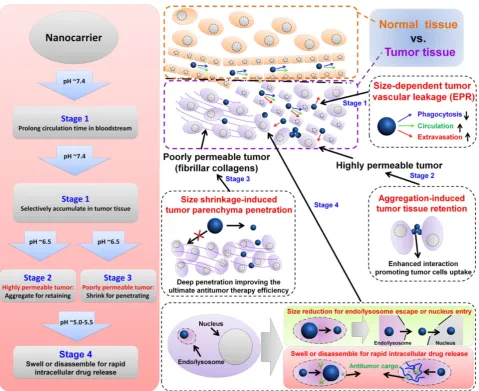
Abstract
Nanotechnology-based antitumor drug delivery systems, known as nanocarriers, have
demonstrated their efficacy in recent years. Typically, the size of the nanocarriers is around 100 nm.
It is imperative to achieve an optimum size of these nanocarriers which must be designed uniquely
for each type of delivery process. For pH-responsive nanocarriers with programmable size, changes
in pH (~6.5 for tumor tissue, ~5.5 for endosomes, and ~5.0 for lysosomes) may serve as an
endogenous stimulus improving the safety and therapeutic efficacy of antitumor drugs. This review
focuses on current advanced pH-responsive nanocarriers with programmable size changes for
anticancer drug delivery. In particular, pH-responsive mechanisms for nanocarrier retention at
tumor sites, size reduction for penetrating into tumor parenchyma, escaping from endo/lysosomes,
and swelling or disassembly for drug release will be highlighted. Additional trends and challenges of
employing these nanocarriers in future clinical applications are also addressed.
Key words: nanocarriers, endogenous pH-responsive, size change, targeted drug delivery, tumor therapy
1. Introduction
Over the last decade, tremendous progress has
been made in the development of nanocarriers as
effective antitumor drug delivery agents. These
carriers are highly attractive due to their nanometer
size and versatile surface properties, which enhance
their pharmacokinetics and bio-distribution while
reducing systemic toxicity [1-5]. To date, a broad
range of nanocarriers has been designed and tested,
including polymer nanoparticles, micelles, liposomes,
dendrimers, star polymers,
and inorganic
nanoparticles made of iron oxide, quantum dots,
silica, gold, and metal oxide frameworks [1, 6-18].
Intravenously injected nanomedicine undergoes a
multistep process before reaching solid tumors [19,
20]. Therefore, for efficient drug delivery in the
complex
in vivo
environment, it is important to
develop multiple stimuli-responsive nanocarriers that
can minimize nonspecific interactions with and
uptake by non-targeted cells or the immune system [6,
21-24].
Specifically designed intelligent
stimuli-sensitive nanocarriers can increase the
concentration of effective anticancer drugs by
targeting their delivery to tumor locations, thus
allowing for greater therapeutic efficacy and reducing
undesired side effects.
Although much work has been carried out on the
development of drug-delivery systems affected by
external stimuli, various limitations have greatly
Ivyspring
tissues and organs. The magnetic-associated
preparations (used as contrast agents in MRI) are
limited by the stringent requirements of carrier
materials. The ultrasound-associated formulations are
usually used for synergistic therapy. Considering the
limitations of external stimulant sources (
e.g.
light,
ultrasound,
magnetic
fields),
endogenous
stimulations, such as enzymes [25-29], glucose
concentrations [30, 31], redox reactions [32-38],
temperatures [39-41] and pH differences [34, 42-51],
are more useful when designing safe, efficient, and
intelligent carriers. Among these endogenous stimuli,
pH differences have been the most widely used
control parameter for tumor-targeted drug delivery
and controlled intracellular drug release. This is
possible due to the pH differences between healthy
and diseased tissues as well as between various
cellular compartments [6, 52-56]. Given the
vulnerability of acid-base homeostasis, acidic
microenvironment changes are associated with
pathological tissues, such as in ischemia,
inflammatory diseases, infections, rheumatoid
arthritis, or solid tumors [6, 52, 57-60]. Especially in
solid tumor tissues, the substantial energy
requirement for their intensive growth results in
increased lactate and hydrogen ions (H
+) produced by
catabolism of glucose. Consequently, the tumor
microenvironment becomes acidic with a pH ~6.5 in
tumor tissues versus pH ~7.4 in normal tissues [57,
61-63]. This difference can then be exploited for
targeting the tumor tissue or triggering drug release
in the tumor’s extracellular matrix [60, 64-68]. At the
cellular level, the intracellular acidic components (pH
~5.5 for endosome, pH ~5.0 for lysosome) can also be
used to trigger drug release and promote the escape of
the carrier into the cytoplasm [6, 69-72]. Therefore,
these endogenous pH differences provide a strong
option for targeted antitumor drug delivery and
controllable release kinetics.
In general, the physicochemical characteristics of
nanocarriers, including size, architecture, and surface
properties determine their fate by affecting one or
more of the above steps in the antitumor drug
delivery process [1, 47, 49, 52, 60, 64, 73-75].
Size-dependent enhanced antitumor findings have
received much attention because of the variable and
strict size requirements of the entire cascade
processes, including their biodistribution, immune
activation, blood circulation, tumor target
accumulation/retention, tumor tissue penetration,
tumor cell uptake, and final cargo release (
Figure 1
)
[52, 76-81]. For example, small nanocarriers (typically
nm) tend to be rapidly taken up by the mononuclear
phagocytic system (MPS) and subsequently
accumulate in the liver, spleen, and to a lesser extent
in the bone marrow [1, 83-88]. Compared to the tight
endothelial junctions of normal vessels (5-10 nm), the
pores of tumor vascular walls have typical junction
sizes ranging from 200 nm to 1200 nm allowing
extravasation of appropriately sized nanocarriers into
solid tumors [1, 19]. Also, the lack of functional
lymphatic drainage in tumor tissues prevented the
reentry of accumulated nanocarriers into blood
circulation. This phenomenon, termed the enhanced
permeability and retention (EPR) effect, is the rational
basis for passive targeting in antitumor drug delivery
[1, 7, 89]. However, proliferating tumor cells compress
the intratumor blood and lymphatic vessels, leading
to a uniformly eleva
ted interstitial fluid pressure.
Additionally, increased fibrillar collagens (such as
type I and type III) in some tumor interstitial matrix
can greatly hinder larger (>60 nm) nanocarriers from
penetrating deeply into the tumor parenchyma [19,
90, 91]. Therefore, pH-responsive nanocarriers with
programmable size changes can be automatically
altered during each stage of the anti-tumor drug
delivery.
Recently,
remarkable
advances in
size-dependent smart nanocarriers have attracted
great interest for their advanced and efficient
antitumor drug delivery as well as their reduced
systematic side effects. Although pH-responsive [6,
92-102] and size-related [1, 20, 83, 103, 104] antitumor
studies have been amply discussed in previous
review articles, there is a need to review several
aspects of endogenous pH-responsive nanocarriers
for antitumor therapy. Considering the unique
advantages of nanocarriers’ size characteristics for
antitumor drug delivery, this review focuses on
pH-responsive nanocarriers with programmable size
changes in antitumor applications, especially
highlighting recently advanced design strategies.
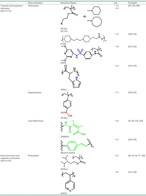
2. Endogenous pH-responsive carriers
with programmable size changes for
antitumor drug delivery
target [105]. To achieve safe and efficient antitumor
drug delivery, pH-responsive nanocarriers with
programmable size changes based on the
tumoral/intracellular phase-transition (protonation/
deprotonation and cleavable bond) (
Table 1
) offer a
feasible approach for overcoming many contradictory
requirements during various phases.
2.1. pH-responsive aggregation for tumor
retention
2.1.1. Design principle
The long-term stability in the circulatory system
necessitates nanocarriers with a different size than
that required for uptake by tumor cells [8, 76, 90,
177-179]. From previous anti-tumor studies, the
well-accepted size for long-term blood circulation is
around 100 nm due to the size-dependent balance
between organ filtration and tumor vessel
extravasation [8, 76, 83]. After intravenous injection,
carriers circulating for a longer time in the blood have
a better chance of accumulating in the tumor tissue
via
EPR effects.
Therefore, for prolonging the blood circulation
time, it is extremely important to avoid nonspecific
interactions with blood components and other
off-targeted cells. Also, nanocarriers undergo a series
of screening events by the reticuloendothelial system
(RES) and organs such as the liver, spleen, lung, and
kidney. Improvement of their “stealth” capabilities is
a key aspect for prolonging blood circulation time.
Furthermore, once accumulation begins in tumor
tissue, it is ideal for nanocarriers to “stick” to the
tumor cells.
structures (pH 6.5-7.2)
(PC6A) (PC7A)
~ 6.9
(PAE)
~ 7.0 [109-112]
(PSD)
~ 7.0 [113-115]
(PHis)
~ 6.5 [116-119]
Deprotonation
(PVBA)
~ 7.1 [120-123]
Acid-labile bond
(DMMA)
~ 6.8 [53, 60, 124, 125]
(Benzoic-imine)
~ 6.5 [126-129]
Endo/lysosome acid responsive structures (pH 4.5-6.5)
Protonation
(PDPA)
~ 6.3 [49, 52, 64, 77, 130]
(P4VP)
(P2VP)
~ 5.0 [135-137]
Deprotonation
(PMAA)
~ 6.3 [138-141]
(PEAA)
~ 5.6 [142, 143]
(PAsp)
~ 5.0 [144-147]
Acid-labile bond
(Citraconic amide)
~ 5.5 [55, 148-150]
(Hydrazone)
~ 6.0 [117, 151-153]
(Imine)
~ 6.5 [154-157]
(β-Thiopropionate)
~ 5.5 [158-161]
(Ketal)
~ 5.0 [162-165]
(Acetal)
~ 5.0 [166-169]
(Cyclic acetal)
~ 5.0 [170-172]
(Ortho ester)
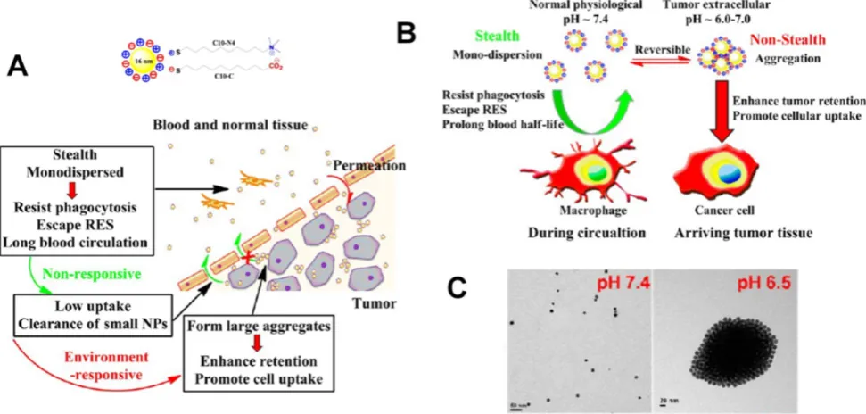
Figure 2.
(A)
Schematic illustration of pH-responsive aggregation of gold nanoparticlesenhancing targeted retention and cellular uptake in response to the tumor
extracellular acidic stimuli.
(B)
Illustration of the size-dependent prolonging of blood circulation time and targeted tumor retention.
(C)
TEM images of
pH-responsive aggregation. Reproduced with permission from [89], copyright 2013 American Chemical Society.
As a result of the time-
and
concentration-dependent internalization process
caused by enhanced interactions between
nanocarriers and cells, carriers can have increased
chances of tumor cells uptake and retention in tumor
tissue [1, 89, 180]. Cytotoxicity of aggregated
nanocarriers compared to non-aggregated ones has
also been reported [179]. All in all, nanocarriers must
be “stealthy” in blood circulation to evade
undesirable RES recognition or protein/cell adhesion
but must be “sticky” to interact with tumor cells. To
this end, nanoparticles with sizes of 20-200 nm are
appropriate to avoid undesirable clearance through
the nonspecific adsorption and urinary excretion.
However, the design strategy employed may need to
take into account the opposing requirements
depending on the intercellular space. For the highly
permeable tumor parenchyma with large intercellular
space, the aggregated nanoparticles can be retained in
the tumor tissue to enhance tumor cell endocytosis.
This is one of the areas where the tunable properties
of nanocarriers are advantageous as their surfaces can
be functionalized appropriately to fulfill size
requirement.
2.1.2. pH-responsive aggregation based on
protonation/deprotonation
pH-responsive aggregation triggered by slightly
acidic stimuli of the extracellular tumor matrix is a
suitable mechanism for designing nanocarriers with
low uptake by normal cells but high uptake by tumor
cells. Based on the protonation/deprotonation
transition triggered by mild acidic stimuli, Ji and
coworkers prepared a series of mixed-charge
zwitterionic monolayer-modified gold nanoparticles
(
Figure 2
) [89, 181]. Owing to zwitterionic monolayer
protonation/deprotonation balance, the optimized
nanoparticles were stable at the physiological pH of
the bloodstream and normal tissues but rapidly
aggregated within a few seconds in response to a
slight pH change from 7.4 to 6.5. This work
demonstrated that controllable aggregation at tumor
sites could enhance retention efficiency and cellular
uptake of nanoparticles in tumors for photo-thermal
tumor-targeted diagnosis and therapy. Inspired by
this study on the surface charge conversion resulting
from ultra acid-labile amide bond breakage, we
fabricated a pH-responsive polymeric nanocarrier
with tumor-targeted aggregation properties for
enhanced retention and endocytosis (
Figure 3
) [52].
The doxorubicin-loaded smart nanocarrier was
constructed from succinic anhydride-
modified poly
mechanism of pH-sensitive surface charge transition,
Chiu and coworkers constructed a nanocarrier using
N
-acetyl histidine-modified D-
α
-tocopheryl
poly-ethylene glycol succinate on the surface of poly
(lactic-
co
-glycolic acid) [182]. The increased
protonation of imidazole groups from histidine
residues resulted in the lack of sufficient interparticle
electrostatic repulsion for maintaining stable
individual particles at pH 6.5, and the DOX-loaded
nanoparticles with an almost neutral surface charge
aggregated into large clusters. Both
in vitro
and
in vivo
results confirmed that tumor acidity-triggered surface
charge neutralization and aggregation could
significantly enhance tumor cell uptake. Ji’s group
also reported a pH-responsive supra-molecular
prodrug micelle based on the host-guest inclusion
[183]. The size of this supra-assembly could be tuned
by pH changes showing significant aggregation
triggered by slight acidity of the tumor extracellular
matrix. Benefitting from aggregation-induced
accumulation in tumor tissues, this supra-assembly
exhibited desirable antitumor effect by efficiently
inhibiting tumor proliferation. In these design
strategies, the pH-responsive size aggregation based
on the protonation/deprotonation is reversible, so the
nanoparticles can disperse as single nanoparticle once
the pH condition recovers, which is convenient for its
preparation, storage, and further functionalization.
2.1.3. pH-responsive aggregation based on cleavable
bond at acidic pH
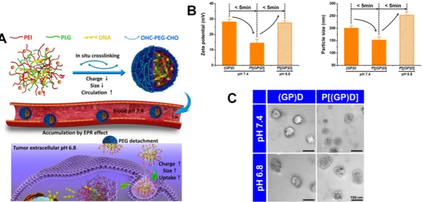
Recently, a nanocarrier with programmable size
changes was designed to satisfy the different phases
of the drug delivery process by Chen’s groups [184].
An ideal smart polymeric gene complex was prepared
from pH-responsive cleavable polyethylene glycol
(PEG) bonded in situ on the surface of a primary
complex of polyethyleneimine (PEI), poly-
L
-
glutamate and therapeutic genes
via
the Schiff base
reaction (
Figure 4
). At the physiological pH 7. 4, the
PEG cross-linked complex could shield the surface
positive charges and enhance its stability to reduce
side effects, prevent premature gene release, and
prolong blood circulation time. However, once the
complexes accumulated in tumor tissues by EPR, the
ultra pH-sensitive Schiff base bonds were responsive
to the slightly acidic environment of tumor tissue. The
PEG surface layer rapidly detached from the complex.
The complex without PEG corona exhibited a higher
positive charge potential and larger size facilitating
tumor cellular uptake and enhancing antitumor gene
delivery. In this design, the cross-linking PEG corona
could shield the surface positive charge and compress
the nanoparticles tightly, exhibiting prolonged blood
circulation, enhanced stability, and reduced
cytotoxicity. The PEG layer could be rapidly detached
by the acid labile cleavable bond in response to the
slight acidity of the tumor tissue exposing the surface
positive charge and inflated size to promote tumor
cell endocytosis. However, compared to the
protonation/deprotonation transition, pH-responsive
size change induced by the acid labile cleavable bond
is generally irreversible. Therefore, the acid labile
nanoparticles have a potential risk of instability in
their preparation, storage, and application.
2.2. pH-responsive size shrinkage for deep
tumor penetration
2.2.1. Design principle
The size of nanocarriers is a significant factor in
promoting their accumulation and penetration in
tumor tissues [1, 76, 185]. To sufficiently illustrate the
important role of size in drug delivery, Tang et al.
have fabricated micelles of the same chemical
structures and physical properties from a single
copolymer but ranging in size from 20 nm to 300 nm.
Their results indicated that the optimal size of
nanocarriers through the cascade processes was
different at different stages,
i.e.
the optimal size range
for suitable blood circulation time and tumor
accumulation was between 100 and 160 nm. However,
greater accumulation of the large micelles (100 nm) at
tumor sites did not result in significantly improved
Figure 3. (A)
Scheme and
(B)
Digital image of the tumor-targeted aggregation of pH-responsive polymeric nanomicelles for enhanced retention at tumor tissue and
rapid intracellular drug release in tumor cells. Reproduced with permission from [52], copyright 2014 Royal Society of Chemistry.
Figure 4. (A)
Schematic of the ultrasensitive pH-triggered charge/size dual-rebound gene delivery system for efficient antitumor applications.
(B)
Fast hydrodynamic
charge/size dual-rebound property and
(C)
TEM of the gene delivery system. (GP)D and P[(GP)D] represent (PLG/PEI)/DNA and PEG[(PLG/PEI)/DNA], respectively.
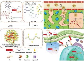
Figure 5.
Schematic illustration of a multistage pH-responsive stepwise size reduction and charge-reversal polymeric nanocarrier. (
A
) negatively charged nanocarrier
at neutral pH to enhance blood retention, (
B
) passive accumulation of larger size nanocarriers in the tumor via EPR effect, (
C
) the first
-stage size reduction into
smaller linear copolymers for tumor tissue deep penetration, (
D
) increased nanocarrier uptake by tumor cells mediated by concomitant R8NLS, (
E
) the second-stage
size reduction into therapeutic drug for intracellular release in the endo/lysosomal compartment, (
F
) endo/lysosome escape and nuclear targeted delivery.
Reproduced with permission from [189], copyright 2015 Wiley-VCH.
2.2.2. pH-responsive size shrinkage based on cleavable
bond at tumor extracellular acidity for tumor
parenchyma penetration
The pH-responsive size shrinkage due to the
acidic environment of the extracellular tumor matrix
provides a feasible strategy to facilitate deep
penetration of nanocarriers into the tumor’s dense
collagen parenchyma. Thayumanavan and coworkers
constructed a series of ultra pH-sensitive
size-changeable nanogel clusters [42]. Large
nanoclusters were obtained by using a pH-sensitive
dynamic covalent imine bond to cross-link individual
nanogels. Under physiological pH conditions,
nanoclusters showed a relatively large size and less
positive surface charge to prolong the blood
circulation time. Because of the breakage of
cross-linked dynamic imine bonds triggered by the
tumor-acidity at pH 6.5, nanoclusters transformed
into the original nanogel, exhibiting small size and a
more positive surface charge to promote deeper
penetration into the tumor matrix. Wang’s group
reported pH-responsive clustered nanoparticles,
comprising the platinum-poly(amidoamine)
den-drimer prodrug-conjugated at the terminal of the
amphiphilic poly(ethylene glycol)-
b
-poly(
Ɛ
-caprol-actone) block polymer using the acid-labile bond [187].
These smart clustered nanoparticles showed an initial
diameter of ~100 nm, and discharged the small size
(~5 nm) prodrug by cleaving its conjugation in the
acidic environment within the tumor for deep
penetration and enhanced endocytosis, followed by
further intracellular reduction to kill tumor cells. Ge’s
group reported a supramolecular polymeric nanogel
based on the host-guest interaction between
adamantanine and
β
-cyclodextrin moieties [188]. Due
to the cleavage of the benzoic imine linkage in
tumor-acidic conditions, this nanogel could
reorganize into smaller nanoparticles from ~220 nm to
~25 nm. For targeted drug delivery into the tumor cell
nucleus, Huang and colleagues synthesized a
multistage nanovehicle with pH-responsive stepwise
size reduction and charge reversal using an ultra
pH-sensitive charge-reversal 2,3-dimethylmaleic
amine modification and hydrazone linkage (
Figure 5
)
[189]. The neutrally charged nanovehicle with
relatively large size (~55 nm) showed good blood
persistence and excellent accumulation in tumor sites
at pH 7.4. Responding to the acidic stimulus of tumor
tissue, the nucleus-homing cell-penetrating
peptide-modified drug conjugate was separated from
the disassembled large nanovehicle, and had a size of
~10 nm in diameter, which allowed deeper tumor
matrix penetration and better cellular internalization.
Subsequently, the antitumor drug was cleaved from
the conjugate in the acidic microenvironment of the
endosome, and efficiently entered the nucleus
via
HeLa tumor growth in nude mice by 75%.
2.2.3. pH-responsive size shrinkage based on
protonation at tumor extracellular acidity for tumor
parenchyma penetration
Another study by Ge and co-workers used ultra
pH-sensitive charge reversal micelles with a large size
(114 nm at pH 7.4) to introduce pH-responsive
hydrophobicity to hydrophilicity transition of tertiary
amine. Under slightly acidic condition (pH 6.8), this
specifically accelerated the release of an encapsulated
smaller poly(amidoamine) dendrimer-conjugated
prodrug (several nanometers) for deep tumor
penetration, and subsequently released the active
drug by cleaving the conjugate in the intracellular
reduction environment [190]. Recently, Wang and
colleagues have developed a class of pH-responsive
to the rapid pH-responsive protonation of the
ionizable tertiary amine group, this smart
superstructure had an initial size of ~80 nm at the
physiological pH of 7.4 during its circulation in the
blood, but undergoes a dramatic and sharp size
reduction to less than 10 nm in the slightly acidic
microenvironment of the tumor. The ultra
pH-sensitive size-changeable superstructures did not
only facilitate enhanced targeted tumor accumulation
by EPR by utilizing the long-circulation benefit of
large nanoparticles but also promoted deeper tumor
penetration by taking advantage of the increased
penetration capability of smaller nanoparticles, which
ultimately improved efficiency of antitumor therapy
in the poorly permeable BxPC-3 pancreatic tumor
model.
Figure 7.
pH-responsive size-shifting cross-linked micelle nanoclusters for enhanced tumor targeting and deep penetration. Reproduced with permission from [91],
copyright 2016 American Chemical Society.
2.2.4. pH-responsive size shrinkage based on cleavable
bond at tumor intracellular acidity for endo/lysosome
escape
pH-responsive size shrinkage triggered by acidic
stimuli of endo/lysosomes is helpful for facilitating
the escape of nanocarriers from these subcellular
compartments. Sha and colleagues prepared a
size-shifting micelle nanocluster based on a
cross-linked framework interspersed with small
micelles using the emulsion-evaporation method
(
Figure 7
) [91]. The cross-linked nanocluster was
stable with a size of around 104 nm and prolonged
half-life for increased tumor accumulation by EPR.
Due to the proton sponge effects of polyetherimide in
an acidic environment, the cross-linked framework of
nanoclusters was able to swell and disintegrate, thus
accelerating the release of individual micelles (14 nm)
and penetration into the cytoplasm. To ensure
delivery of antitumor drug into the nucleus, Chen and
coworkers designed a large compound nanoparticle
with pH-activated size reduction for
in vivo
nucleus-targeted gene delivery (
Figure 8
) [191]. This
large compound nanoparticle with detachable PEG
shell and folate decoration had a size of ~150 nm
under neutral conditions. Once internalized by tumor
cells, breakage of the benzoic imine bond of PEG
conjugate in response to lower pH led to dissociation
of the PEG shell resulting in the formation of smaller
entities (~40 nm) exposing a previously shielded
nuclear localization signal oligo-
L
-lysine. The
resultant smaller nanoparticles could enter into the
nucleus
via
the nucleopores, causing a 20-fold higher
cytotoxicity
in vitro
and greater tumor suppression in
animal models compared with the native drug. For
endo/lysosome escape and nuclear entry, the size
shrinkage in intracellular acidic conditions could
greatly improve the therapeutic efficacy due to the
reduced drug loss.
Figure 9.
Illustration of DOX-loaded micelles for rapid intracellular drug release in response to the acidic stimulus of endo/lysosome in anti-cancer therapy.
Reproduced with permission from [64], copyright 2014 Royal Society of Chemistry.
2.3. pH-responsive swell or disassembly for
burst drug release
2.3.1. Design principle
pH-responsive size change nanocarriers can also
be constructed for encapsulated drug release
intracellularly. After being endocytosed by tumor
cells, the instantaneous pH-responsive swell
accelerates release of encapsulated drugs from the
loose nanocarriers
and
the pH-responsive
disassembly results in the burst drug release [30, 49,
64, 77, 192]. The controllable pH-responsive drug
release determines the therapeutic efficacy and side
effects of the drug [193-195]. The ideal nanocarrier
must tightly retain the encapsulated drug without
premature release during transport but must show
burst release once inside tumor cells to overcome
multidrug resistance and subsequently efficiently
induce tumor cells apoptosis [70, 105, 196-200].
However, nanocarriers do not conform to the ideal
drug release profile; there is burst release of up to 30%
during the first several hours and sustained slow drug
release over the following several days [196]. The lost
drug not only leads to reduced therapeutic efficiency
but also increases toxicity to healthy cells and organs.
Therefore, improvements in release kinetics are still a
tremendous challenge. To match drug release as close
to the ideal profile as possible, the tight encapsulation
of the drug in the nanoparticles is required and their
rapid disassembly to accelerate the release of the
payload drug in response to the intracellular acidic
environment. Many mechanisms including
appropriate cross-linking, reversible conjugation, or
specific physicochemical property transformations
based on the pH are adapted for a viable strategy to
modulate release kinetics and reaching an appropriate
therapeutic window. The payload drug would be
ineffective below a certain concentration [119,
201-204].
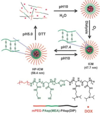
2.3.2. pH-responsive swell or disassembly based on
protonation
interlayer-crosslinked nano-micelles for reduction-
and pH-responsive burst drug release (
Figure 10
)
[196]. Benefitting from the hydrophobicity of the
2-(diisopropylamino) ethyl amine groups in
copolymer at pH 7.4 and disulfide functional
cross-linkage in a normal reduction environment,
these micelles were capable of tightly encapsulating
DOX to avoid drug leakage during storage and blood
circulation. The pH-responsive hydrophilicity
conversion and reduction-responsive cross-linker
breakage in the lysosomes then led to the disassembly
of nano-micelles into separate copolymers;
Subsequently, there was a burst drug release (more
than 95% of loaded DOX released in a
lysosome-simulated aqueous solution of 10 mM
dithiothreitol at pH 5.0) to induce cell apoptosis.
Figure 10.
Highly packed interlayer-crosslinked nanomicelles for reduction-
and pH-responsive collectively triggered burst drug release. Reproduced with
permission from [196], copyright 2011 Wiley-VCH.
2.3.3. pH-responsive swell or disassembly based on
acid-labile cleavable bond
Another pH-responsive intracellular release
strategy is to use acid-labile bond cleavage.
pH-responsive disassembled nanocarriers based on
charge-reversal or degradation mechanisms are
feasible for obtaining a burst release profile. Zhong’s
group improved a number of pH-responsive
degradable polymeric nanocarriers using acid-labile
polycarbonate modified for intracellular drug
delivery (
Figure 11
) [3, 206-212]. The polycarbonate
modification provided the amphiphilic feature for a
remarkably high drug loading content. Notably, these
nanocarriers are stable at pH 7.4 but swell and
eventually disassemble for fast hydrolysis of the
polycarbonate in the acidic conditions of
endo/lysosomes.
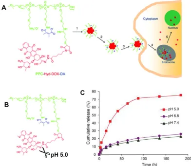
Given the high drug-carrying capacity and
tunable drug-release kinetics, the pH-responsive
prodrug conjugates provide a platform for further
increasing drug delivery efficiency [69, 70, 213, 214].
Zhong and coworkers developed a pH-sensitive
nano-micelle based on the acid-labile acetal bridging
polymer and drug conjugates for accelerated
intracellular antitumor drug release [215]. In release
studies performed
in vitro
, the acetal-link breakage
resulted in paclitaxel (PTX) monomer recovery and
swelling of prodrug nanoparticles, showing that PTX
release was highly pH-dependent,
i.e.
86.9%, 66.4%
and 29.0% in 48 h at pH 5.0, 6.0, and pH 7.4,
respectively. Wang and colleagues developed a dual
tailor-made pH-responsive polymer-drug conjugate
to inhibit drug-resistant cancer stem cells (
Figure 12
)
[53]. This advanced polymeric prodrug could respond
to both extracellular and intracellular acidic stimuli
through chemically defined mechanisms. It was
expected to have a prolonged blood circulation,
accumulation in the tumor
via
EPR, and endocytosis
through pH-responsive surface charge conversion
from negative to positive in response to the mild acid
conditions in tumor tissue. Interestingly, nanosized
prodrug rapidly disassembled into the drug monomer
due to the acid-labile hydrazone bond cleavage in
endo/lysosomes. This multifunctional nanosized
prodrug greatly enhanced antitumor efficacy
providing an advanced guide for antitumor drug
delivery research and clinical applications.
Gu et al. improved a potential theranostic
reagent using a transformable liquid-metal
nanomedicine with a polymeric shell for
encapsulating DOX and a liquid inorganic core for
fusion and subsequent degradation in a mildly acidic
environment (
Figure 13
) [216]. Within 4 h, the
cumulative amount of drug released in the acidic
buffer of pH 5.0 was significantly higher than that in
the neutral condition of pH 7.4 (> 50% for DOX and
44% for paclitaxel). The increased drug release at pH
5.0 resulted from the liquid-metal core fusion
mediating the disruption of the polymeric shell and
subsequent dissociation of the drug-loaded surface
ligand. Besides, this liquid-metal nanoformulation
exhibited a contrast-enhancing capability for imaging
applications, and eventually degraded under mildly
acidic condition showing excellent biocompatibility.
3. Advanced endogenous pH-responsive
carriers with programmable size changes
for tumor imaging
Besides being useful for antitumor therapy, the
pH-responsive size-changeable design strategy has
also been exploited for tumor diagnosis [217-219]. For
example, to overcome the low optical absorbance in
biological tissues’ near-infrared optical window,
Chang et al. developed novel melanin-like
nanoparticles by modifying the hydrolysis-
susceptible surface citraconic amide for photoacoustic
imaging (
Figure 14
) [55]. These improved
pH 7.4) in the near-infrared window of biological
tissues without absorption tuning. The unique
characteristic of photoacoustic amplification in
melanin-like nanoparticle clusters might allow their
use as a contrast agent for highly sensitive
in vivo
tumor target imaging. This innovative targeted tumor
imaging based on the pH-responsiveness unveiled
great opportunities for many basic research studies
and clinical explorations. Recently, Gao and
colleagues have fabricated a large number of the
ultra-pH-responsive nanosensors for amplifying
signals based on the positive cooperativity of the
noncovalent selfassembly of the fluorescence-labeled
functional block copolymers [66, 67, 71, 106-108, 114,
220-226]. Typically, a hybrid nanotransistor probe,
composed of a molecular mixture of three different
ultra-pH-responsive block copolymers, served as a
binary pH threshold sensor exhibiting dramatic signal
amplification (>30-fold) and sharp (<0.25 pH) acidic
response capabilities [220]. The hybrid nanotransistor
stayed “OFF” at pH 7.4, resulting from the dramatic
fluorescence quenching through the
hetero-/homo-molecular fluorescence energy transfer
in micelle state. After endocytosis, the
ultra-pH-responsive block copolymer components
disassembled and fluoresced sequentially as the pH
value decreased. After each pH transition encoding
with a unique fluorescent reporter, the acidification
kinetics at single-organelle resolution could be
quantified using digitization paradigm by a series of
barcodes with binary (0 and 1) output in each channel
reporting the luminal pH of the individual endocytic
organelle (
Figure 15
). This novel nanoprobe provided
a feasible platform for imaging and new biological
investigations in tumor pathology. The preliminary
exploration of the pH-responsive size changeable
nanoprobes showed exciting perspectives in tumor
imaging diagnosis. For the same design mechanism,
the pH-responsive nanoparticles with programmable
size changes provided a feasible platform to integrate
therapy and imaging for tumor theranostic
improvement.
Figure 13.
Illustration of the transformable liquid-metal nanomedicine for the antitumor application.
(A)
Preparation and
(B)
Main components of liquid-metal
nanomedicine.
(C)
pH-responsive delivery of the DOX-loaded liquid-metal nanomedicine for targeted antitumor therapy.
(D)
Acid-triggered fusion and degradation
process of liquid-metal nanomedicine.
(E)
Chemical structures of the main components. Reproduced with permission from [216], copyright 2015 Nature Publishing
Figure 14.
(A)
Schematic illustration of pH-responsive aggregation-
induced amplification of the photoacoustic signal from
Melanin-like nanoparticles and
(B)
Surface
modification of the bare Melanin
-like nanoparticles and their aggregation under mildly acidic condition. Reproduced with permission from [55], copyright 2016 Royal
Society of Chemistry.
Figure 15.
(A)
Schematic of the ultra-pH-responsive hybrid nanotransistor to digitize organelle pH after receptor-mediated endocytosis in tumor cells.
(B)
The
count rates and normalized fluorescence intensity of the hybrid nanotransistor are plotted at different pH values.
(C)
Representative fluorescence images of
multispectral ultra-pH-responsive hybrid nanotransistor at different pH values. Yellow is the merged color of green and red signals. White is the merged color of blue,
green, and red signals. Reproduced with permission from [220], copyright 2017 Wiley-VCH.
4. Conclusions and perspectives
pH-responsive nanocarriers with programmable
size changes have proven to be a powerful and
flexible platform for designing more effective
anticancer drug delivery systems with higher
therapeutic efficacy and fewer clinical side effects. In
this review, we focused on various advanced
pH-responsive size-changeable design strategies: (a)
Nanocarriers with appropriate size are necessary for
avoiding undesirable clearance during circulation by
nonspecific MPS and for enhancing accumulation and
retention at tumor sites
via
the EPR effect. (b) For
highly permeable tumors (such as C26 colon tumor),
the slightly acidic environment in tumor tissues can
promote uptake of nanocarriers with pH-responsive
aggregation through concentration-
and
required therapeutic dose for inducing tumor cell
apoptosis. (e) pH-responsive nanoprobes with
programmable size changes are expected to be useful
for tumor imaging diagnosis.
Although considerable progress has been made
with pH-responsive nanocarriers with programmable
size changes, some challenges still exist. For example,
rapid mutations of malignant tumors may result in
unpredictable and unstable pathological environment
including extracellular pH gradients, varying tumor
vascular leakage by changing pore diameter, and
tumor parenchyma collagen density. The
synchronous development of real-time technology in
tumor imaging and diagnosis is needed to detect the
precise pathological changes for designing an
accurate and targeted clinical approach. Furthermore,
the strategy in the selection of sizes for nanocarrier
aggregation or deep tumor penetration should be
designed based on the permeability of various tumor
types and location. For example, different size
nanocarriers (with diameters of 30, 50, 70 and 100 nm)
exhibit efficient penetration into the highly permeable
tumor, but only small-sized nanocarriers with a
diameter of 30 nm can penetrate the poorly permeable
pancreatic tumors [227]. For tumors with low blood
supply and mesenchyme-enriched tumors, the size
reduction strategy for deep tumor penetration is a
better choice for enhancing antitumor therapeutic
efficacy. On the other hand, the excessive pursuit of
stimuli-responsiveness may come at the cost of loss of
stability which may cause serious physiological
toxicity. Therefore, it is necessary to achieve a rational
balance between ultra sensitivity and stability.
Although pH-responsive size variation is a major
aspect of designing nanocarriers, it is not the only
criterion that is worthy of consideration. Sophisticated
novel antitumor nanocarriers must navigate a hostile
and complex environment
in vivo
and, therefore, a
variety of mechanisms are needed to integrate
multiple stimuli (temperature, redox, enzyme,
glucose, ion, etc.) as well as different factors (surface
charged potential, hydrophilic-hydrophobic balance,
shape etc.). Also, improving biocompatibility and
degradability as well as functionality is important for
the
successful
implementation of clinical
nanomedicine. In summary, pH-responsive
nanocarriers with programmable size changes
provide an excellent platform for precise and
personalized treatment for current antitumor therapy.
However, a great number of challenges remain for the
future clinical nanomedicine.
Abbreviations
EPR
:
enhanced permeability and retention; DLS:
dynamic light scattering; DMMA: 2,3-dimethylmaleic
anhydride; DOX: doxorubicin; H
+: hydrogen ion;
MPS: mononuclear phagocytic system; P2VP:
poly(2-vinylpyridine); P4VP: poly(4-vinylpyridine);
PAE: poly(
β
-amino ester); PAMA: poly(2-aminoethyl
methacrylate hydrochloride); PAsp: poly(aspartic
acid); PC6A: poly[2-(pentamethyleneimino) ethyl
methacrylate]; PC7A: poly[2-(hexamethylene imino)
ethyl methacrylate]; PDPA:
poly[2-(diisopropyl-amino) ethyl methacrylate]; PEAA: poly(ethylacrylic
acid); PEG: polyethylene glycol; PEI:
polyethyl-enimine; PHis: poly(
L
-histidine); PMAA:
poly(meth-acrylic acid); PLG: poly-
L
-glutamate; PSD:
poly(meth-acryloyl sulfadimethoxine); PTX: paclitaxel; PVBA:
poly(4-vinylbenzoic acid); RES: reticuloendothelial
system; SA: succinic anhydride; TEM: transmission
electron microscope.
Acknowledgments
Financial support from the National Natural
Science Foundation of China (51603023, 11332003), the
National Key R&D Program (2016YFC1102305), the
Fundamental Research Funds for the Central
Universities (106112016CDJXY230002, 106112017CDJ
ZRPY0012), the Chongqing Research Program of Basic
research and Frontier Technology
(cstc2017jcyjAX0186), the China Postdoctoral Science
Foundation (2016M602656, 2017T100682) and the
Chongqing Postdoctoral Scientific Research
Foundation (Xm2016011) as well as the Chongqing
Engineering Laboratory in Vascular Implants and the
Public Experiment Center of State Bioindustrial Base
(Chongqing) are gratefully acknowledged. We also
greatly thank Dr. Xi Lu, Prof. Colm Durkan, Mr. Ali
Maruf, Miss Deti Nurhidayah for editing language
friendly.
Competing Interests
The authors have declared that no competing
interest exists.
References
1. Duan XP, Li YP. Physicochemical Characteristics of Nanoparticles Affect Circulation, Biodistribution, Cellular Internalization, and Trafficking. Small. 2013; 9: 1521-32. 2. King MR, Mohamed ZJ. Dual nanoparticle drug delivery: the future of anticancer
therapies? Nanomedicine. 2017; 12: 95-8.
3. Du YF, Chen W, Zheng M, Meng FH, Zhong ZY. pH-sensitive degradable chimaeric polymersomes for the intracellular release of doxorubicin hydrochloride. Biomaterials. 2012; 33: 7291-9.
4. Luo M, Wang H, Wang Z, Cai H, Lu Z, Li Y, et al. A STING-activating nanovaccine for cancer immunotherapy. Nat Nanotechnol. 2017; 12: 648-54.
5. Xing H, Hwang K, Lu Y. Recent Developments of Liposomes as Nanocarriers for Theranostic Applications. Theranostics. 2016; 6: 1336-52.
6. Mura S, Nicolas J, Couvreur P. Stimuli-responsive nanocarriers for drug delivery. Nat Mater. 2013; 12: 991-1003.
7. Liu X, Li H, Jin Q, Ji J. Surface tailoring of nanoparticles via mixed-charge monolayers and their biomedical applications. Small. 2014; 10: 4230-42.
8. Liang S, Yang XZ, Du XJ, Wang HX, Li HJ, Liu WW, et al. Optimizing the Size of Micellar Nanoparticles for Efficient siRNA Delivery. Adv Funct Mater. 2015; 25: 4778-87.
9. Wu W, Ye C, Xiao H, Sun X, Qu W, Li X, et al. Hierarchical mesoporous silica nanoparticles for tailorable drug release. Int J Pharm. 2016; 511: 65-72.
13. Sosnik A, Menaker Raskin M. Polymeric micelles in mucosal drug delivery: Challenges towards clinical translation. Biotechnol Adv. 2015; 33: 1380-92. 14. Liu Y, Zhi X, Yang M, Zhang J, Lin L, Zhao X, et al. Tumor-triggered drug release
from calcium carbonate-encapsulated gold nanostars for near-infrared photodynamic/photothermal combination antitumor therapy. Theranostics. 2017; 7: 1650-62.
15. Witting M, Obst K, Friess W, Hedtrich S. Recent advances in topical delivery of proteins and peptides mediated by soft matter nanocarriers. Biotechnol Adv. 2015; 33: 1355-69.
16. Hajba L, Guttman A. The use of magnetic nanoparticles in cancer theranostics: Toward handheld diagnostic devices. Biotechnol Adv. 2016; 34: 354-61.
17. Zhai Y, Su J, Ran W, Zhang P, Yin Q, Zhang Z, et al. Preparation and Application of Cell Membrane-Camouflaged Nanoparticles for Cancer Therapy. Theranostics. 2017; 7: 2575-92.
18. Li J, Yang J, Li J, Chen L, Liang K, Wu W, et al. Bioinspired intrafibrillar mineralization of human dentine by PAMAM dendrimer. Biomaterials. 2013; 34: 6738-47.
19. Ruan S, Cao X, Cun X, Hu G, Zhou Y, Zhang Y, et al. Matrix metalloproteinase-sensitive size-shrinkable nanoparticles for deep tumor penetration and pH triggered doxorubicin release. Biomaterials. 2015; 60: 100-10.
20. Stylianopoulos T, Wong C, Bawendi MG, Jain RK, Fukumura D. Multistage Nanoparticles for Improved Delivery into Tumor Tissue. Method Enzymol. 2012; 508: 109-30.
21. Karimi M, Ghasemi A, Zangabad PS, Rahighi R, Basri SMM, Mirshekari H, et al. Smart micro/nanoparticles in stimulus-responsive drug/gene delivery systems. Chem Soc Rev. 2016; 45: 1457-501.
22. Andresen TL, Jensen SS, Jorgensen K. Advanced strategies in liposomal cancer therapy: Problems and prospects of active and tumor specific drug release. Prog Lipid Res. 2005; 44: 68-97.
23. Fleige E, Quadir MA, Haag R. Stimuli-responsive polymeric nanocarriers for the controlled transport of active compounds: Concepts and applications. Adv Drug Deliv Rev. 2012; 64: 866-84.
24. Gao Y, Xie J, Chen H, Gu S, Zhao R, Shao J, et al. Nanotechnology-based intelligent drug design for cancer metastasis treatment. Biotechnol Adv. 2014; 32: 761-77. 25. Wong C, Stylianopoulos T, Cui JA, Martin J, Chauhan VP, Jiang W, et al. Multistage
nanoparticle delivery system for deep penetration into tumor tissue. P Natl Acad Sci U S A. 2011; 108: 2426-31.
26. Nguyen MM, Carlini AS, Chien MP, Sonnenberg S, Luo CL, Braden RL, et al. Enzyme-Responsive Nanoparticles for Targeted Accumulation and Prolonged Retention in Heart Tissue after Myocardial Infarction. Adv Mater. 2015; 27: 5547-52. 27. Hu GL, Chun XL, Wang Y, He Q, Gao HL. Peptide mediated active targeting and
intelligent particle size reduction-mediated enhanced penetrating of fabricated nanoparticles for triple-negative breast cancer treatment. Oncotarget. 2015; 6: 41258-74.
28. Cun XL, Chen JT, Ruan SB, Zhang L, Wan JY, He Q, et al. A Novel Strategy through Combining iRGD Peptide with Tumor-Microenvironment-Responsive and Multistage Nanoparticles for Deep Tumor Penetration. ACS Appl Mater Interfaces. 2015; 7: 27458-66.
29. Hu GL, Wang Y, He Q, Gao HL. Multistage drug delivery system based on microenvironment-responsive dendrimer-gelatin nanoparticles for deep tumor penetration. RSC Adv. 2015; 5: 85933-7.
30. Li X, Shang H, Wu W, Li S, Lin Z, Duan J, et al. Glucose-Responsive Micelles for Controlled Insulin Release Based on Transformation from Amphiphilic to Double Hydrophilic. J Nanosci Nanotechnol. 2016; 16: 5457-63.
31. Chen X, Wu W, Guo Z, Xin J, Li J. Controlled insulin release from glucose-sensitive self-assembled multilayer films based on 21-arm star polymer. Biomaterials. 2011; 32: 1759-66.
32. Li RQ, Wu W, Song HQ, Ren Y, Yang M, Li J, et al. Well-defined reducible cationic nanogels based on functionalized low-molecular-weight PGMA for effective pDNA and siRNA delivery. Acta Biomater. 2016; 41: 282-92.
33. Guo X, Wei X, Jing Y, Zhou S. Size Changeable Nanocarriers with Nuclear Targeting for Effectively Overcoming Multidrug Resistance in Cancer Therapy. Adv Mater. 2015; 27: 6450-6.
34. Guo X, Shi CL, Yang G, Wang J, Cai ZH, Zhou SB. Dual-Responsive Polymer Micelles for Target-Cell-Specific Anticancer Drug Delivery. Chem Mater. 2014; 26: 4405-18.
35. Yang C, Li C, Zhang P, Wu W, Jiang X. Redox Responsive Hyaluronic Acid Nanogels for Treating RHAMM (CD168) Over-expressive Cancer, both Primary and Metastatic Tumors. Theranostics. 2017; 7: 1719-34.
36. Huang MM, Zhao KJ, Wang L, Lin SQ, Li JJ, Chen JB, et al. Dual Stimuli-Responsive Polymer Prodrugs Quantitatively Loaded by Nanoparticles for Enhanced Cellular Internalization and Triggered Drug Release. ACS Appl Mater Interfaces. 2016; 8: 11226-36.
37. Patel A, Sant S. Hypoxic tumor microenvironment: Opportunities to develop targeted therapies. Biotechnol Adv. 2016; 34: 803-12.
38. Meng X, Yang Y, Zhou L, Zhang L, Lv Y, Li S, et al. Dual-Responsive Molecular Probe for Tumor Targeted Imaging and Photodynamic Therapy. Theranostics. 2017; 7: 1781-94.
39. Lin Z, Cao S, Chen X, Wu W, Li J. Thermoresponsive hydrogels from phosphorylated ABA triblock copolymers: a potential scaffold for bone tissue engineering. Biomacromolecules. 2013; 14: 2206-14.
40. Hossann M, Wang TT, Wiggenhorn M, Schmidt R, Zengerle A, Winter G, et al. Size of thermosensitive liposomes influences content release. J Control Release. 2010; 147: 436-43.
41. Wu W, Lin Z, Liu Y, Xu X, Ding C, Li J. Thermoresponsive hydrogels based on a phosphorylated star-shaped copolymer: mimicking the extracellular matrix for in situ bone repair. J Mater Chem B. 2017; 5: 428-34.
44. Qu XZ, Yang ZZ. Benzoic-Imine-Based Physiological-pH-Responsive Materials for Biomedical Applications. Chem Asian J. 2016; 11: 2633-41.
45. Ma S, Zhou J, Zhang Y, He Y, Jiang Q, Yue D, et al. Highly stable fluorinated nanocarriers with iRGD for overcoming the stability dilemma and enhancing tumor penetration in an orthotopic breast cancer. ACS Appl Mater Interfaces. 2016; 8: 28468-79 .
46. Wu W, Liu J, Cao S, Tan H, Li J, Xu F, et al. Drug release behaviors of a pH sensitive semi-interpenetrating polymer network hydrogel composed of poly(vinyl alcohol) and star poly[2-(dimethylamino)ethyl methacrylate]. Int J Pharm. 2011; 416: 104-9. 47. Chen T, Wu W, Xiao H, Chen Y, Chen M, Li J. Intelligent Drug Delivery System
Based on Mesoporous Silica Nanoparticles Coated with an Ultra-pH-Sensitive Gatekeeper and Poly(ethylene glycol). ACS Macro Lett. 2016; 5: 55-8.
48. Wu D, Chen X, Chen T, Ding C, Wu W, Li J. Substrate-anchored and degradation-sensitive anti-inflammatory coatings for implant materials. Sci Rep. 2015; 5: 11105.
49. Wu W, Wang W, Li S, Wang J, Zhang Q, Li X, et al. Physiological pH-triggered morphological transition of amphiphilic block copolymer self-assembly. J Polym Res. 2014; 21: 494.
50. Zhou Q, Hou Y, Zhang L, Wang J, Qiao Y, Guo S, et al. Dual-pH Sensitive Charge-reversal Nanocomplex for Tumor-targeted Drug Delivery with Enhanced Anticancer Activity. Theranostics. 2017; 7: 1806-19.
51. Wilson JT, Keller S, Manganiello MJ, Cheng C, Lee CC, Opara C, et al. pH-Responsive Nanoparticle Vaccines for Dual-Delivery of Antigens and Immunostimulatory Oligonucleotides. ACS Nano. 2013; 7: 3912-25.
52. Wu W, Zhang Q, Wang J, Chen M, Li S, Lin Z, et al. Tumor-targeted aggregation of pH-sensitive nanocarriers for enhanced retention and rapid intracellular drug release. Polym Chem. 2014; 5: 5668-79.
53. Du JZ, Du XJ, Mao CQ, Wang J. Tailor-Made Dual pH-Sensitive Polymer-Doxorubicin Nanoparticles for Efficient Anticancer Drug Delivery. J Am Chem Soc. 2011; 133: 17560-3.
54. Fernandez-Piñeiro I, Badiola I, Sanchez A. Nanocarriers for microRNA delivery in cancer medicine. Biotechnol Adv. 2017; 35: 350-60.
55. Ju KY, Kang J, Pyo J, Lim J, Chang JH, Lee JK. pH-Induced aggregated melanin nanoparticles for photoacoustic signal amplification. Nanoscale. 2016; 8: 14448-56. 56. Li F, Lu J, Kong X, Hyeon T, Ling D. Dynamic Nanoparticle Assemblies for
Biomedical Applications. Adv Mater. 2017; 29: 1605897.
57. Zhang X, Lin Y, Gillies RJ. Tumor pH and its measurement. J Nucl Med. 2010; 51: 1167-70.
58. Swietach P, Vaughan-Jones RD, Harris AL, Hulikova A. The chemistry, physiology and pathology of pH in cancer. Philos Trans R Soc Lond B Biol Sci. 2014; 369: 20130099.
59. Linton SS, Sherwood SG, Drews KC, Kester M. Targeting cancer cells in the tumor microenvironment: opportunities and challenges in combinatorial nanomedicine. Wiley Interdiscip Rev Nanomed Nanobiotechnol. 2016; 8: 208-22.
60. Wu W, Wang J, Lin Z, Li X, Li J. Tumor-acidity activated surface charge-conversion of polymeric nanocarriers for enhanced cell adhesion and targeted drug release. Macromol Rapid CommunMacromol Rapid Commun. 2014; 35: 1679-84.
61. Jain RK. Normalization of tumor vasculature: An emerging concept in antiangiogenic therapy. Science. 2005; 307: 58-62.
62. Baish JW, Gazit Y, Berk DA, Nozue M, Baxter LT, Jain RK. Role of tumor vascular architecture in nutrient and drug delivery: An invasion percolation-based network model. Microvasc Res. 1996; 51: 327-46.
63. Subramanian A, Manigandan A, P.R S, Sethuraman S. Development of nanotheranostics against metastatic breast cancer-A focus on the biology & mechanistic approaches. Biotechnol Adv. 2015; 33: 1897-911.
64. Wu W, Chen M, Wang J, Zhang Q, Li S, Lin Z, et al. Nanocarriers with dual pH-sensitivity for enhanced tumor cell uptake and rapid intracellular drug release. RSC Advances. 2014; 4: 30780-3.
65. Li X, Zheng BY, Ke MR, Zhang Y, Huang JD, Yoon J. A Tumor-pH-Responsive Supramolecular Photosensitizer for Activatable Photodynamic Therapy with Minimal In Vivo Skin Phototoxicity. Theranostics. 2017; 7: 2746-56.
66. Ma XP, Wang YG, Zhao T, Li Y, Su LC, Wang ZH, et al. Ultra-pH-Sensitive Nanoprobe Library with Broad pH Tunability and Fluorescence Emissions. J Am Chem Soc. 2014; 136: 11085-92.
67. Wang YG, Zhou KJ, Huang G, Hensley C, Huang XN, Ma XP, et al. A nanoparticle-based strategy for the imaging of a broad range of tumours by nonlinear amplification of microenvironment signals. Nature materials. 2014; 13: 204-12.
68. Fan F, Yu Y, Zhong F, Gao M, Sun T, Liu J, et al. Design of Tumor Acidity-Responsive Sheddable Nanoparticles for Fluorescence/Magnetic Resonance Imaging-Guided Photodynamic Therapy. Theranostics. 2017; 7: 1290-302.
69. Liu J, Huang YR, Kumar A, Tan A, Jin SB, Mozhi A, et al. pH-Sensitive nano-systems for drug delivery in cancer therapy. Biotechnol Adv. 2014; 32: 693-710.
70. Kanamala M, Wilson WR, Yang M, Palmer BD, Wu Z. Mechanisms and biomaterials in pH-responsive tumour targeted drug delivery: A review. Biomaterials. 2016; 85: 152-67.
71. Zhou KJ, Wang YG, Huang XN, Luby-Phelps K, Sumer BD, Gao JM. Tunable, Ultrasensitive pH-Responsive Nanoparticles Targeting Specific Endocytic Organelles in Living Cells. Angew Chem Int Ed. 2011; 50: 6109-14.
72. Fan B, Kang L, Chen L, Sun P, Jin M, Wang Q, et al. Systemic siRNA Delivery with a Dual pH-Responsive and Tumor-targeted Nanovector for Inhibiting Tumor Growth and Spontaneous Metastasis in Orthotopic Murine Model of Breast Carcinoma. Theranostics. 2017; 7: 357-76.