T
Th
he
er
ra
a
no
n
os
st
ti
ic
cs
s
2015; 5(10): 1122-1143. doi: 10.7150/thno.11543Review
MicroRNAs: New Biomarkers for Diagnosis, Prognosis,
Therapy Prediction and Therapeutic Tools for Breast
Cancer
Gloria Bertoli, Claudia Cava, and Isabella Castiglioni
Institute of Molecular Bioimaging and Physiology (IBFM), National Research Council (CNR), Milan, Italy.
Corresponding author: Institute of Molecular Bioimaging and Physiology of the National Research Council, IBFM-CNR, Via F.Cervi 93- 20090 Segrate (Mi), Italy. Email: isabella.castiglioni@ibfm.cnr.it
© 2015 Ivyspring International Publisher. Reproduction is permitted for personal, noncommercial use, provided that the article is in whole, unmodified, and properly cited. See http://ivyspring.com/terms for terms and conditions.
Received: 2015.01.09; Accepted: 2015.06.17; Published: 2015.07.13
Abstract
Dysregulation of microRNAs (miRNAs) is involved in the initiation and progression of several human cancers, including breast cancer (BC), as strong evidence has been found that miRNAs can act as oncogenes or tumor suppressor genes. This review presents the state of the art on the role of miRNAs in the diagnosis, prognosis, and therapy of BC. Based on the results obtained in the last decade, some miRNAs are emerging as biomarkers of BC for diagnosis (i.e., miR-9, miR-10b, and miR-17-5p), prognosis (i.e., miR-148a and miR-335), and prediction of therapeutic outcomes (i.e., miR-30c, miR-187, and miR-339-5p) and have important roles in the control of BC hallmark func-tions such as invasion, metastasis, proliferation, resting death, apoptosis, and genomic instability. Other miRNAs are of interest as new, easily accessible, affordable, non-invasive tools for the personalized management of patients with BC because they are circulating in body fluids (e.g., miR-155 and miR-210). In particular, circulating multiple miRNA profiles are showing better di-agnostic and prognostic performance as well as better sensitivity than individual miRNAs in BC. New miRNA-based drugs are also promising therapy for BC (e.g., miR-9, miR-21, miR34a, miR145, and miR150), and other miRNAs are showing a fundamental role in modulation of the response to other non-miRNA treatments, being able to increase their efficacy (e.g., miR-21, miR34a, miR195, miR200c, and miR203 in combination with chemotherapy).
Key words: Breast cancer, microRNA/miRNA, circulating biomarker, theranostic, diagnosis, prognosis, pre-diction and therapy.
1. Introduction
In 1993, Lee et al. [1] described that a small non-coding RNA in Caenorhabditis elegans was able to regulate the expression and function of another pro-tein-coding mRNA. The discovery of microRNAs (miRNAs or miRs) had a profound impact on the understanding of many gene regulation processes in the following years. Since they were first discovered, the physiological relevance of miRNAs in regulating plant and animal gene expression has been estab-lished.
The primary repository for miRNA sequences
and annotations, miRBase (www.mirbase.org), de-buted in 2006 with just 218 miRNA loci [2-4]. Since then, novel high-throughput sequencing techniques applied to miRNA analysis have allowed the discov-ery of more than 28000 mature miRNAs (miRBase release June 21, 2014). MiRNAs participate in the post-transcriptional regulation of gene expression in almost all key cellular processes [5], such as regula-tion of cell proliferaregula-tion, differentiaregula-tion, angiogenesis, migration, and apoptosis.
Significant evidence has accumulated in the last
Ivyspring
few years, showing a fundamental role of miRNAs in the development of many diseases [6-9]. In particular, in cancer, aberrations in miRNA expression levels have been linked to the onset and progression of var-ious types of cancer [10].
Breast cancer (BC) is the second most common cancer in the world, and by far, the most frequent cancer among women, contributing to an estimated 25% of all new cancers or cases diagnosed in 2012 [11]. Several biological features are routinely used for the diagnosis and prognosis of patients with BC and for determining the therapy, e.g., histological grade [12], lymph node status, hormone receptor status, and human epidermal growth factor receptor type 2 (HER2) status [13]. Some of these factors have been associated with the survival rate of patients and their clinical outcome after treatment [14]. However, some patients, with a similar combination of BC features, have been found to have different clinical outcomes. Thus, the role of these factors in determining diagno-sis and prognodiagno-sis and in predicting therapeutic out-comes in BC remains limited [15].
New affordable methods are therefore needed to help diagnosis and prognosis and to suggest the most appropriate treatment for patients with BC on an in-dividual basis. As a solution, miRNAs have been proposed as promising biomarkers of BC because they can be readily detected in tumor biopsies (non-circulating miRNAs) [16, 17] and are also stably found in body fluids (circulating miRNAs), particu-larly in blood, plasma, serum, and saliva [18, 19]. These circulating miRNAs are highly reliable and protected from endogenous RNAse activity, being bound to lipoproteins such as HDL, associated with Argonaute 2 (Ago2) protein [20], or packaged into microparticles (such as exosome-like particles, mi-crovescicles, and apoptotic bodies.) [19, 20].
Recently, miRNA profiling has been assessed to improve BC classification and to differentiate patients with BC as responding or not responding to therapies, with promising results [21]. It is now clear that these tools have the potential to provide new diagnostic, prognostic, and predictive biomarkers for BC, with a great impact on the clinical management of patients with BC [15].
In this review, we focused on the recent findings related to the role of miRNAs in BC and on how miRNAs have the potential to answer actual clinical needs, such as identification of biomarkers for early and differential diagnosis, prognosis, and prediction of response to specific therapies. New therapeutic strategies represented by miRNA-based theranostic approaches in BC are also introduced and could be-come a starting point for the future development of novel therapeutic tools.
2. miRNA biogenesis and mechanisms of
action
2a. miRNA biogenesis
miRNAs are small, evolutionarily conserved, non-coding RNAs that are approximately 18–25 nu-cleotides in length and constitute the dominating class of small RNAs in most somatic tissues. Other small RNAs in animals include silencing RNAs (siRNAs), PIWI-interacting RNAs (piRNAs), which are typical of germinal cells [22], and non-coding mitochondrial RNAs (ncmRNAs) [23]. Although many aspects of the miRNA biogenesis pathway and repressive mecha-nisms are still obscure, the key processes have been fully characterized.
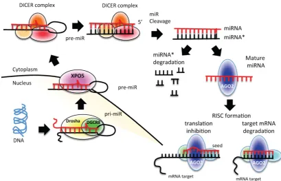
miRNAs are transcribed from individual genes containing their own promoter, or intragenically from spliced portions of protein-coding genes [24]. Like protein-coding genes, miRNAs with their own pro-moters are almost exclusively transcribed by RNA polymerase II in a primary transcript called pri-miRNA [24] (Figure 1). This long transcript con-tains a 7-methylguanosine cap at the 5′ end, a 3′
poly-(A) tail, and sometimes also introns. To be pro-cessed, pri-miRNAs are recognized by Drosha ribo-nuclease and its partner, the double-stranded RNA binding protein DGCR8, through interaction with a stem–loop structure within the miRNA in which the sequences are not perfectly complementary [25, 26]. Processing of pri-miRNAs gives rise to precursor miRNAs (pre-miRNAs) of approximately 70 nucleo-tides [24] (Figure 1). Some intronic miRNAs, called mirtrons, could bypass Drosha processing and use the splicing machinery to generate pre-miRNAs [24]. The generated pre-miRNAs are then exported from the nucleus to the cytoplasm by exportin 5 (XPO5) [27-29], where they are cleaved by the RNase III enzyme Dicer 1 in union with transactivation-responsive RNA-binding protein 2 (TARBP2) and AGO2 (DICER complex). The processing generates a dou-ble-stranded miRNA–miRNA* duplex [30]. The 2 strands are then separated: the mature miRNA (the guide strand) is incorporated into the RNA-induced silencing complex (RISC), whereas the passage miRNA* strand can be loaded in the RISC as well or degraded [31-33]. The mature miRNA guides the AGO protein of the RISC to the complementary mRNA sequence on the target to repress its expres-sion [24] (Figure 1).
2b. miRNA mechanisms of action
The major determinant for miRNA binding to its target mRNA is a 6–8-nucleotide sequence at the 5′
miRNA and the seed region triggers a detectable de-crease in target mRNA expression levels. Seed matches can occur in any region of the mRNA but are
more likely to be present in the 3′ untranslated region (3′ UTR) of a mRNA [34, 35]. Several lines of evidence indicate that miRNAs can also bind to other regions in the target mRNA [36]. Depending on the degree of
homology to the 3′ UTR target sequence, miRNAs can
induce the translational repression or degradation of mRNAs. Given that each miRNA is capable of regu-lating the expression of many genes, each miRNA can simultaneously regulate multiple cellular signaling pathways.
Apart from the “traditional” mechanism of ac-tion of miRNAs described above, other “non-canonical” mechanisms have been proposed recently. Some evidence indicates that miRNAs could increase the translation of a target mRNA by
recruit-ing protein complexes at the AU-rich region of the target mRNA or they could indirectly increase target mRNA levels by interacting and modulating re-pressor proteins that block the translation of the target mRNA [37]. Other evidence suggests that miRNAs could enhance ribosome biogenesis, thereby modu-lating protein synthesis, or skip cell cycle arrest, thereby activating target gene repression [34, 38].
3. Methods for miRNA target prediction
and miRNA–target interaction validation
3a. Methods for miRNA target prediction
Uncovering of miRNA-regulated networks needs large-scale and unbiased methods for miRNA target identification. For instance, the differential ex-pression of a single miRNA would be followed by downstream gene or proteome-wide analysis. A sin-gle miRNA could regulate a set of genes responsible for a particular malignant phenotype. The silencing of that single miRNA can alter the entire set of genes.
To overcome this complexity and to predict the target genes, several algorithms have been developed. The main difficulty in miRNA target prediction is to detect the specific sequences within genes where one miRNA is fully or partially complementary [39], con-sidering the small size of miRNAs and their low specificity.
A collection of tools is available, each with a dis-tinct approach to miRNA target prediction and dif-ferent features [40]. The suitable tool can be decided depending on the requirements [12].
The major features of computational target pre-diction are as follows: sequence composition (e.g., seed match), conservation, and thermodynamic sta-bility (e.g., free energy).
i) Seed match is the start of many computational methods for miRNA target prediction. A seed match usually consists of Watson–Crick (WC) complemen-tarity between the miRNA and miRNA target nucleo-tides. WC complementarity occurs when adenosine
(A) pairs with uracil (U) and guanine (G) pairs with
cytosine (C). The seed is a sequence from the 1st to the
8th nucleotide at the 5′ end of the miRNA. However,
algorithms based only on WC complementarity show low accuracy and a high number of false-positive re-sults [26].
Other sequence compositions can be used as features for miRNA target prediction tools. Bartel et al. [31] showed that the AU residues in target sites
improve the accessibility of miRNAs to form duplex-es. Recent studies have suggested that coding regions of mRNAs can also include target sites for miRNAs [41]. In addition, it has been demonstrated that a transcript can contain multiple target sites for a single miRNA; however, when the target sites show over-lapping sequences, miRNA–mRNA pairing can be compromised [42].
ii) Conservation analysis was introduced in or-der to reduce false-positive results. Conservation re-fers to the maintenance of sequence homology across species [40]. In general, there is higher conservation in the miRNA seed region than in the non-seed region [43]. However, the limit of this approach was demon-strated by Bentwich et al., who showed that several non-conserved miRNAs were missing [43].
iii) Free energy (or Gibbs free energy) can be used as a feature for miRNA target prediction [44, 45]. The thermodynamic stability of the miRNA–mRNA duplex shows the strength of the binding between a miRNA and its target by predicting how the miRNA and its candidate target will hybridize. The free en-ergy is related to duplex formation between the miRNA and its target site. In particular, pairing can be determined by removing existing secondary struc-tures [46]. The free energy is established by the dif-ference between the energy expended in opening the target site structure and that gained by forming the duplex [46, 47].
Many computational algorithms have been de-veloped and implemented as software tools for miRNA target prediction using some of the described features. These packages are very useful to select pu-tative miRNA targets for further biological validation. The most common classifiers are based on machine learning algorithms, e.g., support vector machine (SVM), neural networks, hidden Markov model (HMM), and Naive Bayes (NB). These machine learning methods are trained on a so-called “training” dataset that contains a set of known miRNA quences (positive training dataset) and a set of
se-quences that do not contain miRNAs, such as mRNAs, tRNAs, and rRNAs (negative training da-taset), which represent the limit of this approach [47]. Several studies have tried to overcome this problem with the use of only true/positive models [48-50]. However, the results are worse than those obtained with approaches that utilize both positive and nega-tive training sets [49]. Many tools of machine learn-ing-based approaches for miRNA target prediction are currently available, e.g., HHMMiR [51], PicTar [52], MiRFinder [53], RNAmicro [50, 54], ProMiR [55], MiRRim [56], BayesMiRNAFind [57], and SSCprofiler [58].
Other computational algorithms use approaches different from machine learning. The TargetScan al-gorithm was the first miRNA target prediction tool for human genome [40]. It searches for perfect comple-mentarity in the seed region, and all seed sequences outside complementarity are filtered out. Predictions are ranked by a combinatorial score on the basis of sequence composition (seed sequence), conservation, and thermodynamic stability (free energy).
Diana-microT uses a larger frame for scanning complementarity. It focuses on orthologous human
and mouse 3′ UTRs from the mRNA Reference S e-quences (RefSeq) database and 94 miRNAs conserved in human and mouse. It applies a modified dynamic programming algorithm to calculate the minimum free energy for each segment with a miRNA [59].
The miRanda algorithm gives scores for seed complementary regions. The results are evaluated for free energy. Each target that has a predicted free en-ergy below a threshold is then passed to the last step, i.e., conservation [60].
These algorithms are summarized in Table 1, together with their main characteristic approaches and features.
3b. miRNA–target interaction validation
Many experimental technologies for validating miRNA–mRNA interactions have been developed [61, 62]. In general, the effects of differential miRNA ex-pression on the target gene obtained through trans-fection of miRNA mimic or miRNA inhibitor oligo-nucleotides or constructs [63] are established at the protein level by western blotting and at the mRNA level by quantitative real-time PCR (qRT-PCR), with a specific probe for the target gene [61, 62]. The most important disadvantage of these techniques is that they are not able to distinguish between direct and secondary miRNA–target interactions.
3b.1 Luciferase assay
such as receptor activity or intracellular signal trans-duction of protein–protein interactions. To analyze direct miRNA–mRNA interactions, the firefly lucif-erase-based assay is widely used because the reporter activity is available immediately upon translation, the assay is very rapid and sensitive, and no background luminescence is found in the host cells (Figure 2). To be used as a reporter assay for validation of the
in-teraction of a miRNA with the 3′ UTR of a gene of
interest (GOI), the luciferase-based assay needs
clon-ing of the 3′ UTR of the GOI, where the mi R-NA-recognized sequence is supposed to be present, downstream of the luciferase gene in the reporter
vector (Figure 2). The cells are then transfected with this construct in the presence or absence of the miR-NA mimic oligonucleotide. If the miRmiR-NA is able to
recognize the seed in the 3′ UTR of the GOI, the level
of luciferase expression is decreased, thus causing a diminished bioluminescence emission (Figure 2B); on the other hand, if the miRNA does not interact with
the 3′ UTR, the emission of light is unaffected (Figure
2A). The disadvantages of this type of reporter assays are that they are laborious, expensive, sensitive only
for the 3′ UTR chosen for cloning, and difficult to use
for transfection [62, 63].
Table 1. The main algorithms for computational miRNA-target prediction
Algorithm Features Approach References
HHMMiR Seed match, and conservation HMM [51]
PicTar Seed match HMM [52]
MiRFinder Seed match, and conservation SVM [53] RNAmicro Sequence composition, conservation, and thermodynamic stability SVM [54] ProMir Sequence composition, conservation and thermodynamic stability. HMM [55] MiRRim Sequence composition, conservation, and free energy. HMM [56] BayesMiRNAFind Sequence composition and free energy. Naïve Bayes Classifier [57] SSCprofiler Sequence composition, conservation and free energy. HMM [58] Diana-microT Seed match, conservation, and free energy Dynamic programming algorithm [59] TargetScan Seed match, conservation, and free energy Combinatorial score [40] MiRanda Seed match, conservation, and free energy Score [60]
3b.2 RISC immunoprecipitation
Another biochemical method to identify and isolate direct miRNA–target complexes is based on the immunoprecipitation of RISC components (such as AGO and TNRC6). This method is able to capture low-abundant and transient miRNA–mRNA pairs. Target mRNAs undergoing direct miRNA regulation are co-immunoprecipitated along with the RISC and are identified by qRT-PCR, microarray, or deep se-quencing [64]. The successful pull-down of the entire complex relies on the strong interaction between the miRNA–target complex and RISC and on the ability of the used antibody to precipitate AGO2, the core RISC protein usually used for complex immunopre-cipitation. Some companies have developed a domi-nant negative mutant of an RISC protein subunit to trap the miRNA–target complex into the RISC, thus limiting further processing [65]. This strategy allows the recovery of transient and low-abundance mRNA targets that would otherwise be lost. A FLAG epitope is then used for the capture of the entire complex [65]. qRT-PCR or next-generation sequencing techniques are used to confirm the interaction between the miRNA and the target mRNA.
4. miRNAs and BC
Advanced technologies, such as microarray pression data, have shown that aberrant miRNA ex-pression is the rule rather than the exception in BC [66, 67]. The tight integration of miRNAs in physio-logical circuits could become a problem, because the dysregulation of a small number of miRNAs could profoundly affect the expression profile of the cells, driving them toward transformation [68]. BC miR-NAs, which have an important role in the patho-physiology of the disease, facilitating invasion, me-tastasis, epithelial to mesenchymal transition (EMT), and maintenance of BC stem cells, have become an interesting topic in BC management.
4a. Mechanisms altering miRNA expression levels
Because of amplification, each miRNA can in-crease the control over its target gene. If the target gene is an oncogene, the cancer does not develop (oncosuppressor-miRs); if the target gene is a tumor suppressor, the cancer develops (oncomiRs). Due to deletion, each miRNA can reduce the control over its target gene. If the target gene is an oncogene, the cancer develops (oncomiRs); if the target gene is a tu-mor suppressor, the cancer does not develop ( on-cosuppressor-miRs).
Several mechanisms can influence miRNA ex-pression levels (Figure 3). Tumors often present al-tered levels of mature miRNAs [101] as a consequence of the following:
1. Epigenetic mechanisms (Figure 3, section 1). A large proportion of miRNA loci on the genome are associated with CpG islands, giving strong bases for their regulation by methylation (Figure 3, section 1) [69]. A recent critical review on aberrant DNA meth-ylation of miRNAs in BC showed that although aber-rant DNA methylation is a well-described mechanism for gene silencing, an actual demonstration of the link between miRNA expression and gene methylation was still missing in several of the analyzed studies [70]. However, Castilla et al. have clearly demon-strated in 70 BC cases that a relationship exists be-tween miR-200 family expression, gene methylation, and metastatic potential of the tumors [71]. A map-ping-based study has identified miRNA promoters silenced in BC [72], and different patterns of methyla-tion have been observed in the miR-200b cluster pro-moter in different BC sub-types [72]. Aure et al., fo-cusing their attention on let7e-3p miRNA, found that the genomic region that encodes for this miRNA be-longs to a hypomethylated, and thus silenced, chro-mosome [73]. The researchers have associated
let-7e-3p downregulation with poorer BC prognosis [73]. Another epigenetic phenomenon altered in BC is histone acetylation. Studies with deacetylase inhibi-tors have revealed that the reduction of acetylated histones could diminish the expression of an-ti-oncogenic miRNAs [74, 75].
2. A genetic alteration (Figure 3, sections 1 and 2), i.e., frameshift mutations resulting from microsat-ellite instability. Such genetic alternations can affect the expression of several mRNAs, e.g., the mRNA of TARBP2 (Figure 3, section 5), the Dicer stabilizing protein. This has been found, for example, in colorec-tal and gastric cancer [76] and in BC [77]. Moreover, more than half of the known miRNAs are located in cancer-associated region, such as fragile sites, mini-mal regions of loss of heterozygosity, minimini-mal regions of amplification (minimal amplicons), or common breakpoint regions [78]. In the literature, some miR-NA families emerge to be overall more involved in tumor development [79], such as the let-7 miRNA family. In BC, several let-7 family members, together with miR-125b, miR100, and miR34a, have been found to be located at fragile sites of human chromosomes (11q23–q24D), potentially contributing to aberrant
Figure 3: Altered steps in miRNA biogenesis lead to cancer. A schematic representation of altered steps of the miRNA biogenesis pathway, commonly deregulated in cancer: 1. miRNA genes contain upstream regulator elements (enhancers/repressors) and promoter regions, indicating that miRNAs are subjected to CpG methylation (CpG promoter met); 2. The alteration in the copy number of miRNA (due to genomic amplification or deletion, activating or repressing mutation, loss of epigenetic silencing and transcriptional activation) could increase the oncogenic miRNAs or decrease the tumor suppressor miRNAs; 3. Alteration in the miRNA processing machinery, i.e. downreg-ulation of Drosha, could decrease the cropping of pri-miR to pre-miR; 4. XPO5 mutation could prevent pre-miR export to the cytoplasm; 5. Mutation of TARBP2 or down-regulation of DICER1 decrease mature miRNA levels, causing finally a loss on tumor suppressor miRNAs; 6 and 7. Accumulation of oncogenic miRNAs or loss of tumor suppressor miRNAs could finally lead to cancer development.
3. Defects in the miRNA biogenesis pathway (Figure 3, sections 3–5): each step of miRNA biogene-sis could be affected, thus altering miRNA expression levels and making the cell suitable for oncogenic changes. Reduced Dicer and Drosha expression (Fig-ure 3, sections 3 and 5) have been associated with high-grade BC and shorter metastasis-free survival or with higher-grade BC and shorter disease-free sur-vival [80-83]. Reduced Dicer expression (Figure 3, section 5) has been also found in many other human tumors [84], e.g., in prostate [85], gastric [86], or squamous cell carcinoma [87]. In BC, reduced Dicer expression has been associated with the tri-ple-negative phenotype [83, 88]. Moreover, in BC, nucleolin (NCL), a component of the Drosha/DGCR8 microprocessor complex, has been demonstrated to promote the maturation of a set of metasta-sis-promoting miRNAs (miR-221/222 cluster, miR-21,
miR-103, and miR-15a/16) [89, 90]. Furthermore, XPO5, a key protein for pre-miRNA export to the cytosol, has been suggested as a possible prognostic biomarker for BC [91] (Figure 3, section 4).
4. Transcriptional repression by other upstream proteins (Figure 4). A plethora of transcription factors can influence the expression levels of a single miRNA. Several lines of evidence suggest that miRNAs and transcription factors work cooperatively. miRNAs are
involved in the functional feedback loop, in which transcription factors influence miRNA expression levels and vice versa [92-94]. Thus, tumorigenic miRNA expression alterations could be due to the activity of tumor-related transcription factors, such as SMAD [90, 95], p53 protein family (p53, p63, and p73) [96], ataxia telangiectasia mutated (ATM) [97], and Myc [98]. In BC, the BC 1, early onset (BRCA1) tran-scription factor [99] and the epidermal growth factor receptor (EGFR/HER1), a hypoxic transcription factor involved in the regulation of the RISC [100], are able to inhibit miRNA maturation, thus enhancing cell survival and invasiveness.
4b. miRNAs and BC progression models
Figure 4: Contribution of transcription to miRNA level alteration in cancer. Several transcription factors are able to control the level of expression of miRNAs. In particular, as described in the text, SMAD, Myc, ATM, BRCA1/2 and p53 influence miRNA transcription. P53 can regulate onco-suppressor miRNAs, which are involved in the control of p53 turnover. SMAD, ATM, BRCA1/2 and Myc could influence the transcription levels of miRNAs involved in cell plasticity, cell proliferation and survival, and cell invasion control. Moreover, SMAD is also involved in miRNA processing, by Drosha expression levels control. Ex: example of miRNA regulated by transcription factors.
1. 2D cell culture. 2D cell culture studies in the oncogenic field have played a pivotal role in further-ing our understandfurther-ing of the disease mechanisms and drug discovery. The majority of scientific studies on miRNAs use 2D cell cultures for modulation of single miRNA expression and validation of the interaction between a single miRNA and its predicted targets via protein or gene expression analyses. This culture condition is easy to be manipulated, less expensive than the other approaches, and particularly suitable when a small number of miRNAs have to be studied.
Recently, particular attention has been given to emerging inadequacies associated with 2D culture systems, such as their inability to fully emulate in vivo
tumor growth conditions and to provide physiologi-cal relevance. In fact, in the body, nearly all cells re-side in an extracellular matrix (ECM) consisting of a complex 3D architecture, and interact with neighbor-ing cells through biochemical and mechanical cues. These features cannot be obtained in 2D culture con-ditions. Cell–cell and cell–ECM interactions establish a 3D communication network that maintains the specificity and homeostasis of the tissue and influ-ences tumor growth and its interaction with the whole organ. This approach has been extensively used in many works on the assessment of miRNAs in BC [101-103].
2. 3D cell culture. To overcome some shortcom-ings of 2D cultures, 3D cell cultures have been de-veloped, with the use of specific matrix (such as nat-ural ECM-based hydrogels, 3D spheroids, and
trans-well inserts) that are able to support the growth of tumor cells for the establishment of physiological cell–cell and cell–ECM interactions of the native tis-sues. These matrix supports can mimic the environ-mental conditions in which the tumor cells grow with greater physiological relevance than conventional 2D cultures. The development of new biological supports is further fueled by the optimism that 3D models may significantly accelerate translational research in can-cer biology. For example, use of 3D tumor cell culture is emerging as an important tool to characterize the morphogenesis of mammary epithelial cells and to elucidate the tumor-modulating actions of ECM. Fo-cusing on miRNAs, the comparative analysis of 2D and 3D cell cultures has revealed a profound differ-ence in miRNA profiles between the 2 culture condi-tions, particularly for BC cells and lung adenocarci-noma [104, 105]. In particular, the miRNA profiles in 2D and 3D cultures of 2 BC cell lines were compared. The findings revealed that the 3D culture exhibited a greater discrimination between the miRNA profiles than the 2D culture [105]. For example, the lower ex-pression of miR-429 was highlighted in the 3D cul-ture-specific miRNA profile better than that in the 2D culture-specific profile, correlating with the 3D inva-sive capacity of the MDA-MB-231 BC cell line.
method can be used to obtain a miRNA profile during ongoing tumor development. Otherwise, tu-mor-xenografted mice are used for the therapeutic study of miRNA modulation. In fact, several reports have examined the effect of miRNA modulation by treating xenografted animals with oligonucleotides that increase (miRNA mimic) or decrease (antagomiR) the expression levels of a specific, single miRNA or by using an expression vector for miRNA level modula-tion [106]. Thus, the effects of miRNA modulamodula-tion are analyzed by measuring the growth of the tumor, its invasive capacity, presence of metastatic masses, and vascularization of the tumor [107].
Several models to study miRNA involvement in BC progression are based on BC cell lines or patient specimens implanted into mouse mammary fat pads. For example, use of the triple-negative MDA-MB-231 BC cell line to generate a xenografted BC model al-lows identification of miR-124 as the key regulator of the myc/p27/phospho-Rb pathway, which is usually altered in BC and ovarian cancer [108]. Another BC model, obtained by the orthotopic implantation of xenografted human BC specimens into NOD/SCID mouse mammary fat pads (called patient-derived human-in-mouse breast tumor xenograft model or PDX model), was used to study the spontaneous generation of BC-derived lung cancer metastasis. With a combined approach of gene expression arrays and global miRNA analysis, miR-138 was demon-strated to be a key regulator of tumor invasion in lung, targeting the EMT process of BC cells [109]. Another xenografted mouse model was used to demonstrate the anti-metastatic potential of the pep-tide nucleic acid (PNA)-modified antagomiR-21 oli-gonucleotide on BC cells. Yan et al. [110] demon-strated that use of the antagomiR-21 oligonucleotide is able to block proliferation, cell migration, and in vivo tumor growth of 2 BC cell lines (MCF7 and MDA-MB-231) implanted in BALB/c-nude mice, proposing the use of this oligonucleotide for potential therapeutic applications in BC treatment.
4. Engineered mouse models have been widely used. These models could be of 2 types. The first type, the genetically engineered mouse model, obtained by oncogene amplification or tumor suppressor gene deletion to characterize a specific cancer, is used to generate an expression profile of miRNAs to clarify which miRNAs are involved in the development of that specific tumor. In BC, this model has been used to identify a miRNA profile associated with 8 different mammary-engineered mouse models [111]. In the second type, knocking-in or -out of specific miRNAs in the mouse germline allows to study the influence of miRNAs on tumor progression [112]. This approach also involves using genetic constructs to induce
miRNA overexpression or downregulation in a par-ticular tissue, at a parpar-ticular development stage, or under pharmacological control. For example, several strains of mice lacking or overexpressing can-cer-associated miRNAs have been developed and characterized. These include germline transgenic or knockout mice for the following: miR-155, which, if overexpressed in B cell lineage, induces B cell malig-nancy [113]; miR-21, which leads to lung tumorigene-sis if ubiquitously deleted [114]; miR-17-92 and its paralogs, whose overexpression in lymphocytes in-duces lymphoproliferative disease and autoimmunity [115]; miR-15 and miR-16, whose deletion induces lymphoproliferative disorders [116]; miR-146, which causes myeloid sarcomas and lymphomas when de-leted [117]; and miR-29, whose deletion causes B-cell lymphoma [118].
To our knowledge, no germline transgenic miRNA-engineered mouse models have been pro-posed yet to study BC onset and development.
5. miRNAs and cancer stem cells (CSCs)
5a. miRNAs involved in CSCs
si-lencing of specific genomic regions; alteration in chromatin remodeling, which controls the accessibil-ity of chromatin to transcription factors; and alteration in specific miRNA expression levels) [121].
The miRNA profile of CSCs is remarkably dif-ferent from that of non-stem cancer cells, and many miRNAs have been shown to regulate the self-renewal and differentiation properties of CSCs [122, 123], such as the let-7 miRNA family [124]. Being a tumor suppressor, the tumorigenic potential of the
let-7 family is due to its downregulation in many tu-mors, such as lung cancer or BC [125]. Cancer initia-tion, progression, and aggressiveness are driven by CSCs [126-128]. The let-7 miRNA family appears to play a substantial role in the CSC phenotype. In fact, it seems that each tumor, being either hematologic or solid, includes a minor population of CSCs, capable of tumor initiation [129]. These TICs have downregu-lated let-7 expression and, having tumor stem cell properties, can also undergo asymmetric division, thereby sustaining differentiated tumor proliferation [130]. In BC, let-7 is found to be downregulated. In normal tissue, it plays the role of a regulator of self-renewal, acting as a pro-differentiation miRNA,
whereas in BC it is repressed by the Wnt/β-catenin pathway [124]. Thus, its loss in BC leads to an increase in the CSC population.
In addition to let-7, miR-34 has been described as a regulator of the Notch signaling pathway, necessary for stem cell maintenance, in colon CSCs [131]. Asymmetric cell division, a characteristic of CSCs required for self-renewal, is directed toward sym-metry by the presence of miR-146a, which targets Numb to stabilize β-catenin expression and leads to symmetrical division [132]. In BC, expression of
miR-34 leads to cell cycle arrest [133], whereas its downregulation increases the invasive capacity and metastatic potential of BC cell lines in vitro and in vivo
[134]. All the discussed miRNA as summarized in Table 2.
5b. miRNAs and EMT
Emerging evidence demonstrates that miRNAs play an essential role in controlling stem cell
proper-ties, such as self-renewal and differentiation, by reg-ulating the expression of certain key stem cell regu-latory genes [135-137] and by regulating EMT [138, 139]. EMT refers to the process in which tumor epi-thelial cells acquire mesenchymal features, with high invasiveness and metastatic abilities. In fact, EMT is associated with the loss of intracellular junctions and epithelial polarity and increase in cell motility, which are fundamental characteristics for tumorigenesis, invasion, and metastasis that allow cancer cells to infiltrate adjacent stroma and metastasize to distant sites. These phenotypic changes appear to be induced by several miRNAs, such as let-7, miR-10, miR-34, miR-200, and miR-205 [139]. In BC, miR-155 and
miR-21, described as oncomiRs, are implicated in EMT, cell migration, and invasion control. A well-known target of miR-21 is PTEN, a tumor sup-pressor, which negatively regulates the PI3K pathway [133, 140]. Growing evidence suggests that BC cell plasticity, necessary for the spread of a tumor, arises because of partial reactivation of EMT in a mature cancer cell in order to give the cell pluripotency and a stem-like phenotype.
All the discussed miRNAs are summarized in Table 2.
6. Potential of miRNAs as BC biomarkers
BC is a heterogeneous disease with several morphological appearances, molecular features, be-haviors, and response to therapy [143, 144]. Thera-peutic management of BC is based on the availability of strong diagnostic, prognostic, and predictive fac-tors to guide the decision and the choice of different treatment options [145-147].The current in vivo diagnostic tools for BC, e.g., mammography and ultrasound, are used for the de-tection of early-stage BC. However, several technical limitations exist for these techniques, such as breast density or calcification detection. Other imaging mo-dalities, e.g., magnetic resonance imaging (MRI), have been proposed as complementary diagnostic modali-ties, with limited sensitivity.
Table 2. Examples of miRNAs involved in CSC phenotype, and EMT process. This table focuses on few examples of miRNAs described in the text, with a particular attention on their function and the type of cancer where they have been found.
miRNA annotation Function Tumor Ref.
let-7 Regulator of self-renewal, cell proliferation and EMT Lung, BC [124]
miR-34, miR-146a Symmetric and asymmetric division of CSCs colon [132]
miR-34, Cell cycle control, invasion capacity and metastatic potential BC [133, 134]
let-7, miR-10, miR-34, miR-200, miR-205; miR-30 Stemness and EMT regulation BC [139, 141, 142]
Some proteins have been associated with BC by the analysis of expression levels of specific mRNAs, e.g., carcinoembryonic antigen (CEA) and CA-125 [148]. For BC diagnosis and prognosis, several mRNA-based genetic tests are currently available, such as the PAM50 assay (based on the NanoString technology), MammaTyper assay (based on the qRT-PCR technology), MammaPrint test (based on the microarray technology), Oncotype DX test (based on the qRT-PCR technology), Endopredict (based on the qRT-PCR technology), and Genomic Grade Index (based on the microarray technology) [149]. Use of
independent cores for gene expression testing in BC, coming from different gene signatures, may be a suc-cessful strategy to overcome tumor heterogeneity and sampling error.
Although direct measurements of tissue gene biomarkers have greatly improved BC diagnosis, the invasive and unpleasant nature of the diagnostic procedures limits their application. Isolation and subsequent characterization of circulating miRNAs provide the opportunity to bypass the problems as-sociated with tissue biopsy, which is required as per the currently available genetic tests. In fact, circulating miRNAs are small molecules, found in body fluids (blood, plasma, serum, saliva, urine…). Being im-portant regulators of gene expression and being dysregulated in several types of cancer diseases [150], circulating miRNAs have become interesting in new cancer biomarker research. They have been found to be stably and specifically expressed in mammary tis-sues and in body fluids when the disease is ongoing [151, 152]. These features enable them to respond to the current clinical needs, allowing them to be used as easy, affordable, and clinically accessible molecular biomarkers in the retrospective analysis of large tissue collections and for diagnosis, prognosis, and predic-tion of therapeutic outcomes in BC.
6a. miRNAs dysregulated in BC
Several studies have looked at possible specific miRNAs dysregulated in BC with a diagnostic pur-pose [153, 154]. Dysregulated miRNAs could be di-vided into 2 groups, being either upregulated or downregulated (Table 3).
Increased expression of miR-21 has been found in vitro in human BC cell lines and tissues, playing a key role in all phases of BC pathogenesis [141, 155], alt-hough it also appears to be able to monitor early BC onset[156]. miR-21 activity controls cell proliferation, G2/M check point, and metastasis diffusion [157-159] and the expression of many anti-oncogenes, including TPM1, programmed cell death 4, maspin, and Bcl-2, to support the metastasis and hyperplasia of BC cells [160].
Several other miRNAs have been validated to be overexpressed in BC; these include the miR-221/222
cluster [161], miR-9, miR10b, miR-29a, miR-96,
miR-146a, miR-181, miR-373, miR-375, miR-520c, and
miR589 [162], highlighting their potential use for BC diagnosis, prognosis, and therapeutic studies [80, 137, 163-165].
Some upregulated miRNAs could cooperate in controlling a network of functional genes to help tu-mor development or metastasis. Figure 5 shows ex-amples of miRNA regulatory networks in BC that promote metastasis through their ability to coordi-nately target multiple genes [166]. Ma et al. [167] proved the role of miR-10b as a driver of metastasis:
miR-10b, under the control of the TWIST transcription factor, binds HOXD10 gene, enhancing cell migration and invasion. HOXD10, in turn, inhibits the Ras homolog gene family, member C (RHOC) protein, favoring metastatic diffusion of the tumor (Figure
5A). The miR-10b locus also encodes for
miR-10b*/miR10b-3p. miR-10b*, although considered functionally irrelevant, was very recently demon-strated to be important for BC insurgence and devel-opment [168]. Hence, if miR-10b-5p upregulation leads to the induction of ECM remodeling factors for meta-static invasion, miR-10b-3p downregulation is in-volved in primary BC onset and development, as its overexpression inhibits the proliferation of BC cell
lines by targeting cell cycle regulator proteins (BUB1,
PLK1, and CCNA2) [168, 169] (Figure 5A).
Among the downregulated miRNAs, miR-30a,
miR-31, miR34, miR-93, miR-125, miR-126, mR-146a,
miR-195, miR-200, miR-205, miR-206, miR-503, and
publication, we proposed let-7c as a possible bi-omarker of a 4-miRNA signature, capable of distin-guishing between grade 1 and grade 3 BC samples [21].
miR-92a is another possibly downregulated miRNA. This miRNA belongs to the miR-17-92 family, which can promote tumor proliferation by controlling the PI3K/Akt/mTOR pathway [187]. Moreover, this miRNA has Bcl-2 interacting mediator of cell death (Bim) and p53 proteins as targets, thus inhibiting tu-mor cell apoptosis and cell cycle arrest and promoting tumorigenesis [98, 188, 189]. In addition, it is involved in promoting tumor invasion and metastasis by modulating the TGF-β signaling pathway [190, 191].
Another downregulated miRNA, miR-206, has
been found to be underexpressed in estrogen receptor (ER)α-positive BC, both in patient samples and BC cell lines [192, 193], and in lymph node metastatic BC [194, 195]. With regard to its functions, it has been
recently demonstrated to regulate the 3′ UTR of cyclin
D1, inducing G1 arrest and a decrease in cell prolifer-ation in BC cells [196], suggesting a potential role as a tumor suppressor. It has been shown that miR-206
regulates ERα via interaction with its 3′ UTR [193], demonstrating a specific role in most aggressive types
of BC.
Other downregulated miRNAs, typical of BC tissues, are a group of miRNAs usually expressed in stem cells. This group includes the miR-200 family [197], miR-15/16, miR-103/107, miR-128b,miR-145, and
miR-335 [137]. All these miRNAs are downregulated in CSCs, targeting common genes (Bmi1 and Suz12 component, Zeb1/2, and Klf4), all belonging to a reg-ulatory circuit that sustains the breast CSC state [137].
6b. miRNAs as biomarkers of diagnosis, prognosis, and therapy prediction in BC
Several attempts have been made to identify af-fordable BC signatures for diagnosis, prognosis, and prediction of the therapeutic response (Table 4).
With respect to diagnosis, Iorio et al. [164] iden-tified a 13-miRNA signature that could differentiate BC from normal breast tissues with 100% accuracy. Blenkiron et al. [202] identified 133 miRNAs that dis-played aberrant expression levels in breast tumor tissues compared with normal breast tissues. Despite the identification of miRNA with aberrant expression in BC tissues, there remain discrepancies among the different reported miRNA signatures. This is proba-bly because of the intrinsic heterogeneity in BC and
because of clinicopathological variables such as the tumor stage, vascular invasion, prolif-eration index, and expression of HER2, ER, or progesterone re-ceptor (PR). Thus, an attempt has been made to develop a miRNA signature that reflects the histopathological features of the tumor. At the simplest level, BC comprises 3 different histo-logical subtypes: hormone re-ceptor-positive (ER+, PR+)
tu-mors, which cover approxi-mately 60%–70% of diagnosed BCs; HER2+ tumors, which
cov-er 15%–20% of diagnosed BCs; and triple-negative (ER−, PR−, HER2−) tumors [66]. Genomic mRNA profiling has subdivided BC into 4 different classes: lu-minal A (ER+ and low grade),
luminal B (ER+ and high grade),
HER2+, and basal like (mainly
triple negative) [202].
Table 3. BC deregulated miRNAs: an overview.
miRNA annotations Samples type Ref.
Up-regulated miRNAs miR-21 BC cell lines [141, 155-157, 163]
miR-221/222 cluster BC cell lines [161]
miR-9, miR10b, miR-29a, miR-96, miR-146a, miR-181, miR-373,
miR-375, miR-520c, miR589 BC cell lines [162-165, 167, 198, 199]
miR-10b BC cell lines [167]
miR-155 BC cell lines [158, 163, 200]
miR-210 BC cell lines [197]
Down-regulated
miR-NAs miR-30, miR-31, miR-34, miR-93, miR-126, miR-146a, miR-195, miR-205, miR-206, miR-503 BC cell line; TNBC cell lines [163, 170-175, 201]
let-7 family BC cell lines [163, 175, 182]
miR-92a cluster BC cell lines; TNBC cell lines [187-191]
miR-200 family BC cell lines [163, 197]
miR-15/16 cluster, miR-103/107, miR-145, miR-335, miR-128b BC cell lines [137]
miR-10b* BC cell lines and xenografted tumor [168, 169] TNBC= triple negative BC.
Table 4. Circulating and non-circulating miRNAs as BC biomarkers. All the reported miRNAs have been validated on BC patients. For each miRNA, we indicated whether they have a role in diagnosis, prognosis or in prediction of therapy response in BC. For all groups we indicated the biological samples used for the validation, the validation assay, the cohort of data, the main results and the references. This table focused on few examples of single miRNA or miRNA signatures described in the text. GGI=gene expression grade index: TAM=tamoxifen; H= herceptin; N= normal tissue; T= tumor tissues; TNBC= triple negative breast cancer.
Type of miRNAs miRNA annotation Role Biological
Samples Technique/cohort Results Ref.
Non-circulating
miRNAs 13 miRNAs (miR-9-1, miR-21, miR-34, miR-29b/102, miR-10b, miR126, miR125a/b1/b2, miR-140as,
miR-145, miR-155, miR-194, miR-204, miR-213)
Diagnosis Tissue Microarray and north-ern blot/
76 BC vs 10 N
4/13 are downregulated (5 miR-NAs are the most constantly de-regulated in BC)
[164]
133 miRNAs Diagnosis Tissue Microarray / 99 BC vs
5 N and 33 BC cell lines 31 miRNAs are associated with tumor subtype or clinical patho-logical fators
[202]
15 miRNAs
miR-342, miR-299, miR-217, miR-190,
miR-135b, miR-218.;
miR-520g, miR-377, miR-527-518a,
miR-520f-520c;
miR-520d, miR-181c, miR-302c,
miR-376b, miR-30e
Diagnosis Tissue Microarray / 95 BC vs
17 N ER+; PR+; HER2/neu+: [203]
6 BC-miRNAs signature
miR-21 miR-17-5p miR-29b-2 miR-146 miR-155 miR-181b-1
Diagnosis Tissue Microarray/ 363 T vs
177 N 31% of the total miRNAs varied among T and N tissues; they iden-tified the most commontly altered miRNAs in solid tumors
[184]
miR-7,
miR-128a, miR-210, and miR-516–3p; miR-210
Diagnosis Tissue TaqMan /
185 ER+ vs 114 ER- BC 4-miRNA signature associated with tumor aggressiveness in ER+ BC and miR-210 associated with early relapse in
ER-[204]
let-7c, miR320dmiR567, miR139-5p Diagnosis Tissue Microarray /
42 BC G1 vs 42 BC G3 4 miRNA signature [21]
let-7a
miR-335 Diagnosis and prognosis Tissue TaqMan/ 60 BC vs 60 N Both miRNAs are decreased in BRCA mutant; miR-335 could be used as prognostic marker
[66]
miR-155, miR-493, miR-30e, miR-27a Diagnosis and
prognosis Tissue Microarray/ 80 high risk vs 80 low risk 2 upregulated, ‘protective’ miR-NAs (miR-155, miR-493); 2 down-regulated risk-associated miRNAs (miR-30e , miR-27a )
[205]
miR-210
miR-148a Prognosis Tissue qRT-PCR/ 89 ER+ BC +TAM vs 56 N
miR-210 and miR-148a are associ-ated with relapse free survival; [206]
miR-190b miR-339-5p miR-520c-3p /g/h miR-139-3p miR-204 miR-502-5p miR-365 miR-363 miR-7
Prediction of therapy re-sponse (TAM)
Tissue Microarray/ 26 patients with re-currence vs 26 patients without recurrence
miR-7 correlates with tumor grade [212]
miR-30c miR-422a miR-30a-3p miR-187
Prediction of therapy re-sponse (TAM)
Tissue qRT-PCR/ 38 ER+ BC vs 15 BC+TAM
Higher miR-30a-3p, miR-30c, and miR-182 are associated with treat-ment benefits
miR-182 miR-21, miR-181b,
miR-26a/26b, miR-27b miR-23b, let-7 family,
miR-125a-5p/b-5p
Prediction of therapy re-sponse (TAM)
Cell lines
and tissue qRT-PCR/ i) BC cell lines ± ER activation ii) 15 ER+ BC + ex-emestane and TAM
All miRNAs are upregulated upon anti-estrogen treatment [214]
miR-26a, miR-30b, let-7 family,
miR-125a/b Prediction of therapy re-sponse (H)
Tissue Microarray/ 83 BC + H vs adiacent stromal microdissected cells
With SVM technique, they devel-oped a 35 miRNA signature for H treatment response
[216]
Circulating
miRNAs miR-155 Diagnosis Serum qRT-PCR/89 BC vs 29 N miR-155 is increased, both in pri-mary and metastatic BC [208]
miR-195 Diagnosis Blood qRT-PCR/
83 BC patients vs 44 N miR-195 is increased +19.25 fold [209]
miR-29a
miR-21 Diagnosis and prognosis Serum qRT-PCR/20 BC sera vs 20 N both miRNAs are increased; miR29a correlates with tumor stage [210]
miR-16 miR-25 miR-222 miR-324-3 p
Diagnosis and
prognosis Serum TaqMan /48 BC vs 48 N All are increased in high risk pa-tients [211]
let-7 miR-21 miR-202
Diagnosis and
prognosis Blood and serum RT-PCR/ 136 BC vs 60 non BC All miRNAs are increased in BC patients; [224]
miR-18b miR-103 miR-107
miR-652 Diagnosis, prognosis Serum RT-PCR/ 33 primary TNBC vs 33 N 4 miRNA signature predict tumor relapse and overall survival [225]
miR-210 Prediction of therapy re-sponse (H)
Plasma TaqMan / 18 BC+ H vs 11 not responding BC
miR-210 is higher in patients with residual BC (+2 fold) [222]
miR-155 Prediction of therapy re-sponse (taxane)
Serum RT-PCR/
103 BC+taxane vs 55 N miR-155 expression correlates with the treatment course [223]
Blenkiron et al. [202] tried to associate a miRNA profile with each of these genomic classes. Among the 309 miRNAs identified in 93 BCs, 9 miRNAs (miR-15b,
miR-99a, miR-100, miR-103, miR-107, miR-126*,
miR-130a, miR-136, and miR-146b) could discriminate luminal A from luminal B BC [202].
Subsequently, Lowery et al. [203] identified a 15-miRNA predictive signature corresponding to the expression of ER (miR-135b, miR-190, miR-217,
miR-218, miR-299, and miR-342), PR (miR-377,
miR-520f, miR-520g, and miR-527-518a,), and the HER2 receptor (miR-30e, miR-181c, miR-320c, miR-376b, and
miR-520d). The same approach was used by Volinia et al. [184], corresponding to the identification of a 17-miRNA signature for the status of ER (miR-30d and
miR-30e), PR (miR-19a, miR-29c, miR-30a-5p, and
miR-106b), HER2+ (let-7f, let-7g, miR-10b, miR-107, miR-126, miR-154, and miR-159), and ER/PR (miR-25, miR-142-5p, miR-200a, and miR-205,).
Finally, Foekens et al. [204] described a subset of miRNAs significantly associated with an ER+ luminal
signature, identifying particularly 4 miRNAs associ-ated with BC aggressiveness. The discrepancies among the different miRNA signature studies could result from the fact that miRNAs identified in each study were not examined in the others, besides other variables such as clinicopathological parameters of the tumor (tumor size, grade, etc.) or the use of dif-ferent detection platforms (RT-PCR, next generation sequencing, etc.).
Hence, in our recent publication, starting from public BC databases containing gene expression
pro-files, copy number information, and miRNA propro-files, we have described new 4 miRNA-based signatures, identifying a small group of miRNAs typical of BC, which could distinguish BC with different grades [21]. Several other small signatures or single miRNAs have been proposed with a diagnostic or prognostic aim [66, 205, 206].
All the described miRNAs are summarized in Table 4, section “Non-circulating miRNAs.”
The observation that miRNAs could be secreted by a solid tumor into the surrounding environment and that they are stable in body fluids make miRNAs promising targets easily found in blood, plasma, and serum [207]. Some attempts have been made to iden-tify single circulating miRNA or small circulating miRNA signatures with a diagnostic or prognostic purpose (Table 4, section “Circulating miRNAs”). Few examples of single circulating miRNAs proposed as diagnostic or prognostic tools have been suggested by Roth et al. [208], who found miR-155 in the serum of patients with BC and not in healthy controls, and by Heneghan et al. [209] who found elevated miR-195
expression in the blood of only patients with BC. Other miRNAs have been detected in the serum of patients with BC, such as miR-29a and miR-21 [210] or the 4-miRNA signature of Hu et al. (miR-16, miR-25,
miR-222, and miR-324-3p) [211] (Table 4).
[ta-moxifen (TAM)], taxanes (taxol or paclitaxel and docetaxel), 5-fluorouracil (5-FU), and cyclophosph a-mide. Despite advances in treatment achieved by the combination of some of these compounds, a large number of patients do not respond to chemotherapy. In this context, miRNAs that are able to predict the therapeutic response of a given patient could help clinicians in the choice of the correct therapeutic ap-proach. Some signatures have been developed in search for miRNAs able to predict the therapeutic response of patients with BC [206, 212-214] (Table 4). TAM, one of the main molecules used in BC treat-ment, is a drug that reduces or eliminates circulating estrogen or blocks the interaction of ER with genomic targets. In some studies, it was evaluated whether TAM could be a successful treatment for ER+ BC. The
analyzed population had already developed metasta-sis prior to the onset of treatment and the benefit of treatment was measured as an objective response ac-cording to the REMARK criteria [215].
Other miRNA signatures have been studied for predicting the response of BC to Herceptin (or trastuzumab, H) [216]. This molecule is a recombinant humanized monoclonal antibody against HER2 pro-teins that blocks the HER2-mediated activation of intracellular kinases and effectors [66]. Although H treatment prolongs survival in adjuvant and meta-static settings, a majority of women with HER2+
met-astatic disease will develop resistance to the therapy within a year of treatment. Identification of a miRNA signature that predicts patient risk, disease outcome, and tolerability to H therapy would greatly improve the personalized management of HER2+ BC. A few
studies have successfully identified a prognostic miRNA signature for the response of BC tissue sam-ples to H therapy [217]. Combining the results of non-circulating miRNA signatures of BC cell lines treated with TAM or H, in order to identify miRNAs with predictive ability, only the let-7 family and
miR-125a-5p/b-5p emerge as important predictors of therapeutic response [214, 217]. miR-125, whose ex-pression correlates with the HER2 status [218], has been found to be significantly downregulated in pa-tients with BC [183, 219]. Experimentally, the overex-pression of miR-125 reduces ERBB2 and ERBB3, de-creasing cell motility and the invasiveness of numer-ous cancers, including BC [219]. The let-7 regulatory network suppresses metastasis by directly targeting the chromatin-remodeling protein HMGA2 and the transcription factor BACH1 [180, 220] (Figure 5B). Both targets promote the transcription of pro-invasive genes that suppress cell invasion and metastasis to the bone [180, 220].
Almost all publications of circulating miRNA profile and HER2+ BC response to therapy have used
BC cell lines to identify single miRNAs or groups of miRNAs whose expression is altered after prolonged H treatment [217, 221]. One miRNA possibly involved in the response to H therapy is miR-210, which is present both in tissue and in body fluids of patients with BC [222]. Circulating miR-210 has been associ-ated with H sensitivity, tumor presence, and lymph node metastasis, suggesting a possible use of miR-210
to monitor the response of HER2+ BC to H-based
therapies [222]. Another miRNA, miR-155, has been used to monitor the effect of taxane treatment on BC. Sun et al. observed the decreased expression of
miR-155 in serum after chemotherapy, which reached levels comparable to those of healthy subjects [223].
6c. miRNAs and hallmarks of BC
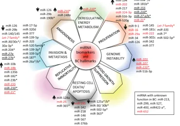
We have depicted an overview of miRNAs that can already be considered as BC biomarkers. This is outlined in Table 4. We have tried to classify all cir-culating and non-circir-culating miRNAs with diagnos-tic, prognosdiagnos-tic, and predictive capacity in relation to their function as described in the literature. In partic-ular, we have related them to altered pathways, the hallmarks of BC [226], generating a daisy-shaped fig-ure (Figfig-ure 6) in which each of the petal represents one of the hallmark function altered in BC. The major group of miRNAs (27 miRNAs) affects genes belong-ing to the proliferation pathway, although some of these miRNAs (miR-155, miR-210, miR-21, and let7 family) are also involved in directing the BC invasion and metastatic pathways, controlled by other 24 miRNAs. Resting cell death and apoptosis are the targets of the third larger 14-miRNA group. Five miRNAs are responsible for the control of angiogene-sis, whereas 2 miRNAs control genomic instability.
miR-210 is the miRNA with a wider activity, being involved in energy metabolism, angiogenesis, and genomic instability beside the already described role in invasion and proliferation. Other miRNAs, such as
miR-21, miR-27a/b, and miR-155, have been demon-strated to have multiple functions. This feature may be partially explained by the fact that the action of certain miRNAs is dependent upon the cellular model or environmental context in which they have been studied. Only 5 miRNAs have not yet been charac-terized in vitro for their function in BC development (miR-213, miR-299, miR-422a, miR-493, and miR-527).
With respect to the clinical use of the depicted miRNAs, the majority of them (33/59) can be used as diagnostic tools; a small number of them (7/59) (in-dicated in italics in Figure 6) also have prognostic ability (members of the let-7 family, miR-27a, miR-30,
miR-7, miR-21, miR-23b, miR-26a/b, miR-27b, miR-30b/c,
miR-125 a/b, miR-139-3p, miR-181b, miR-182, miR-187,
miR-204, miR-210, miR-339-5p, miR-363, miR-365,
miR-502-5p, and miR-520 family); 10/59 (marked in red in Figure 6) are circulating miRNAs, showing different functions in BC, and 2/59 (miR-155 and
miR-210) are circulating miRNAs with both diagnos-tic, prognosdiagnos-tic, and predictive role in BC.
7. miRNA for therapeutic use in cancer
Use of miRNAs for the development of new
therapeutic strategies is based on 2 approaches: 1) use of miRNAs as drug molecules, based on the synthesis and delivery of specific oligonucleotides, able to in-crease or dein-crease miRNA levels in BC or 2) modula-tion of miRNAs in combinamodula-tion with non-miRNA-based therapies to increase the efficacy of the conventional treatments.
7a. Methods for miRNA modulation
There are 2 main approaches for developing miRNA-based therapies: antagonist and mimic oli-gonucleotides. MicroRNA antagonists or antagomiRs are generated to inhibit miRNAs that acquire a gain of function in human disease.
The most common strategy to ablate the function of miRNAs is achieved by single-stranded oligonu-cleotides with miRNA complementary sequences. In
contrast, miRNA mimics are used to restore miRNAs that show a loss of function, as in the traditional gene therapy. This approach, also known as miRNA re-placement therapy, has attracted much interest as it provides a new opportunity to therapeutically exploit tumor suppressors. The low molecular weight of miRNAs permits the delivery of therapeutic miRNAs as short double-stranded oligonucleotides [227]. To improve the efficiency of miRNA/anti-miRNA
de-livery in vivo, modified miRNA molecules, both
miRNA mimics and antagomiRs, with longer half-lives and increased efficiency have been devel-oped, such as anti-miRNA oligonucleotides (AMOs) [228], locked nucleic acid (LNA)-modified oligonu-cleotides [229], cholesterol-conjugated antagomiRs [230], and the recently developed 2′-O-methoxyethyl-
4′-thioRNA (MOE-SRNA) [231].
In recent years, a method has been described to inhibit miRNA function using synthetic mRNAs con-taining multiple binding sites for a specific miRNA, called miRNA sponges [232, 233]. In bladder cancer cell lines it has been demonstrated that the forced expression of a miRNA sponge designed to inhibit
miR-21 leads to a reduction in tumor aerobic glycoly-sis, i.e., the ability of the cells to metabolize glucose even under aerobic conditions [234]. miRNA sponges have been validated even in an SUM149-epithelial BC cell xenografted mouse model, where inhibition of
Myc-driven miRNA-9
using a synthetic mRNA containing sev-eral miR-9 binding sites reduced the develop-ment of lung metastases [198]. Inhibition of the BC cell proliferation effect has been ob-served even in another xenografted mouse model implanted with the MDA-MB-231 BC cell line, where a
miR-150 sponge-based inhibition led to a re-duction in tumor mass proliferation via in-crease in the P2X7 re-ceptor [235].
Figure 6: miRNA biomarkers and BC hallmarks. miRNAs have a role as diagnostic miRNA, prognostic miRNAs (italics),
7b. miRNA-targeted therapies
Use of miRNAs alone in anti-cancer therapy to inhibit BC proliferation and development is still a challenge, although some promising results have al-ready been obtained in both ex vivo and in vivo ex-periments. For instance, miR-145 has been chosen as a target therapy in BC cells because it is usually found to be downregulated in BC [236]. Use of mimic or
inhibitor miRNA oligonucleotides has been exploited in in vivo experiments. For instance, miR-21 has been found to be of particular interest in BC because chemically modified anti-miRNA oligonucleotides have already been developed for the in vivo treatment of xenografted BC mouse models [110].
To avoid rapid degradation and excretion of miRNAs, study of new delivery systems, which could enhance the stability and the delivery to target tissues, is necessary.
Recently, some studies focused on the potential use of nanomaterials to facilitate the delivery of bio-molecules inside tumors. In particular, gold nanopar-ticles, with their high affinity for biomolecules, re-duced cytotoxicity, easy size control, and well-developed surface chemistry, have been modi-fied to increase their complementarity for nucleic ac-ids [237], allowing the effective delivery of silencing RNAs inside cells [238, 239]. The same approach has been used for the delivery of miR-145 into BC cells [236]. Therefore, gold nanoparticles have been suc-cessfully used for the delivery of miR-145 oligonucle-otides inside BC cell lines [240].
The question regarding the effect of the delivery on non-target organs and systemic toxicity of the compound remains unanswered. An obstacle for the use of miRNAs in therapy is the fact that miRNA modulation can affect hundreds of transcripts in dif-ferent tissues, being potentially capable of shutting down entire pathways. Thus, till date, few companies have used miRNAs to develop a new class of cancer therapeutics. MRX34, a miR-34a mimic compound, is probably one of the first miRNA replacement agents to undergo clinical trials. At the time of writing this review, Mirna Therapeutics is recruiting participants to a phase I study of MRX34 (NCT01829971) (http://clinicaltrials.gov/ct2/show/NCT01829971). Similarly, Regulus Therapeutics is developing anta-go-miR-221 for hepatocellular carcinoma treatment
and antagomiR-10b for glioblastoma treatment
(http://www.regulusrx.com/therapeutic-areas/#On cology). All these companies suggest that antago-miR oligonucleotides can be easily administered through local or parenteral injection routes with sufficient up-take of the agent to achieve sustained target inhibition in tissues and organs without the need for a formula-tion. Nevertheless, miRNA features, such as their
sta-bility and widespread activity on several targets, lead us to think that before using miRNA in therapies, much work is required to obtain more detailed and comprehensive knowledge about miRNA therapeutic potential, such as miRNA tissue distribution and systemic toxicity.
7c. miRNAs and chemoresistance
miRNAs can potentially be used to increase the response of BC to a therapeutic intervention. As an example, BC has been shown to be chemoresistant when some miRNAs are dysregulated (e.g., miR-125b, [239]). miRNA mimic oligonucleotides, which in-crease the levels of a given miRNA in a BC in which this miRNA is lost, can be used in combination with conventional therapy to obtain an increased benefit for patient outcomes. An example of this approach has been used in the study by Yang et al. [241], where the upregulation of miR-195, obtained by mimic oli-gonucleotides, supplied to ADR-resistant MCF7 BC cell lines increased the sensitivity of the cells to the treatment, leading to apoptosis through downregula-tion of Raf-1 and Bcl-2. In addidownregula-tion, the combined treatment of a patient with BC using antago-miR oli-gonucleotides to shut down the increased miRNA expression levels in a specific patient could poten-tially increase the effect of conventional therapy. This approach has been used, for example, to demonstrate
in vitro that miR-21 antisense oligonucleotides could be used in combination with H to kill resistant BC cells in xenografted mouse models [242].
For a more detail review on the role of miRNAs in chemoresistance modulation, see [243].
7d. miRNAs, CSCs, and chemoresistance