ISSN 2449-8955
EJBRAT 6(2) 2016
Volume 6
Number 2
April-June 2016
European Journal
of Biological Research
formerly
Journal of Biology and Earth Sciences
MNiSW points 2015:
11
Index Copernicus 2014:
96.49
European Journal of Biological Research, Volume 6, Issue 2, April-June 2016 European Journal of Biological Research
ISSN 2449-8955
Editor-in-Chief
Tomasz M. Karpiński
Poznań University of Medical Sciences, Poznań, Poland
Co-Editors (Thematic Editors)
Artur Adamczak – biological sciences
Institute of Natural Fibres and Medicinal Plants, Poznań, Poland
Anna K. Szkaradkiewicz – medical sciences
Poznań University of Medical Sciences, Poznań, Poland
Statistical Editor
Paweł Zaprawa, Lublin, Poland
Language Editor
Dominik Piechocki, London, UK
Scientific Editorial Board
Tamara Bayanova, Apatity, Russia
Alexander Ereskovsky, Marseille, France
Agnieszka Gałuszka, Kielce, Poland
Vittorio Gentile, Naples, Italy
Stanisław Hałas, Lublin, Poland
Fadi Hage Chehade, Beirut, Lebanon
Afaf M. Hamada, Stockholm, Sweden
Sven Herzog, Tharandt, Germany
Liviu Holonec, Cluj-Napoca, Romania
Miłosz A. Huber, Lublin, Poland
Shri Mohan Jain, Helsinki, Finland
Wouter Kalle, Wagga Wagga, Australia
Tomasz Klepka, Lublin, Poland
Nikolaos Labrou, Athens, Greece
Igor Loskutov, Sankt Petersburg, Russia
Ákos Máthé, Sopron, Hungary
Ahmed El-Mekabaty, Mansoura, Egypt
Artem V. Mokrushin, Apatity, Russia
Shahid M. Mukhtar, Birmingham, USA
Robert Pal, Pécs, Hungary
Amal K. Paul, Kolkata, India
Rajiv Ranjan, Narkatia Ganj, India
Antonio Tiezzi, Viterbo, Italy
Timotej Verbovšek, Ljubljana, Slovenia
Vladimir K. Zhirov, Apatity, Russia
List of Peer-Reviewers
http://www.journals.tmkarpinski.com/index.php/ejbr/pages /view/reviewers
Author Guidelines
http://www.journals.tmkarpinski.com/index.php/ejbr/about /submissions
More information
www.journals.tmkarpinski.com/index.php/ejbr
DISCLAIMER
The Publisher and Editors cannot be held responsible for errors and any consequences arising from the use of information contained in this journal; the views and opinions expressed do not necessarily reflect those of the Publisher and Editors, neither does the publication of advertisements constitute any endorsement by the Publisher and Editors of the products advertised.
Cover: http://openwalls.com/image?id=20115, Licence Creative Commons Attribution 3.0 Unported (CC BY 3.0)
Copyright: © The Author(s) 2016. European Journal of Biological Research © 2016 T.M.Karpiński. All articles and abstracts are open-access, distributed under the terms of the Creative Commons Attribution Non-Commercial 4.0 International License, which permits unrestricted, non-commercial use, distribution and reproduction in any medium, provided the work is properly cited.
European Journal of Biological Research, Volume 6, Issue 2, April-June 2016
Contents
64-73
74-81
82-91
92-102
103-111
112-118
119-126
127-134
Prevalence of intestinal parasitic infections among school children of Al-Mahweet Governorate, Yemen
Gawad M. A. Alwabr, Ebtisam E. Al-Moayed
The decline of the white-tailed jackrabbit (Lepus townsendii): carbohydrate and soil texture analysis
Kelsey Gilcrease, Kayla Inman, Ashley Preston, Gary Bolinger
Effects of crude plant extracts on wounded Ricinus communis plants
Suzan A. Sayed, Mohamed A. A. Gadallah
Enzyme producing capabilities of some extremophilic fungal strains isolated from different habitats of Wadi El-Natrun, Egypt. Part 1: Protease, lipase and phosphatase
Abdel-Aal H. Moubasher, Mady Ahmed Ismail, Nemmat A. Hussein, Hassan A. Gouda
Enzyme producing capabilities of some extremophilic fungal strains isolated from different habitats of Wadi El-Natrun, Egypt. Part 2: Cellulase, xylanase and pectinase
Abdel-Aal H. Moubasher, Mady Ahmed Ismail, Nemmat A. Hussein, Hassan A. Gouda
Nutritive values of some edible forest tree seeds in Makurdi-Benue, Nigeria
Henry Japheth Dau, E. D. Kuje, S. A. Dawaki
Allellopathic effects of Mesembryanthemum forsskalii Hochst. ex Boiss. on seed germination and seedling growth of Malva parviflora L. and Plantago ovata Forssk.
Hediat Mohamed Salama, Mona Soliman Al Whibi
Protective role of supplemental vitamin E on brain acetylcholinesterase activities of rabbits fed diets contaminated with fumonisin B1
ISSN 2449-8955 European Journal
of Biological Research Research Article
European Journal of Biological Research 2016; 6 (2): 64-73
Prevalence of intestinal parasitic infections among school
children of Al-Mahweet Governorate, Yemen
Gawad M. A. Alwabr
1*, Ebtisam E. Al-Moayed
21
Sana'a Community College, Sana'a, P.O. Box 5695, Yemen
2Al-Nasser University, Sana'a, P.O. Box 4365, Yemen
* Corresponding author: Gawad M. A. Alwabr; Phone: 00967 777160932; Email: [email protected]
ABSTRACT
Intestinal parasitic infection is one of the ten top major public health problems in developing countries, including Yemen. Epidemiological para-sitology study of the prevalence and distribution of intestinal parasitic infections among primary schools pupils (aged 7-15 years), was conducted in six primary schools in the period between March and November 2012 in Al-Mahweet Governorate, Yemen in order to determine the prevalence of intestinal parasitic infections and associated factors among primary school children. 200 pupils were selected by using multi-stages sampling technique with the targeted schools in the study area. Stool samples were collected and examined by the Kato-Katz technique and direct method. A semi-structured questionnaire was administered to the study subjects and microscopic examination of stool was done. Chi-square was used to determine if there was any relationship between age and sex on the occurrence of the intestinal parasitic infections. The overall prevalence rate in the present study was 90%. Nine species of intestinal parasitic were identified. The most common diagnosed were
Entamoeba histolytica cysts (64%), Schistosoma mansoni (36.5%), amorphous amoebae (22.5%), Trichuris trichiura (18%) and Enterobius vermicu-laris (13%). Multiple intestinal parasitic infections
were recorded (75.5%) having the highest preva-lence among the children. Male (46.5%) were more infected than female (43.5%). Also, there was a difference in the percentage of infections observed among the different age groups of the studied children. The study revealed that poor hygienic practices and unsanitary condition were responsible for the high prevalence of intestinal parasites. Deworming of the primary school children and health education on proper hygiene are recom-mended.
Keywords: Prevalence; Schoolchildren; Intestinal
parasitic infections; Yemen.
1. INTRODUCTION
Intestinal parasitic infections are endemic worldwide and have been described as constituting the greatest single worldwide cause of illness and disease [1]. These infections are one of the major health problems in several developing countries [2], including Yemen. Rates of the infection prevalence in these countries range from 30-60%, as compared to < 2% in the developed countries [3].
WHO has estimated about 3.5 billion people to be affected with these parasites worldwide, and 450 million people fall ill as a result of these infections, with the majority being children [4].
Received: 21 January 2016; Revised submission: 29 February 2016; Accepted: 07 March 2016
Copyright: © The Author(s) 2016. European Journal of Biological Research © T.M.Karpiński 2016. This is an open access article licensed under the terms of the Creative Commons Attribution Non-Commercial 4.0 International License, which permits
65 | Alwabr & Al-Moayed Prevalence of intestinal parasitic infections among schoolchildren in Yemen
European Journal of Biological Research 2016; 6 (2): 64-73
These infections represented more than 40% of the burden of all the tropical diseases, excluding malaria [5].
Several environmental and socioeconomic factors have been identified to be responsible for the continued persistence of intestinal parasite infections in children [6]. These infections continue to be a global health problem, particularly among children in poor communities in developing countries [7-9]. Also, in Yemen, intestinal parasitic infections are common varying from one area to another, depending on the degree of personal and community hygiene sanitation and climatic factors [10].
School-age children are the group with the highest prevalence and infection intensities and are also very vulnerable to the effects of worm infection, including nutritional deficiencies which aggravate malnutrition and worse the rates of anemia and impaired physical and mental development contributing significantly to school absenteeism [11-14]. About 400 million school-age children around the world are infected with roundworm, whipworm and hookworm [15]. In low-income countries, children aged 5-14 years has 12% of the total disease burden of intestinal worms infections [16]. Peak levels of these infections typically occur in hosts aged between 10 and 14 years in endemically infected communities [11]. This study was designed to determine the prevalence of intestinal infections and to identify risk factors associated with intestinal infections among the schoolchildren of Al-Mahweet Gover-norate, Yemen.
2. MATERIAL AND METHODS
2.1. Study area
This study was carried out during March and November 2012 in Al-Mahweet Governorate (Fig. 1). Al-Mahweet Governorate located to the north-west of the capital Sana'a, between longitude 43-44 to the east and latitude 15-16 to the north and rises from sea level 2100 m. It is away from the capital Sana'a a distance of 111 km.
Figure 1. Geographic maps of Al-Mahweet Governorate
(study area) in Yemen.
2.2. Study population and sample size
For the present study, six schools were selected on the basis of their location in the four administrative divisions (Al-Mahweet city, Shebam-Kaokaban, Al-Taweela, and Al-Rogum). Two repre-sentative primary schools were randomly selected from the division of Al-Mahweet city, two repre-sentative schools were randomly selected from the division of Shebam-Kaokaban. One representative school was randomly selected from the division of Al-Taweela and one representative primary school was randomly selected from the division of Al-Rogum. The study population included school- children attending years 1-9 at the selected schools. Children aged between 7 and 15 were selected randomly from a list provided by each school.
66 | Alwabr & Al-Moayed Prevalence of intestinal parasitic infections among schoolchildren in Yemen
European Journal of Biological Research 2016; 6 (2): 64-73
calculating the sampling interval. Further discus-sions were held with an administration of the selected schools and with local health officials to secure their approval and cooperation. A random cluster sample of urban and rural schoolchildren were chosen separately. The total sample size was 200 children, 100 children of the total sample was from rural areas and the others 100 children was from urban areas.
2.3. Data collection
After obtaining written consent from the parent of the child, a pre-tested interviewer-administered structured questionnaire was used to collect data, such as students' age, sex, school year, personal hygiene and social determinants. Students from grades 1 to 3 were given the questionnaire to be completed by their parents.
At the beginning of the study, each student was weighed and height measured. The students were measured wearing light uniforms, without shoes, belts, caps or any other material that could tamper with their actual heights and weights. Each student was assigned an identification number.
2.4. Collection and analysis of stool sample
Stool samples were collected using a sterile, labeled, clean, dry, wide-mouthed plastic containers with identifying marks, which were given to students on the day of the study, after thoroughly explaining the way of collection. Each container was labeled to correspond with the number of the questionnaires given to them. Collected stool samples were transported to the laboratory of the medical centers under the ministry of health in each area, as soon as possible.
The examinations of stool samples were performed immediately by experienced personnel in the laboratory, for analysis by using the Kato-Katz technique [17, 18]. Duplicate Kato-Kato-Katz slides were prepared from each stool specimen. By a systematic manner, the Kato-Katz slides were examined microscopically for intestinal parasitic eggs.
2.5. Data quality control and microscopic exami-nation
To assure the reliability of data collected in the study the questionnaire was prepared and before the questionnaire was used in the actual data collection it was pre-tested at 2 schools. Stool sample collection and investigation was made according to a standard procedure without any delay for more than 30 min after collection of the samples. Microscopic reading was made by
laboratory technologists and results were confirmed by them a microscopic examination.
2.6. Statistical analysis
The collected data were tabulated and analy-zed through computer facilities using the Statistical Package for Social Science (SPSS) for windows version 11.5.
3. RESULTS
Of the 200 children, 180 (90%) were infected. 87 (43.5%) of them were females while 93 (46.5%) of them were males. The infected children of age group 7-9 years were recorded (11.5%). While the age group (10-12 years was recorded (43%), and the age group 13-15 were recorded (35.5%). The positive infection was high in almost of the different areas, and the high positive infection was in the children who use stream water as a source of drinking water (Table 1).
Eleven types of intestinal parasitic species were encountered in the study, these are:
Entamoe-ba histolytica cyst (64%), Schistosoma mansoni
(36.5%), amorphous amoebae (22.5%), Trichuris
trichiura (18%), Enterobius vermicularis (13%), Ascaris lumbricoides (10%), Hymenolepis sp.
(5.5%), E. histolytica trophozoite (3.5%), Giardia
lamblia (3%), Taenia saginata (1%) and Giardia lamblia trophozoite (0.5%) (Fig. 2).
infec-67 | Alwabr & Al-Moayed Prevalence of intestinal parasitic infections among schoolchildren in Yemen
European Journal of Biological Research 2016; 6 (2): 64-73
tion. Cases of multi-infection were higher in both of male and female children (Table 2).
3.1. Comparison of prevalence for each species in
all the four locations
Table 3 shows the prevalence of intestinal parasitic infection among children in the four locations. The frequency of infection with each Parasites and Helminths is shown separately. In general, the prevalence of intestinal parasites at Al-Taweela and Al-Mahweet city were present in appreciable numbers. The prevalence of intestinal parasites at Al-Rogum and Shebam-Kaokaban were present in lowest numbers. At Al-Taweela location the prevalence of Schistosoma mansoni is highest of others parasites, and at Al-Mahweet city location the prevalence of E. histolytica cyst, amorphous amoebae and Trichuris trichiura, are highest of others parasites. While at Al-Rogum and Shebam-Kaokaban locations the prevalence of E. histolytica cyst is highest of the others intestinal parasites. The prevalence of Giardia lamblia trophozoite and
Taenia saginata were very low at all the four
locations.
The data in locations where intestinal parasitic infections are highly endemic, it is common to find a significant proportion of subjects who are infected with two or more of the parasites. That was true of the children in Al-Taweela and Al-Mahweet city locations as shown in Table 3.
To identify levels of childhood personal hygiene that contribute to the risk of Yemeni children acquiring infections with intestinal para-sites, every child was interviewed by a member of the research team and each was observed and checked with personal hygiene. The combined results enabled a search for evaluating a level of each children' personal hygiene, that most likely placed a child at risk of becoming infected. The findings of this study showed that 30.5% of the sample children were in the level of bad personal hygienic and 7.5% were to the level of so bad personal hygiene. While 62% of their were in the level of a good compare to other levels.
Table 1. Association between socio-demographic characteristic and intestinal parasitic infection. Factor
Parasites positive Parasites
negative Total Coefficient association value Gender: Male Female Total 93 (46.5%) 87 (43.5%) 180 13 (6.5%) 7 (3.5%) 20 106 94
200 0.269
Age group: 7-9 years 10-12 years 13-15 years Total 23 (11.5%) 86 (43%) 71 (35.5%) 180 0 (0) 8 (4%) 12 (6%) 20 (10%) 23 94 83 200 0.727 Administrative divisions: Al-Mahweet city Shebam-Kaokaban Al-Taweela Al-Rogum Total 50 (25%) 34 (17%) 50 (25%) 46 (23%) 180 (90%) 0 (0%) 16 (8%) 0 (0%) 4 (2%) 20 (10) 50 50 50 50 200 0.401
Source of drinking water at home:
68 | Alwabr & Al-Moayed Prevalence of intestinal parasitic infections among schoolchildren in Yemen
European Journal of Biological Research 2016; 6 (2): 64-73
Figure 2. Types and percentages of intestinal parasites
that appeared amongst the sample.
Table 2. Association between gender and kind of
infection.
Total Kind of infection
Gender
Multi Single
93 (46.5%) 78 (39%)
15 (7.5%) Male
87 (43.5%) 73 (36.5%)
14 (7%) Female
180 (90%) 151 (75.5%)
29 (14.5%) Total
Nature of a toilets facilities used by study sample showed that the children who used pit latrines had the highest prevalence (57.5%) of intestinal parasitic infection, followed by children who used modern latrines (32%), while children that used pit, recorded the least prevalence (9.5%) of intestinal parasitic infection.
3.2. Effects of intestinal parasitic infections on children growth and development
In order to evaluate the effect of intestinal parasitic infections on the growth and development of schoolchildren, the body weights and heights were measured. Weight (kg) and height (m or cm) data were analyzed separately as well as combined to compute weight/height ratios (W/H) and body mass index (BMI) (kg/m2). Our results have shown that 22% of schoolchildren were in the level of a stunting and 67.5% were in the level of an underweight body while only 10.5% were in the level of the normal body.
4. DISCUSSION
The overall prevalence of intestinal parasites in the present study was 180 (90%) of all stool samples examined. Such a combined rate was not reported in any previous literature in the country. However, rates exceeding 50% were reported from Haja town (54% Trichuriosis), Al-Mahweet (61%) and Maitam (53%) both for Ascariosis [19].
Table 3. Prevalence of intestinal parasitic infections among children in all the four locations of Al-Mahweet Governorate. Parasites Al-Taweela Al-Rogum
Shebam-Kaokaban
Al-Mahweet city
Total
No %
E . histolytica trophozoite 3 3 0 1 7 3.5
Giardia lamblia trophozoite 1 0 0 0 1 0.5
Hymenolepis 4 4 2 1 11 5.5
Schistosoma mansoni 50 14 7 2 73 36.5
Taenia saginata 1 0 1 0 2 1
Trichuris trichiura 1 8 1 26 36 18
Amorphous amoebae 0 2 0 43 45 22.5
E. histolytica cyst 10 44 27 47 128 64
Giardia lamblia 4 0 2 0 6 3
Enterobius vermicularis 12 8 6 0 26 13
Ascaris lumbricoides 2 12 3 3 20 10
69 | Alwabr & Al-Moayed Prevalence of intestinal parasitic infections among schoolchildren in Yemen
European Journal of Biological Research 2016; 6 (2): 64-73
The high prevalence of intestinal parasites recorded in the study area could be attributed to exposure of the children to predisposing factors to intestinal parasitic infections; such as (poor sewage disposal system, unsafe sources of water, poor sanitary conditions, poor housing and lack of awareness on the part of the parents and children). This finding agreed with the study conducted in Kenya showed that the overall, 91.6% of the children were infected [20]. And also, was close to findings of other studies. A study conducted in Amhara region, North West Ethiopia showed that the overall intestinal parasite in the present study was (84.3%) [21]. The study conducted in South-eastern Nigeria reported that schoolchildren (75.7%) were infected with at least one helminth parasite [6]. The others studies reported the opposite finding. A previous study conducted in Jeddah, KSA reported that the overall prevalence of the parasitic infection was 48% [22]. A study conducted in Morocco showed that the mono- or poly-parasitism was detected in 34.5% of the children [23]. A study conducted in Ethiopia showed that the overall prevalence of intestinal helminths was 51.5% [24]. A study conducted in South Ethiopia reported that the overall prevalence of intestinal helminthic infection was 26.9% [25]. A study conducted in Nigeria showed that only 15.75% were positive for parasitic infection [26]. Another study conducted in Nigeria showed that 45.5% were positive for parasitic infection [13]. Another study conducted in Ondo state, Nigeria showed that 48% were observed to be infected [27]. A study conducted in Nepal reported that 23.71% of the rural public school children were found to be harboring one or more intestinal parasites [4]. Another study conducted in Eastern region of Nepal showed that the overall intestinal protozoan infection was found to be 18.5% [28]. Possibly the difference might be due to the geographical difference, the living and the socioeconomic nature of the study subjects.
The present study findings showed that the infection rate of intestinal parasites were different between male (46.5%) and female (43.5%). A previous study conducted in Al-Mahweet, Yemen mention that, the infection rates were significantly higher among the boys than in the girls [19]. This finding agreed with previous studies in different
countries. A study conducted in Jeddah, KSA showed that the infection in females (48.7%) more than male (47.8%) [22]. A study conducted in Nepal reported that the prevalence of intestinal parasitic infections among boys (28.2%) was higher com- pared to that of girls (20.2%), but the difference was not statistically significant (P-value = 0.191) [4]. Another study conducted in Nepal reported that the Protozoan infections were in 18.4% of males and 18.6% of females, but the difference was not significant (P-value = 0.984) [28]. A study conducted in Nigeria reported 52.3% of infected females, while 47.7% of males [27]. Another study conducted in Nigeria reported that the helminthic infection rate was higher among males (18.5%) than female pupils (13.2%) [26]. Another study conducted in Ebonyi State, Nigeria reported that the helminthic infection rate was higher among males (27.7%) than females (23.6%) [29].
Whereas, other studies have indicated the opposite finding. A study conducted in Yemen reported that the infection rate of protozoa bet- ween male (33%) and female (9%) has a signifi- cant (P-value = 0.001) [10]. A study conducted in Morocco showed that the females showed a higher prevalence of intestinal parasitic infection (41,3%) than the males (26,4%), this is statistically signi-ficant (P-value = 0.02) [23]. A study conducted in South Ethiopia reported that the overall prevalence of infection was 7.7% for girls and 17.1% for boys and the difference was statistically significant (P-value = 0.006) [25]. This indicated that the gender may or may not play a role in Parasitosis depending on the region and other environmental or behavioral factors.
70 | Alwabr & Al-Moayed Prevalence of intestinal parasitic infections among schoolchildren in Yemen
European Journal of Biological Research 2016; 6 (2): 64-73
children of Yemen showed that the age interval, 9-13 years, was the most affected group in Al-Mahweet area, whereas it was 10-12 years in the Taiz area [19].
Other studies have agreed with this study result. A study conducted in Morocco showed that the age distribution of the prevalence of infection showed that the infection rate was highest among the children aged more 10 years (54.5%), this observed difference in prevalence by age was statistically significant (P-value = 0.02) [23]. A study conducted in Nepal showed that the highest among children aged less than 6 years (37.5%) followed by children aged 6-10 years (31.6%) and children aged more than 10 years (16.8%), this prevalence of intestinal parasites was found to be statistically different among the age groups [4]. A study conducted in Nigeria showed that the pupils within the group 8 to 9 years had the highest prevalence (42.0%) which decreased with increase in age [26]. Another study conducted in Nigeria showed that pupils between 8 and 10 years of age were the most infected [13].
The present study results also showed that the
E. histolytica cyst (64%), Schistosoma mansoni
(36.5%), amorphous amoebae (22.5%), Trichuris
trichiura (18%) and Enterobius vermicularis (13%)
infections, were the highest prevalence among schoolchildren in Al-Mahweet Government area, Yemen. Other studies have indicated the opposite finding. A study conducted in Egypt showed that identified the prevalence of intestinal parasitic infections are Entamoeba coli (19.3%), Ascaris
lumbricoides (3.8%), Hymenolepis nana (12.5%), Enterobius vermicularis (5.7%) and Giardia lamblia
(12.5%) in school pupils [15]. A study conducted in India reported that the predominant parasite detected was Ascaris lumbricoides (54.9%) followed by
Trichuris trichiura (32.5%), Taenia saginata
(9.1%), Enterobius vermicularis (2.6%) and H. nana (2.05%) [30]. A study conducted in Nigeria showed that four different types of helminths were encountered namely: 33 (52.4%) Ascaris
lumbri-oides, 14 (22.2%) hookworm, 12 (19.0%) Taenia
spp. and 4 (6.3%) Schistosoma mansoni [26]. Another study conducted in Ondo state, Nigeria reported that the parasite species encountered in it were: Ascaris lumbricoides (45.5%), Strongyloides
stercoralis (26.1%) and hookworm (21.6%) [27]. A
study conducted in southeastern Nigeria showed that the intestinal helminth parasites including
Trichuris trichiura (34.5%), hookworms (33.7%), Ascaris lumbricoides (22.7%) and Strongyloides stercoralis (3.6%) were encountered in the faecal
samples examined [6]. Another study conducted in Zamfara state, Nigeria reported that the common intestinal worms in the area are Ascaris
lumbri-coides (32.23%), Enterobius vermicularis (21.05%), Trichuris trichiura (20.39%), hookworm (13.81%)
and Taenia spp. (12.50%) [31]. Another study conducted in Nigeria showed that Ascaris
lum-bricoides were encountered in 46% of the infected
specimens, hookworms in 23%, Trichuris trichiura in 9% and Strongyloides stercoralis in 11% [13]. A study conducted in Amhara region, north west Ethiopia reported that the most prevalent intes- tinal parasites were hookworm (71.2%), Entamoeba
histolytica/dispar (6.7%) and Strongyloides sterc-oralis (2.4%) [21].
The reason for the difference might be the geography of the place or the socioeconomic condition of the study area and the habit of the study participants in relation to hygienic circumstances.
This study results also showed that the anthropometrical measurements revealed that the children of Al-Mahweet Governorate are shorter and lighter than WHO standards. Which, this study results showed that more than 67% of the schoolchildren were found to be underweight and 22% stunting.
71 | Alwabr & Al-Moayed Prevalence of intestinal parasitic infections among schoolchildren in Yemen
European Journal of Biological Research 2016; 6 (2): 64-73
In this study, the prevalence and distribution of intestinal parasites among the schoolchildren of different locations varied according to where the children lived. The prevalence of intestinal parasites at locations of Al-Taweela and Al-Mahweet city were present in appreciable numbers. While the prevalence of intestinal parasites at locations of Al-Rogum and Shebam-Kaokaban, were present in lowest numbers. Therefore, knowledge of the prevailing distribution and intensity of intestinal parasitic infection in each local area was essential for planning, implementing, and evaluating intervention programs.
5. CONCLUSIONS
The study revealed a high level of intestinal parasitic infection burden, a situation which is not too good for the physical, mental and cognitive development of the children.
This study highlights the need for periodical school deworming interventions to control child morbidity associated with intestinal parasitic in-fections. Appropriate health education regarding hygienic practices along and de-worming interven-tions are recommended to reduce worm burden among schoolchildren in Al-Mahweet Governorate. Yemen government should embark on measures to control the spread of intestinal parasitic infection among schoolchildren in Al-Mahweet Governorate. Also, Non-Governmental Organizations (NGOs)
should be practically involved in the control of intestinal parasitic infection through public
enlightenment on the undesirable consequences of the infection in children. The use of free chemo-therapy, health education campaigns in the commu-nities and improved socioeconomic conditions will no doubt enhance the control of intestinal parasitism and morbidity caused by these worms. There is a necessity for further study to investigate in large sample allowing a better understanding of the risk factors associated with intestinal parasites.
The major limitation of the current study was a low sample. But, it was thought that the results were still important because there is little know-ledge on the data of the region.
ACKNOWLEDGEMENTS
The investigators would like to express their gratitude to the team working on this project (Khaled Awwad, Ismaeel AL-Ashwal, Khatwan AL-Katwane, Nabel Sooilh, Nabel A. AL-Kaiate, Mogahed AL-Romaim and Nabeel AL-SAeede). Many thanks to Mr. Hemyar Alrobb of health office in Al-Mahweet, for securing a space and laboratory facilities. Many thanks to Mr. Hameed AL-Sadeek and Mr. Adnan Abbas for offering residence for the team and for assistance throughout the study time. Also, many thanks to Dr. Ahmad Al-Wadaf for his assistant.
AUTHORS’ CONTRIBUTION
Both authors have equally contribution in conducted studies and manuscript preparation. The final manuscript has been read and approved by both authors.
TRANSPARENCY DECLARATION
The authors declare no conflicts of interest.
REFERENCES
1. Sehgal R, Gogulamudi VR, Jaco JV, Atluri VSR. Prevalence of intestinal parasitic infections among schoolchildren and pregnant women in a low socioeconomic area, Chandigarh, north India. J Rev Infect. 2010; 1(2): 100-103.
2. Shrihari N, Kumudini T, Marira J, Krishna S. The prevalence of intestinal parasitic infections in a tertiary care hospital - a retrospective study. J Pharmac Biomed Sci. 2011; 12(13): 1-4.
3. Hussein RA, Shaker MJ, Majeed HA. Prevalence of intestinal parasitic infections among children in Baghdad city. J Essent Educ Coll. 2011; 71: 139-147.
4. Pradhan P, Bhandary S, Shakya PR, Acharya T, Shrestha A. Prevalence of intestinal parasitic infections among public schoolchildren in a rural village of Kathmandu valley. Nepal Med Coll J. 2014; 16(1): 50-53.
72 | Alwabr & Al-Moayed Prevalence of intestinal parasitic infections among schoolchildren in Yemen
European Journal of Biological Research 2016; 6 (2): 64-73
6. Wosu MI, Onyeabor AI. The prevalence of intestinal parasite infections among schoolchildren in a tropical rainforest community of southeastern Nigeria. J Animal Sci Adv. 2014; 4(8): 1004-1008. 7. Usip ELP, David NC. The prevelance of human
intestinal helminths and the efficacy of anthelmentic levamisole drug in Abak local government area of Akwa Ibom state Nigeria. Basic Res J Med Clin Sci. 2013; 2(5): 52-58.
8. Azam SSA, Bhuiyan RMM, Choudhury ZM, Miah AK. Intestinal parasites and sanitary practices among the rural children. J Teachers Assoc. 2007; 20(1): 1-5.
9. Seid M, Dejenie T, Tomass Z. Prevalence of intestinal helminths and associated risk factors in rural schoolchildren in Were-Abaye sub-district, Tigray region, northern Ethiopia. Acta Parasitol Globalis. 2015; 6(1): 29-35.
10. Raja’a YA, Sulaiman SM, Mubarak JS, El-Bakri MM, Al-Adimi WH, El-Nabihi MT, et al. Some aspects in the control of schistosomosis and soil-transmitted helminthosis in Yemeni children. Saudi Med J. 2001; 22(5): 428-432.
11. Amenu D. Assessment of soil-transmitted helminths infections, malnutrition, and anemia among primary schoolchildren. World J Life Sci Res. 2014; 1(1): 1-6.
12. Avhad SB, Hiware CJ. Soil-transmitted helmint-hiasis among school-age children Aurangabad district, Maharashtra state, India. DAMA Int Trends Parasitol Res. 2012; 1(2): 31-34.
13. Emeka LI. Prevalence of intestinal helminthic infection among schoolchildren in rural and semi urban communities in Nigeria. IOSR J Dent Med Sci. 2013; 6(5): 61-66.
14. El-Mekki MA, Abd Elmajed HE, Elhassan MM. Prevalence rate of intestinal parasites with interaction of other factors among displaced people in Khartoum state. J Nat Med Sci. 2014; 15(2): 53-59.
15. Ibrahium FAA. Prevalence and predisposing factors regarding intestinal parasitic infections among rural primary school pupils at Minia Governorate, Egypt. J Public Health Afr. 2011; 2(29): 123-126.
16. Mwanthi MA, Kinoti MK, Wamae AW, Ndonga M, Migiro PS. Prevalence of intestinal worm infections among primary schoolchildren in Nairobi city, Kenya. East Afr J Public Health. 2008; 5(2): 86-89. 17. Kato K, Miura M. Comparative examinations of
faecal thick smear techniques with cellophane paper covers. Japan J Parasitol.1954; 3: 35-37.
18. Katz N, Chaves A, Pellegrino J. A simple device for quantitative stool thicksmear technique in Schistosomiasis mansoni. Rev Inst Med Trop Sao Paulo. 1972; 14: 397-400.
19. Raja’a YA, Assiragi HM, Abu Luhom AA, Mohammed ABS, Albahr MH, Ashaddadi MA, Al Muflihi ARN. Schistosomes infection rate in relation to environmental factors in schoolchildren. Saudi Med J. 2000; 21(7): 635-638.
20. Brooker S, Miguel EA, Moulin S, Luoba AI, Bundy DAP, Kremer M. Epidemiology of single and multiple species of helminth infection among schoolchildren Busia district, Kenya. East Afr Med J. 2000; 77(3): 157-161.
21. Workneh T, Esmael A, Ayichiluhm M. Prevalence of intestinal parasitic infections and associated factors among Debre Elias primary schoolschildren, East Gojjam zone, Amhara region, north west Ethiopia. J Bacteriol Parasitol. 2014; 15(1): 1-5. 22. Al-Malki JS. Factors associated with high
prevalence of Entamaeba histolytica/dispar infec-tion among children in Jeddah, KSA. Am Euras J Agric Environ Sci. 2014; 14(1): 50-56.
23. Messaad SA, Laboudi M, Moumni M, Sarhane B, Belghyti D, El Kharrim KH. Children intestinal parasites related to socioeconomic factors in Salé hospital, Morocco. Int J Innov Appl Stud. 2014; 8(2): 833-840.
24. Abera B, Alem G, Yimer M, Herrador Z. Epide-miology of soil-transmitted helminths, Schistosoma
mansoni, and haematocrit values among
school-children in Ethiopia. J Infect Dev Countr. 2013; 7(3): 253-260.
25. Alemu M, Hailu A, Bugssa G. Prevalence of intestinal schistosomiasis and soil-transmitted helminthiasis among primary schoolchildren in Umolante district, South Ethiopia. Clin Med Res. 2014; 3(6): 174-180.
26. Garba DD, Jatau ED, Inabo HI, Thomas HZ. Prevalence of intestinal helminths among primary schoolchildren in Chikun and Kaduna south local Government areas of Kaduna state, Nigeria. J Med Med Res. 2014; 2(2): 6-11.
27. Simon-Oke IA, Afolabi OJ, Afolabi TG. The prevalence of soil-transmitted helminthes among school children in Ifedore local Government area of Ondo state, Nigeria. Eur J Biol Med Sci Res. 2014; 2(1): 17-22.
73 | Alwabr & Al-Moayed Prevalence of intestinal parasitic infections among schoolchildren in Yemen
European Journal of Biological Research 2016; 6 (2): 64-73
among the schoolchildren of Itahari, eastern region of Nepal. J Chitwan Med Coll. 2013; 3(3): 32-36. 29. Uhuo AC, Odikamnoro OO, Ani OC. The incidence
of intestinal nematodes in primary schoolchildren in Ezza north local Government area, Ebonyi state Nigeria. Adv Appl Sci Res. 2011; 2(5): 257-262. 30. Lone R, Syed K, Lone A. Recent patterns and risk
factors of intestinal helminths infection among schoolchildren in Kashmir, India. iMedPub J. 2011; 2(3:2): 1-4.
ISSN 2449-8955 European Journal
of Biological Research Research Article
European Journal of Biological Research 2016; 6 (2): 74-81
The decline of the white-tailed jackrabbit (
Lepus
townsendii
): carbohydrate and soil texture analysis
Kelsey Gilcrease
1*, Kayla Inman
1, Ashley Preston
1, Gary Bolinger
21
Department of Chemistry and Applied Biological Sciences, South Dakota School of Mines and Technology, 501 E. St. Joseph Street, Rapid City, South Dakota, 57701 USA
2
Department of Interdisciplinary Sciences, South Dakota School of Mines and Technology, Rapid City, USA * Corresponding author: Kelsey Gilcrease; Phone: 605-394-2624, Fax: 605-394-1232;
E-mail: [email protected]
ABSTRACT
A decline of the white-tailed jackrabbit, (Lepus
townsendii), has been occurring throughout the
species natural range. This has provoked the need for research and a greater understanding of the reasons behind the decline. Literature suggests that the white-tailed jackrabbit forage quality may not be sufficient, which is important for pre-natal nutrition and further, that the metabolism of the jackrabbit is higher in the winter; however, the amount of carbohydrates available to jackrabbits has not been investigated. Prairie grasses and soils from white-tailed jackrabbit inhabited areas in central and western South Dakota, were sampled from three counties from the fall of 2013 until the spring of 2015. The results of this study suggest that the carbohydrate concentration (glucose and fructose) of grasses are low during the fall and winter when pre-natal nutrition for the first litter is important and the concentrations of glucose, fructose, and soil texture between all three counties were significant (p<0.001). Jackrabbits were also found in areas with a higher clay concentration for soils. Jackrabbit biochemical studies coupled with physiological research is needed to help portray a better
understanding of white-tailed jackrabbit popula- tion health.
Keywords: White-tailed jackrabbit; Lepus townsendii;
Glucose; Fructose; Soil texture; Vegetation.
1. INTRODUCTION
The white-tailed jackrabbit (Lepus townsendii) is an endemic species located in the north-central to north-western United States to as far south as northern New Mexico to upwards into Canada [1]. This species has diffused dispersal into eastern Iowa and Wisconsin; however, the range has retracted [2-4]. The white-tailed jackrabbit is listed as a species of “special concern” for several states including Iowa, Wisconsin, Nevada, Washington and Oregon. However, the jackrabbit is listed as a predator or varmint status in other states such as Wyoming and South Dakota. Also called the Prairie Hare, the white-tailed jackrabbit preferred habitat includes pasture, cropland [5], prairie [3], sagebrush steppe [6]. To date, it has been thought that jack-rabbit declines are due to land use intensification [3] human intrusion, predators [7], while others have suggested fragmentation of habitat and monocul-tures of crop plants [4].
Received: 07 January 2016; Revised submission: 17 February 2016; Accepted: 9 March 2016
Copyright: © The Author(s) 2016. European Journal of Biological Research © T.M.Karpiński 2016. This is an open access article licensed under the terms of the Creative Commons Attribution Non-Commercial 4.0 International License, which permits
75 | Gilcrease et al. The decline of the white-tailed jackrabbit (Lepus townsendii)
European Journal of Biological Research 2016; 6 (2): 74-81 Past literature suggests that the jackrabbit
distribution was associated with cultivation or settlement activities (e.g. [2, 8, 9]) and glaciated soils [5]. However, edaphic conditions and soil moisture can impinge on the fecundity of animals [10]. This underpins our assumptions that jack-rabbits could be associated with vegetation and soils that are most like fresh cleared land converted to cultivation (see [11] for discussion). In addition, [12] and [13] say that moisture, soil elements, and chemistry impinge on the distribution of fauna. The moisture levels, chemistry, and soil elements create different environments where various species of vegetation grows and further, the vegetation determines which fauna will be in the area. The different soils also play a factor in the distribution of the vegetation which then in turn, affects the fauna. However, soil texture has not been analyzed in areas where jackrabbits are located.
Studies have been put forth regarding black-tailed (Lepus californicus) and white-black-tailed jack-rabbit nutrition and preferred vegetation (e.g. [14-18]). The study completed by [18] identified white-tailed jackrabbits coming into Canadian city limits to forage on spike plants (Cordyline australis) during the winter. As [18] point out, eating the spike plant could also indicate a lack of sufficient supply of jackrabbit preferred vegetation, or perhaps lack of sufficient nutrition for the jackrabbit. Further, [17] indicated that late winter forage quality may impinge on prenatal mortality for the first litter of white-tailed jackrabbits.
Other studies have highlighted the body condition of the white-tailed jackrabbit. For example, [19] conducted South Dakota state-wide research on the white-tailed jackrabbit and indi-cated seasonal changes of kidney fat index (KFI) on the jackrabbits and [4] also found kidney disease in white-tailed jackrabbits in Iowa. One role of the kidneys is to regulate glucose homeo-stasis [20].
Given that the metabolic rate of white tailed jackrabbits is higher in the winter [21], it is impor-tant to investigate the amount of carbohydrates available to them, especially as the jackrabbits prepare for their first litter of leverets in the spring. To date, the edaphic and vegetation parameters have not been studied with the white-tailed jackrabbit, especially in South Dakota and this is
what we report here. There are specific areas in South Dakota where jackrabbit populations are present and areas where they are not located at all, but could be present. We report the areas where jackrabbits are present.
2. MATERIAL AND METHODS
Vegetation and soils were collected and analyzed from fall 2013 to the winter 2015 and then further analysis such as soil texture and carbohydrate content of vegetation collected were carried out. Collections were taken where jack-rabbits have eaten the plant or direct known presence (by observer or presence of fecal pellets). Soil depth collection was between 0-13 cm deep. Soil collec-tion was dependent upon the weather and how firm the ground was to collect the soil. GPS locations were taken with a Garmin Oregon 550. Additional tests on soil and vegetation were completed with a “Chemical Composition of soil kit” and A LaMotte® Plant Tissue Test kit was used to determine the presence or absence of nitrogen, phosphorus, and potassium in plant stems (see Appendix 1 for data). Single factor and two-way ANOVA in Excel was used for statistical analysis as there were more than two study areas and more than two means to compare. Two-way ANOVA was used to determine any significance between the three sites and four seasons. In addition, these areas were similar to each other in that they all had areas where jackrabbits were inhabited.
2.1. Soil parameters
Soil pH was measured with an Accumet Basic AB15. In addition, soil texture was analyzed by a LaMotte® soil texture unit. The procedures were followed with standard procedure.
2.2. Carbohydrate Assays
76 | Gilcrease et al. The decline of the white-tailed jackrabbit (Lepus townsendii)
European Journal of Biological Research 2016; 6 (2): 74-81
2.3. Study locations
Butte County, South Dakota: the soils there are Grummit clay (GrE), Manvel silty clay loam (McB), and Enning-Minnequa silty clay loams (PMd). This was a recreational area which contains species of wheatgrass (Agropyron spp.) clover (Trifolium spp.), sagebrush (Artemisia spp.), and smooth bromegrass (Bromus spp.). This is a grassland area with little shrubs and forbs.
Hughes County, South Dakota: the soil types are Highmore-Eakin silt loams and Onita silt loams. Some of the most predominant vegetation includes species of wheatgrass, ragweed (Ambrosia spp.), and alfalfa (Medicago spp.). This was an agri-cultural area.
Bennett County, South Dakota: the soil types are Keith-Rosebud silt loams and Oglala-Canyon loams. This was an agricultural area which contains species of wheatgrass and turf grasses.
3. RESULTS
3.1. Soil pH and Texture
Soil pH averaged to 7.36 between Butte, Hughes, and Bennett Counties. For sand content of soils ( = 37.4% SD=18.4) (Fig. 1), for silt,
= 13.3% SD=11.4) (Fig. 2), and for clay soils,
= 48.5%, SD=26.6) (Fig. 3). Single factor ANOVA
revealed no significant difference in soil texture between sites where jackrabbits inhabit (F=0.00015, p<0.001).
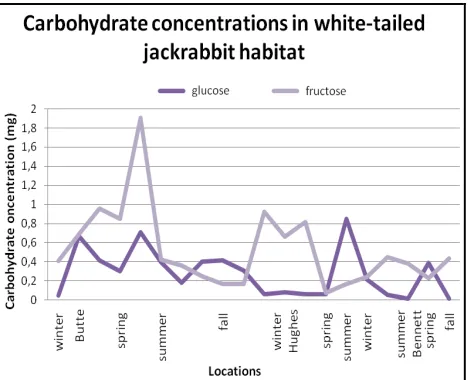
3.2. Glucose concentration
Single factor ANOVA revealed no significant difference in glucose concentrations between sites where jackrabbits inhabit (F=0.2817, p<0.001).Two way ANOVA also showed no significant difference in glucose concentrations between sites (F=0.2170, p<0.001) where jackrabbits are and season: fall, winter, spring, summer (F=0.3107, p<0.001) (Fig. 4). See Table 1 for data.
Figure 1. Percent sand content in Butte, Hughes, and
Bennett Counties with standard error. For each county, there are four bars which represent winter, spring, summer, and fall left to right.
Figure 2. Percent silt content in Butte, Hughes, and
Bennett Counties with standard error. For each county, there are four bars which represent winter, spring, summer, and fall left to right.
Figure 3. Percent clay content in Butte, Hughes, and
77 | Gilcrease et al. The decline of the white-tailed jackrabbit (Lepus townsendii)
European Journal of Biological Research 2016; 6 (2): 74-81 Table 1. Glucose concentrations between Butte, Hughes, and Bennett Counties.
Glucose
Butte (mg) Hughes (mg) Bennett (mg)
GrE McB PMd PMd*
Winter 0.0533 0.0489 0.6702 NA 0.8510 NA Spring 0.3874 0.4149 0.3010 NA 0.0806 0.0170 Summer 0.7125 0.3982 0.1803 0.5661 0.0535 0.5431 Fall 0.4022 0.4174 0.3119 0.1304 NA 0.3923 *denotes second sample at PMd.
Table 2. Fructose concentrations between Butte, Hughes, and Bennett Counties. Fructose
Butte (mg) Hughes (mg) Bennett (mg)
GrE McB PMd PMd*
Winter 1.378 0.4083 0.6908 NA 0.1719 NA Spring 0.9578 0.8535 0.4851 NA 0.6631 0.4359 Summer 1.903 0.4236 0.3622 0.2885 0.4476 0.7846 Fall 0.2517 0.1720 0.1720 0.2517 NA 0.2271 *denotes second sample at PMd.
3.3. Fructose concentration
Single factor ANOVA revealed no significant difference in fructose concentrations between sites where jackrabbits inhabit (F=0.4047, p<0.001). Two way ANOVA also showed no significant difference in fructose concentrations between sites (F=0.5517, p<0.001) where jackrabbits are and season: fall, winter, spring, summer (F=2.089, p<0.001) (Fig. 4). See Table 2 for data.
Figure 4. Glucose and fructose concentrations in
white-tailed jackrabbit habitat.
4. DISCUSSION
One study had assembled a list of seasonal preferred foods for the white tailed jackrabbit [15]; however, the carbohydrate content of various forbs, grasses, and shrubs that the white-tailed jackrabbits eat at various seasons had not been attempted until this study. Our vegetation species identified where jackrabbits forage and inhabit were similar to the findings of [15]. These include a mixture of exotic and native grasses and shrubs (e.g. smooth brome, wheatgrass species, and sagebrush).
The results of this study also demonstrate the similarity of soil texture and glucose and fructose concentrations on the three white-tailed jackrabbit habitat sites in western South Dakota; however, for the majority of our plant species (except fall thickspike wheatgrass and winter ragweed), fructose was usually higher than glucose concentrations. This was also a similar result to [22] with higher fructose concentrations in vegetation species such as smooth brome grass. Since [8] describes the
78 | Gilcrease et al. The decline of the white-tailed jackrabbit (Lepus townsendii)
European Journal of Biological Research 2016; 6 (2): 74-81 between higher fructose diets and kidney fat
accumulation in rats. However, this study was for rats and the dietary fructose concentration within jackrabbits vs. kidney fat has not yet been deter-mined.
When it comes to peaks of carbohydrate concentrations, our results were comparable to [25], who found that carbohydrate concentrations peaked in the summer for brome grass. Further, [26] found that brome grass had a commendable metabolizable energy value. If brome grass had a metabolizable energy value and if the metabolic rate of white tailed jackrabbits is higher at lower temperatures in the winter [21], this could indicate a preferable dietary choice for the jackrabbit. Unfortunately, there is no data on the white-tailed jackrabbit that specifies a healthy carbohydrate load for seasons.
Between Butte, Hughes, and Bennett Counties, the white-tailed jackrabbits seemed to prefer clay soils. It is unknown as to why the jackrabbits would have preference with clay soils and lower silt concentrations. Clay soils are one of the most chemically active [27]. One possible explanation with clay soil association could be the nutrient availability as described by [28].
One study demonstrated that the amount of carbohydrates vary in rye grasses during different times of the day with fructose being at the highest concentration at noon [29], while [22] pointed out that the carbohydrates vary at different parts of the grasses and at different maturity stages. While our study analyzed the carbohydrate concentration during various seasons, future studies could focus on analyzing the carbohydrate concentrations of the grasses during various parts of the day with a focus of plant maturity. If one were to determine the time of day and season that jackrabbits foraged more heavily on that vegetation, one could determine if jackrabbits were optimizing their carbohydrate concentrations from the grasses.
Other studies have analyzed the relationship between organismal physiology and dietary prefe-rence. For example, [15] showed seasonal changes with uterine width and size of ovaries [15]. Further, that study showed that jackrabbits chose which vegetation they eat by season (e.g. the preference of shrubs such as Parry’s rabbitbrush during the winter [15]). Further studies examining the relationships between physiology and biochemistry and diet are
needed to help portray a better understanding of white-tailed jackrabbit population health.
5. CONCLUSION
This study quantitatively analyzed the amount of carbohydrates in grasses and soil texture in white-tailed jackrabbit habitats in central and western South Dakota. The results of this study suggest that the carbohydrate concentration (glucose and fructose) of grasses are low during the fall and winter when pre-natal nutrition for the first jackrabbit litter is important. The results of this study also showed the concentrations of glucose, fructose, and soil texture between all three counties were statistically significant (p<0.001). Jackrabbits were also found in areas with a higher clay concentration for soils. It would be beneficial to compare carbohydrate concentrations and soil texture analyses to other states that contain
white-tailed jackrabbits and where they are not located to see if they are statistically significant. Further jackrabbit biochemical studies coupled with physio-logical research is needed to help portray a better understanding of white-tailed jackrabbit population health and population declines from the species.
ACKNOWLEDGEMENTS
We would like to thank the Sophie Danforth Conservation Biology Fund from Roger Williams Park Zoo for funding this study. We would also like to thank Dion Deutscher, Brittany LaDue, Diedre Wolf, Brittany Williams, and Brody Heid for their assistance on this study and the South Dakota Game, Fish and Parks for approvals to gather vegetation and soils.
AUTHORS’ CONTRIBUTION
79 | Gilcrease et al. The decline of the white-tailed jackrabbit (Lepus townsendii)
European Journal of Biological Research 2016; 6 (2): 74-81
TRANSPARENCY DECLARATION
The authors declare no conflicts of interest.
REFERENCES
1. Lim BK. Mammalian species, Lepus townsendii. Am Soc Mammal. 1987; 288: 1-6.
2. DeVos A. Range changes of mammals in the Great Lakes Region. Am Midl Nat. 1964; 71: 210-231. 3. Dumke RT. The white-tailed jackrabbit in
Wiscon-sin. Wisc Cons Bull. 1973; 38(5): 16-18.
4. Tapia II. Genetic diversity and connectivity of white-tailed jackrabbit populations in Iowa with notes on seasonal home ranges. Iowa State University, Graduate Theses and Dissertations. Paper 11273, 2010.
5. Kline PD. Notes on the biology of the jackrabbit in Iowa. Proc Iowa Acad Sci. 1963; 70: 196-204. 6. Schaible DJ. Status, distribution, and density of
white-tailed jackrabbits and black-tailed jackrabbits in South Dakota. Master’s thesis, Brookings, South Dakota State University, 2007.
7. Carter FL. A study in jackrabbit shifts in range in western Kansas. Trans Kan Acad Sci. 1939; 42: 431-435.
8. Palmer TS. The jack rabbits of the United States. Washington, Government Printing Office, 1896. 9. Mohr WP, Mohr CO. Recent jack rabbit populations
at Rapidan, Minnesota. J Mammal. 1936; 17: 112-114.
10. Andrewartha HG, Birch LC. The distribution and abundance of animals. Chicago, University of Chicago Press, 1954.
11. Bowles JB. Distribution and biogeography of mammals of Iowa. The Museum Texas Tech University Special Publications, 1975.
12. Huggett RJ. Fundamentals of biogeography. New York, Routledge, 2004.
13. Brown JH, Lomolino MV. Biogeography. Sunderland, Sinauer Associates, 1998.
14. Riegel A. Some observations of the food coactions of rabbits in Western Kansas during periods of stress. Trans Kan Acad Sci. 1942; 45: 369-375. 15. Bear GD, Hansen RM. Food habits, growth, and
reproduction of white-tailed jackrabbits in southern Colorado. Colorado, Colorado State University, 1966.
16. Fatehi M, Pieper R.D, Beck RF. Seasonal food habits of blacktailed jackrabbits (Lepus californicus) in Southern New Mexico. Southwest Nat. 1988; 33 (3): 367-370.
17. Rogowitz GL. Seasonal energetics of the white-tailed jackrabbit (Lepus townsendii). J Mammal. 1990; 71(3): 277-285.
18. Beaudoin AB, Beaudoin Y. Urban white-tailed jackrabbits (Lepus townsendii) eat spike plants (Cordyline australis) in winter. Can Field Nat. 2012; 126: 157-159.
19. Schaible D, Dieter C. Health and fertility impli-cations related to seasonal changes in kidney fat index of white-tailed jackrabbits in South Dakota. Great Plains Res. 2011; 21: 89-94.
20. Triplitt CL. Understanding the kidneys’ role in blood glucose regulation. Am J Manag C. 2012; 18: S11-16.
21. Rogowitz GL, Gessaman JA. Influence of air temperature, wind and irradiance on metabolism of white-tailed jackrabbits. J Therm Biol. 1990; 15(2): 125-131.
22. Smith D. Carbohydrates in grasses. II. Sugar and fructosan composition of the stem bases of bromegrass and timothy at several growth stages and in different plant parts at anthesis. Crop Sci. 1967; 7: 62-67.
23. Kretowicz M, Johnson RJ, Ishimoto T, Nakagawa T, Manitius J. The impact of fructose on renal function and blood pressure. Int J Neph. 2011; 2011: 315879.
24. deCastro UG, dos Santos RA, Silva ME, de Lima WG, Campagnole-Santos MJ, Alzamora AC. Age-dependent effect of high-fructose and high-fat diets on lipid metabolism and lipid accumulation in liver and kidney of rats. Lipids Health Dis. 2013; 12: 136. 25. Reynolds JH, Smith D. Trend of carbohydrate reserves in alfalfa, smooth bromegrass, and timothy grown under various cutting schedules. Crop Sci. 1962; 2(4): 333-336.
26. Swift RW, Cowan RL, Ingram RH, Maddy HK, Barron GP, Grose EC, Washko JB. The relative nutritive value of Kentucky bluegrass, timothy, brome grass, orchard grass, and alfalfa. J Anim Sci. 1950; 9: 363-372.
27. Bleam WF. Soil and environmental chemistry. Waltham, Academic Press, 2012.
28. Steenwerth KL, Jackson LE, Calderon FJ, Strom-berg MR, Scow KM. Soil microbial community composition and land use history in cultivated grassland ecosystems of coastal California. Soil Biol Biochem. 34: 2002; 1599-1611.
80 | Gilcrease et al. The decline of the white-tailed jackrabbit (Lepus townsendii)
European Journal of Biological Research 2016; 6 (2): 74-81
APPENDIX 1. Macronutrients from Butte, Hughes, and Martin Counties.
Butte
Macronutrient Winter Spring Summer Fall
GrE McB PMd GrE McB PMd GrE McB PMd GrE McB PMd
Vegetation
Phosphorus NA NA 1 NA 1 1 1 1 1 1 1 1 Nitrates NA NA 1 NA 1 1 0 0 1 0 0 1 Potassium NA NA 1 NA 1 1 1 0 1 1 0 1
Soil
Carbonates 1 0 NA NA NA 1 0 1 1 0 1 1 Nitrates 0 0 NA NA NA 0 0 0 0 0 0 0 Sulfates 1 1 NA NA NA 1 0 0 1 0 0 0 Ammonium 0 0 NA NA NA 0 0 0 0 0 0 0 Phosphates 0 NA NA NA NA 1 0 0 1 0 0 0 Magnesium 1 NA NA NA NA 0 0 0 0 0 0 0 Calcium 1 1 NA NA NA 1 1 1 1 1 1 1 Potassium 0 0 NA NA NA 0 1 0 0 1 0 0 Iron 0 0 NA NA NA 0 1 0 0 1 0 0 Note: 0 denotes the lack of the nutrient and 1 denotes presence of the nutrient. “NA” denotes that these values were not examined.
Hughes
Macronutrient Winter Spring Summer Fall
Vegetation
Phosphorous 1 1 NA 1
Nitrates 1 1 0 1
Potassium 1 0 NA 0
Soil
Carbonates 0 NA 1 1
Sulfates 0 NA 0 0
Ammonium 0 NA 0 0
Phosphates 1 NA 1 0
Magnesium 1 NA 1 1
Calcium 1 NA 1 1
Potassium 1 NA 0 1
Iron 1 NA 0 0
81 | Gilcrease et al. The decline of the white-tailed jackrabbit (Lepus townsendii)
European Journal of Biological Research 2016; 6 (2): 74-81 Martin
Macronutrient Winter Spring Summer Fall
Vegetation
Phosphorous NA NA 1 1
Nitrates NA NA 1 1
Potassium NA NA 0 0
Soil
Carbonates NA 0 0 0
Sulfates NA 0 1 0
Ammonium NA 0 1 0
Phosphates NA 0 1 0
Magnesium NA 0 0 0
Calcium NA 1 1 1
Potassium NA 0 1 0
Iron NA 0 0 0
ISSN 2449-8955 European Journal
of Biological Research Research Article
European Journal of Biological Research 2016; 6 (2): 82-91
Effects of crude plant extracts on wounded
Ricinus
communis
plants
Suzan A. Sayed
1*, Mohamed A. A. Gadallah
Botany and Microbiology Department, Faculty of Science, Assiut University, Assiut, 71516, Egypt *Corresponding author: Prof. Suzan Abd El-moneim Sayed; Fax: 0020882342708;
E-mail: [email protected], [email protected]
ABSTRACT
The effects of mechanical wounding with or without crude extracts of neem (Azadirachta indica) and
Citrullus colocynthis (CCT) supplementation on
hydrogen peroxide (H2O2), lipid peroxidation (MDA),
ascorbic acid (Asc A), phenolic compounds, and some enzymes activities in Ricinus communis plants were studied. In response to mechanical wounding
Ricinus plants produced more ascorbic acid, MDA,
free and bound phenolic components and to less extent H2O2. On the other hand, peroxidase, catalase
and SOD activities were declined upon wounding as compared with unwounded plants. Neem and CCT crude extracts application, whether independently or in combination, counteracted in various degree the deleterious effects of wounding stress on the parameters tested. Effectively, their supplementation increased the antioxidant defense ability through enhancement of ascorbic acid, reduction of H2O2
and MDA intensities. In addition, crude extracts reduced membrane injury, increased phenolic com-ponents and improved wounded plants growth. The results clearly indicate that the protection by CCT and neem crude extracts may be mediated through the modulation of cellular antioxidant levels.
Keywords: Antioxidant; Citrullus colocynthis;
En-zyme activities; Leakage; Neem; Phenolic compounds.
1. INTRODUCTION
Plants have evolved a highly sensitive and efficient system for monitoring changes in their environment. They can respond to physical da- mage by an increase in their general metabolism, including respiration rate, and this response appears to be in proportion to the severity of the damage [1]. Wounding is one of the many a biotic stresses that produce signals that migrate through cells into uninjured tissue and induces a number of
physiological responses [2].
In fact, when plants are exposed to wound-causing agents they activate mechanisms directed to healing and further defense [3]. Most of the induced responses include the generation/release, percep-tion and transducpercep-tion of specific signals for the subsequent activation of wound-related defense genes [4].
Citrullus colocynthis (L.) (CCT) is one of the
native plants of the Middle East countries used in traditional medicine and naturally adapted to arid environments and originally from tropical Asia and Africa. Watermelon (Citrullus lanatus var. lanatus (Thunb.) Matsum and Nakai) seed and root exudates inhibit germination and seedling growth of plants and growth of pathogenic fungi and bacteria. The results of Howard et al. [5] indicated that the testa of Citrullus contain at least two compounds that are
Received: 07 February 2016; Revised submission: 15 March 2016; Accepted: 23 March 2016
Copyright: © The Author(s) 2016. European Journal of Biological Research © T.M.Karpiński 2016. This is an open access article licensed under the terms of the Creative Commons Attribution Non-Commercial 4.0 International License, which permits