November 2019 Volume 25 Issue 6 www.zumj.journals.ekb.eg 960
ORIGINAL ARTICLE
VITAMIN D STATUS IN CHILDREN WITH ACUTE LYMPHOBLASTIC
LEUKEMIA
Laila Sherief1, Mohammed Beshir1, Nermin Raafat2, Heba Magdy Elsayed1* 1
Pediatric Department, Faculty of Medicine, Zagazig University, Zagazig, Egypt 2
Medical Biochemistry Department, Faculty of Medicine, Zagazig University, Egypt
*Corresponding author :
Heba Magdy Elsayed
Pediatric Department, Faculty of Medicine, Zagazig
University, Zagazig, Egypt email:
drhebahamza2012@gmail.com
Submit Date 2019-03-20
Accept Date 2019-04-25
ABSTRACT
Background: Skeletal morbidities are a common initial presentation and outcome in acute lymphoblastic leukemia(ALL). Vitamin D plays a vital role in the physiological regulation of calcium and phosphate transport and bone mineralization. Also high (25(OH)) level and high vitamin d intake at the time of diagnosis and initiation of anticancer treatment were associated with improved outcome. Methods: Twenty five ALL patients admitted to hematology and oncology unit, children hospital Zagazig University in one-year .vitamin D (25 (OH)) level was assessed in all patients at diagnosis. Patients were classified into deficient vitamin D (0-20)ng/ml , insufficient vitamin D (20-30)ng/ml and sufficient vitamin D (30-50)ng/ml. Statistical analysis was done to determine relation between different groups and ALL patients characteristics. Results: Vitamin D deficient in 24% of patients, vitamin D insufficient in 48% of patients, and vitamin D sufficient in 28% of patients. There was high statistical difference between different groups regarding bone pain and sex. Conclusions: A non-sufficient level in childhood all (deficiency and insufficiency) is common in childhood ALL and this is related to bone pain as initial clinical presentation. Vitamin D is related to presentation and prognosis of ALL in children.
Key words: Acute lymphoblastic leukemia; Vitamin D, Bone pain
INTRODUCTION
cute lymphoblastic leukemia (ALL) is the most common cancer in childhood. It accounts for 25 % of all pediatrics cancer. Skeletal morbidities are very common with ALL initially and during treatment [1]. Vitamin D plays an important role in regulation of calcium and phosphate transport and bone mineralization. It was considered that vitamin D deficiency involved in development of rickets in
children and osteomalacia in adults [2]. Several studies showed that vitamin D has a role in decreasing the risk of cancer. High 25(OH) D and high vitamin D intake at the time of diagnosis and initiation of anticancer treatment were associated with improved outcome and good prognosis [3].The most reliable way to determine Vitamin D status is by measuring serum 25-hydroxyvitamin D (25(OH)D) [4].
November 2019 Volume 25 Issue 6 www.zumj.journals.ekb.eg 961 The aim of this study was to assess
vitamin D status in pediatric patients with ALL and to identify risk factors for vitamin D deficiency.
METHODS
Our study was a cross sectional study which included all newly diagnosed acute lymphoblastic leukemia patients admitting to hematology and oncology unit, children's hospital, Zagazig University in one year. Inclusion criteria were newly diagnosed acute lymphoblastic leukemia patients age (1-18) years old and both sexes Exclusion criteria were acute lymphoblastic leukemia on therapy, patients with other malignancies and relapse ALL Consent was obtained from relatives of patients and Approval for performing the study was obtained from pediatric Department, Faculty of Medicine, and Zagazig University after Institutional Review Board of Zagazig University (IRB-ZU) approval.
All subjects in the study were subjected to Full medical history and Full physical examinations.
They were also subjected to Laboratory investigations including
-Complete blood count (CBC) by (sysmex KX-21) with examination of leishman stained peripheral smears , serum electrolytes.
-patients were evaluated for calcium(Ca),phosphorus(Ph) and alkalinephosphatase(ALP)
- Serum vitamin D (25(OH)) level assessment after after end of consolidation therapy using an enzyme-linked immunosorbent (ELISA) technique.
Serum [25(OH) D] levels of 20 to 30 ng/ml defined Vitamin D insufficiency, and a level of<20 ng/ml defined vitamin D deficiency, while levels>30 ng/ml defined normal vitamin D level (sufficient) [5].
- Bone marrow aspiratiation with examination of Leishman stained films was
done and examined for morphology and immunophenotype.
The patients were divided to 3 groups: Group (1) included patients with vitamin D deficiency.
Group (2) included patients with vitamin D insufficiency.
Group (3) included patients with vitamin D sufficient.
Statistical analysis:
Relation between vitamin D status and patients characteristics was statistically analyzed by computer using Statistical Package of Social Services version 22 (SPSS), as showing in Table (3). Suitable statistical tests of significance were used after checked for normality.
Continuous quantative variables e.g age were expressed as the mean ± SD & median (range).Comparison of several groups median was done by Kruskall Wallis test.
Categorical qualitative variables were expressed as absolute ferquencies (number)& relative frequencies (percentage) and comparison between several groups median was done by chi square test.
The results were considered statistically significant when the significant probability was less than 0.05 (P < 0.05). P-value < 0.001 was considered highly statistically significant (HS), and P-value ≥ 0.05 was considered statistically insignificant (NS).
RESULTS
November 2019 Volume 25 Issue 6 www.zumj.journals.ekb.eg 962 pain at diagnosis .Regarding bone minerals
mean of calcium was 9.47.±0.609 ,mean of phosphorus was 4.17±0.41 and mean of alkaline phosphatase was 110±16.8.
Table (2) shows that the mean of serum vitamin D (25(OH)) level among the studied group is 25.17±6.9 ng/ml, with a range from (13-35). About %24 of the patients were deficient in vitamin D. %28 of the patients were of insufficient level and (48.0%) were sufficient in vitamin D.
Table (3) shows that mean Age in patients with vitamin D deficiency 7.6±2.7years .84% are males. 67% of them suffered from common ALL. All patients with adeficient level of vitamin d suffered from bone pain.
In patients with vitamin D Insufficiency mean age were 8.8±4.3years. 58 % of them was Male and 42% was female .Common ALL immunophenotype was in 67% and T ALL immunophenotype was in33%. 33%of insufficient vitamin D level patients suffered from bone pain. Patients with vitamin D sufficiency mean age was 7.1±3.8 years. 57% of them were Male and 43 %were female. Common ALL immunophenotype was in %71 of patients and T-ALL immunophenotype was in %29 of patients of patients of patients of patients. Only 14% of them suffered from bone pain.
It was a high statistical significance in patients groups regarding bone pain. P value=0.005.
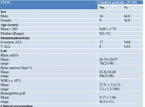
Table 1. Characteristics of all studied patients.
ITEM Studied patients (N=25)
No. %
Sex
Male 16 64.0
Female 9 36.0
Age (years)
Mean ± SD 8.08 ± 3.79
Median (Range) 9(3-15)
Immunophenotype
Common ALL 17 %68
T ALL 8 %32
Lab
Blast cells% Mean range
66.33±20.97 70(25-98) Bone marrow blast %
Mean range
83.8±18.48 90(25-98) WBCs x 109/l
Mean range
27.9 ± 115.13 3.2 ( 1.3-580) Hemoglobin g/dl
Mean range
November 2019 Volume 25 Issue 6 www.zumj.journals.ekb.eg 963
Bone pain 11 %44
Bone minerals Calcium mg/dl Mean
range
9.47.±0.609 9.5(8.16-10.70) Phosphorus mg/dl
Mean range
4.17±0.41 4.2(3-5) Alkaline phosphatase U/L
Mean range
110±16.8 108 (70-141) Table 2.Vitamin D level and status among the studied ALL patients.
Item Studied patients (N=25)
No %
Vitamin D level ng/ml
Mean ± SD 25.17±6.9
Median (Range) 26(13-35)
Vitamin D status ng/ml
Deficiency (0-20) 6 24
Insufficiency (20-30) 12 48
Sufficiency (30-50) 7 28
Table 3. relation between vitamin D status and patient characteristics in our study
Item Group (1)
(N=6)
Group (2)
(N=12)
Group (3) (N=7) p-value
N % N % N %
Age Mean range
7.6±2.7 (5-10)
8.8±4.3 (3-15)
7.1±3.8 (3-14)
0.604 #
Sex
Male 5 %83 7 %58 4 %57 0.526*
female 1 %16 5 %41 3 %42
IPT
Common ALL 4 %67 8 %67 5 %71 0.974*
T ALL 2 %33 4 %33 2 %29
lab Blast% Mean range
63.33 ± 19.66 30-90
62± 26.62 98.00
72.86 ±22.15 30-90
0.518#
November 2019 Volume 25 Issue 6 www.zumj.journals.ekb.eg 964 *chi square #kruskall walles **significant p-value
DISCUSSION
Bone disease has been reported previously in association with ALL in children and adults. But no clear mechanism has been identified. One possibility is direct involvement of bone by disease. In addition the effects of chemotherapy on gastrointestinal and renal handling of nutrients may cause alterations in mineral homeostasis that eventually lead to abnormal turnover of bone mineral[6]. Therefore, our aim of work is to assess vitamin D status in pediatric patients with ALL and to identify risk factors for vitamin D deficiency.
Regarding immunophenotyping results in this work it was found that about 2/3 of studied group diagnosed as common ALL (68%).and (32%) of them are of T-ALL. In most studies that discussed ALL immunophenotype it was reported that common ALL is the most predominant immunophenotype in childhood ALL. In study by Zhai et al, C-ALL phenotype
present 72% of the patients and T-ALL presented 7% of patients [7] and in Veerman et al., C-ALL was in 52 % of patients[8]. No one of patients in our study has B-ALL immunophenotype .This may be explained by low number of studied ALL patients in our study compared to other studies.
In this work bone pain was presented in (44%) of patients. Clinical findings in our study match that in most studies .Study carried out by Sinigaglia concluded that (34.4%) of ALL patients had bone pain [9]. Musculoskeletal pain was present in 36% of patients at the time of diagnosis in the study performed by Halton [10].
In the current work we biochemically evaluated status of bone in children with ALL through measuring of ( calcium,phosphorus and ALP ).The mean serum Ca levels at diagnosis was 9.47.±0.6 mg/dl. This value correlates with that of [9] who reported that mean of Ca at diagnosis 9.48 mg/dl.
Mean range
86.17 ± 7.22 80-97
78.17± 28.27 98.00
87.86 ±19.09 45-98
0.293#
Wbc X109/l Mean range
2.98 ± 1.73 1.3-6
4.87± 3.13 2.20- 12.20
3.26± 0 .99 1.5-4.3
0.354#
Hb g/dl Mean range
9.43± 1.098 8-11
9.16 ± 1.28 6.50 - 11.00
8.99 ±0.63 8.2-10
0.727#
Cinical presentation
Bone pain 6 %100 4 %33 1 %14 0.005**
Bone minerals Ca mg/dl Mean Range
9.5 ±0 .39 8.9- 10
9.47±0.603 8.16-10.30
9.46 ±0 .83 8.19-10.7
0.991#
Ph mg/dl Mean range
4.3±0 .4 4-5
4.115±0.503 3-5
4.17 ±15.67 4-4.4.5
0.840#
Alp U/L Mean range
117.33 ±13.23 101-140
104.58±18.298 70-126
113±0.142 97-141
November 2019 Volume 25 Issue 6 www.zumj.journals.ekb.eg 965 In the present study, we assessed
vitamin D status in ALL pediatric patients. The current study revealed that the mean of vitamin D level at diagnosis among the studied group is 25.17±6.9 ng/ml. while Demisory et al. who reported that the mean of vitamin D level at diagnosis among the studied group is 19.03 ±10.98 ng/ml [11]. Possible explanations for this variation are differences in reference data and methods of vitamin D assessment [12]. Poorer levels of vitamin D were related to the season of evaluation and they decrease with increase of age as stated in different studies [13-15] . According to vitamin D status at diagnosis of ALL among our ALL patients, 24% of children with ALL were deficient in vitamin D level, 48% Insufficient and 28% Sufficienct.This is consistent with the study done by Helou et al who reported that 71% of leukemic children were in abnormal level of vitamin D at diagnosis [16]. It was noticed that there was association between vitamin D statuses and gender with study which revealed also that younger ages were associated with vitamin d deficiency [13], however in our work it was no statistical significance gender and age and vitamin d deficiency. in the present study, there was no significant difference between age, sex and vitamin D levels at diagnosis.Our findings are compatible with Simmons et al. [14] and Choudhary et al.[17] findings that reported there was not enough evidence to suggest treatment type or gender were associated with low serum 25(OH) D levels.
CONCLUSION
More than half of children diagnosed with ALL are with Non-sufficient levels (deficiency and insufficiency) of vitamin D .Vitamin D status is related to bone pain in ALL.
Declaration of interest
The authors report no conflicts of interest. The authors alone are responsible for the content and writing of the paper.
Funding information
None declared
REFERANCES
1 Mandel, K., Atkinson, S., Barr, R. D., & Pencharz, P. :Skeletal morbidity in childhood acute lymphoblastic leukemia. Journal of Clinical Oncology 2004; 22(7), 1215-1221. 2-Deeb KK, Trump DL, Johnson CS. :
Vitamin D signaling pathways in cancer: potential for anticancer therapeutics. Nat Rev Cancer 2007;7:684–700.
3. Goodwin PJ, Ennis M, Pritchard KI, Koo J, Hood N. Prognostic effects of 25hydroxyvitamin D levels in early breast cancer. J Clin Oncol 2009;27:3757– 63.
4- Wagner, D., Hanwell, H.E. and Vieth, R.,. An evaluation of automated methods for measurement of serum 25-hydroxyvitamin D. Clinical biochemistry 2009;42(15), pp.1549-1556.
5-Holick MF. Vitamin D deficiency. New England Journal of Medicine. 2007; 19;357(3):266-81.
6-Strauss, A. J., Su, J. T., Dalton, V. M. K. et al. Bony morbidity in children treated for acute lymphoblastic leukemia. Journal of Clinical Oncology 2001; 19(12), 3066-3072.
7-Zhai, X., Wang, H., Zhu, X. et al. Gene polymorphisms of ABC transporters are associated with clinical outcomes in children with acute lymphoblastic leukemia. Archives of medical science 2002; 8(4), 659.
8-Veerman AJ, Kamps WA, van den Berg H, van den Berg E, Bökkerink JP, Bruin MC et al. Dexamethasone-based therapy for childhood acute lymphoblastic leukaemia: results of the prospective Dutch Childhood Oncology Group (DCOG) protocol ALL-9 (1997–2004). The lancet oncology. 2009 ;10(10):957-66.
9- Sinigaglia, R., Gigante, C., Bisinella, G. e Musculoskeletal manifestations in pediatric acute leukemia. Journal of Pediatric Orthopaedics 2008; 28(1), 20-28.
10- Halton, J.M., Atkinson, S.A., Fraher, L., Webber et al. Mineral homeostasis and bone mass at diagnosis in children with acute lymphoblastic leukemia. The Journal of pediatrics1995; 126(4), pp.557-564.
November 2019 Volume 25 Issue 6 www.zumj.journals.ekb.eg 966 Calcium Supplementation With Bone
Mineral Density in Pediatric Acute Lymphoblastic Leukemia Patients. Journal of pediatric hematology/oncology 2017; 39(4), 287-292.
12- van der Sluis, I. M., van den Heuvel-Eibrink, M. M., Hählen, K., Krenning et al. Altered bone mineral density and body composition, and increased fracture risk in childhood acute lymphoblastic leukemia. The Journal of pediatrics 2002;141(2), 204-210. 13- Sinha, A., Avery, P., Turner, S. et al.
(Vitamin D status in paediatric patients with cancer. Pediatric blood & cancer2011; 57(4), 594-598.
14- Simmons, J. H., Chow, E. J., Koehler, E. et al. Significant 25‐hydroxyvitamin D deficiency in child and adolescent survivors
of acute lymphoblastic leukemia: Treatment with chemotherapy compared with allogeneic stem cell transplant. Pediatric blood & cancer 2011; 56(7), 1114-1119.
15 - Mohan, R., Mohan, G., Scott, J. et al. Vitamin D insufficiency among children with cancer in India. Indian journal of medical and paediatric oncology: official journal of Indian Society of Medical & Paediatric Oncology 2016; 37(1), 14.
16- Helou, M., Ning, Y., Yang, S., et al. Vitamin d deficiency in children with cancer. Journal of pediatric hematology/oncology 2014; 36(3), 212-217.
17- Choudhary A, Chou J, Heller G, Sklar C. Prevalence of vitamin D insufficiency in survivors of childhood cancer. Pediatr Blood Cancer. 2013;60:1237–9.
To Cite This Article: Laila SM, Nermin R, Heba ME. Vitamin D Status In Children With Acute