This is an open access journal, and articles are distributed under the terms of the Creative Commons Attribution-Non Commercial-ShareAlike 4.0 License, which allows others to remix, tweak, and build upon the work non-commercially, as long as appropriate credit is given and the new creations are licensed under the identical terms.
© 2017 Journal of Advanced Pharmacy Education & Research | Published by SPER Publication 404
Augmentation of fenestrated pedicle screws with cement in
patients with Osteoporotic spine
Mahmoud Abousayed
*, Ahmed Sultan, Wael Koptan,
Yasser El Miligui
Orthopedic Department, Kasr Al Ainy Medical School, Cairo University, Egypt.11956
Correspondence: Mahmoud Abousayed, Orthopedic Department, Kasr Al Ainy Medical School, Cairo University, Egypt.11956, E. Mail: [email protected]
ABSTRACT
Study design: Retrospective observational study
Purpose: To evaluate the purchase of fenestrated pedicle screws augmented with cement in patients with osteoporosis.
Background: Backing out and failure of pedicle screws in patients with osteoporosis is becoming a big problem due to wide use of these screws nowadays.
Patients and methods: From May 2015 to January 2016, 25 patients with a poor bone stock condition underwent posterior fixation by fenestrated pedicle screws and cement augmentation. Assessment of pain improvement was done by visual analogue scale (VAS) score while long-term clinical outcome was assessed by Oswestry low back disability questionnaire (Oswestry disability index ODI). Implant stability was evaluated by plain radiography. Complications were evaluated in all cases.
Results: All patients were followed up clinically and radiographically for a mean of 24.84 months. There was significant reduction in pain and improvement of quality of life as detected by VAS scores and ODI questionnaire consecutively (P value<0.001). No radiological loosening or backing out of screws was observed. Cement leakage occurred in five cases.
Conclusion: Augmentation of fenestrated screws with cement provided effective and lasting purchase in patients with osteoporosis. The only clinical complication strictly related to this technique was cement leakage.
Keywords: Fenestrated pedicle screw, polymethylmethacrylate, osteoporotic bone
Introduction
Osteoporosis is a systemic disease characterized by reduction of the bone mass with deterioration of the microarchitecture. The resulting decrease in bone mechanical strength typically manifests as fragility fractures, with about one half of osteoporotic fractures occurring in the spine[1]
. The spinal surgeon may be required to treat direct sequelae of osteoporosis in the form of painful spinal fractures or resultant deformity or to consider osteoporosis as it relates to spinal reconstructive
surgery in the older patient[2].
Pedicle screws are widely used for spine stabilization in the elderly, as they enable easier and faster recovery in different conditions, such as fractures, infections, tumors and degenerative conditions. However, failures due to screw loosening or backing out are becoming a major cause of morbidity in the elderly because of their poor bone quality[3].
Many solutions have been proposed to reduce this risk, including the use of hydroxyapatite-coated screws[4]
, expandable screws[5], longer screws anchoring the anterior cortex, larger
diameter screws[5], and cement augmentation[6, 7].
The use of fenestrated cannulated screws improves the cement augmentation procedures[3]. The aim of this retrospective study
was to evaluate the purchase of fenestrated pedicle screws augmented with cement in patients with osteoporosis.
Patients and Methods
From May 2015 to January 2016, 25 surgical procedures were performed by means of a posterior approach using pedicle screws in the thoracic spine, lumbar spine and the sacrum for the treatment of traumatic, degenerative, or neoplastic
Access this article online
Website: www.japer.in E-ISSN: 2249-3379
How to cite this article:Mahmoud Abousayed, Ahmed Sultan, Wael Koptan, Yasser El Miligui. Augmentation of fenestrated pedicle screws with cement in patients with Osteoporotic spine. J Adv Pharm Edu Res 2017;7(4):404-409.
Source of Support: Nil, Conflict of Interest: None declared.
405
Journal of Advanced Pharmacy Education & Research | Oct-Dec 2017 | Vol 7 | Issue4
conditions. In 25 patients with bone softening caused by osteoporosis, infection or neoplastic conditions, fenestrated screws were used for cement augmentation in order to achieve better purchase. There were 20 women and 5 men, with a mean age of 61.96 years (range 50–78). Indication for the use of cemented screws was confirmed by evaluating the degree of osteoporosis in all patients. T score <-2.5 SD was an indication for this technique, and it was found in eight patients with degenerative disease, nine with traumatic fracture, three with post-traumatic kyphosis, one case of failed previous surgery, two cases with infection and two neoplastic patients.
A total of 97 fenestrated screws were implanted (min 2; max 6), always in combination with standard screws (a total of 24 standard screws were implanted) of the same system. In tumor patients, we performed short fixations without fusion, one level below and above the lesion. Inclusion criteria: Osteoporotic vertebral fractures, Degenerative diseases and deformities in osteoporotic vertebrae, Infections and tumors in elderly spine. Exclusion criteria: Medically unfit patients for anaesthesia, Old patients with T score >-2.5 SD, Sensitivity to screws metal or cement, Patients who refuse or unwilling to do surgery. Patients Were assessed preoperatively using Visual Analogue Score (VAS) and Oswestry Disability Index (ODI). Data were collected from preoperative and postoperative notes as well as follow up notes in outpatient clinic.
Surgical technique
Intravenous antibiotics were given one hour prior to surgery, general anesthesia induction, patient positioned in prone. Posterior midline approach was used for all patients (Fig. 1). Decompression according to the lesion level was performed for patients with degenerative diseases, patients with neurological deficit and patients with spondylodiscitis. Conventional C-arm fluoroscopy was used for the entire procedure. Pedicle screws holes were done above, below the level of the lesion and in the affected level when possible (in infection).
Figure 1: Posterior midline approach
The tract was palpated with a straight sensor probe to make sure the pedicle wall of the tract and the anterior cortex of the vertebral body were not violated. Fenestrated screws used in this study had two oval holes set into the grooves of the distal portion of the thread (Fig.2).
They were available in diameters of 6.5 and 5.5, and only polyaxial uploading models. The cement injected under pressure through the cannulation, is extruded through the holes to fill the spaces inside the osteoporotic cancellous bone, thereby increasing the purchase of the screw. The fenestrated screw is inserted into the pedicle, as done with conventional
screws. The length of the screw and the positions of the holes, located as far as possible from the posterior wall, must be carefully checked in order to prevent possible leakage into the canal.
Figure 2: Cannulated screw with distal fenestrations
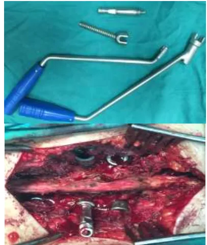
The screw and the cement injector were connected by a specifically designed connector (Fig.3 A & B). Common vertebroplasty cement is delivered through its specific syringe which is connected to the connector.
Figure 3: A) Showing set used including screw, connector and tightening instrument. B) Showing the specifically designed
connector
Journal of Advanced Pharmacy Education & Research | Oct-Dec 2017 | Vol 7 | Issue4 406
defect area and harden in vivo without generating heat. The rod (5.5 diameter) must only be connected to the screws once the polymerisation process had been completed, in order to prevent microfractures at the screw bone interface.
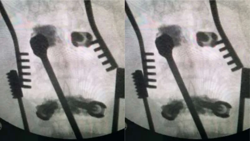
Figure 4: A) Cement injection through its specific syringe B) checked by image intensifier
Cement distribution was checked with fluoroscopic images in AP and lateral projections (Fig. 5 A & B). In case of epidural,
intradiscal or prevertebral cement extravasation, the injection of cement stopped.
After insertion of screws posterior or posterior-lateral fusion was performed by local bone graft, then the muscle and subcutaneous tissue were closed in layers, skin was closed subcuticular in all cases.
Postoperative care:
Immediately after closure ofwound and recovery from anaesthesia inside the operating theatre, neurological assessment was done to avoid any deterioration. Intravenous antibiotics (3rd generation cephalosporins), analgesics and subcutaneous Clexanewere given for 24 to 72 hours. Drain was removed 48 hours postoperative in all patients. Patients were then discharged on oral antibiotics, analgesics, anticoagulant and antacids for two weeks. Patients were allowed to ambulate with protected thoracolumbar-sacral orthosis or lumbar-sacral orthosis second day postoperative. Usually the orthosis was used for 1 to 2 months. Hospital stay was from 2 to 7 days postoperative with a mean of 3.5 days. Supplementary calcium and bisphosphonates were routinely given for treating the general osteoporosis, together with neuromodulators. Physiotherapy was started 2 weeks postoperative for 12 sessions. Hospital stay was from 2 to 7 days postoperative with a mean of 3.5 days.
Figure 5: A) AP view B) Lateral view
Assessment of the outcome
A) Clinical Outcome Measurements
They were measured pre- and postoperatively using the visual analogue scale (VAS) and Oswestry low back disability questionnaire (ODI).
B) Radiographic Assessment
Implant stability and fusion results were evaluated by plain radiography (X-rays in AP and lateral views) performed: Immediately after surgery, monthly in the first three months, every three months thereafter. Assessment was done by a spine consultant independent blinded. Assessment was for 1) Implant stability: by the position of the screws and rods, any loosening or backing out was considered implant instability. 2) Fusion:
standard radiograms were used to assess how the fenestrated screws supported the bone fusion. It was confirmed by the presence of trabecular bone bridging the interspace between the adjacent vertebral bodies.
Statistical Analysis
Mann-407
Journal of Advanced Pharmacy Education & Research | Oct-Dec 2017 | Vol 7 | Issue4
Whitney test to compare Preoperative VAS and ODI to Postoperative VAS and ODI.
Results
From May 2015 to January 2016, 25 surgical procedures were performed by means of a posterior approach using Fenestrated pedicle screws with cement augmentation in the dorsal, lumbar and sacral spine for treatment of traumatic, degenerative, infective and neoplastic conditions in Cairo University Hospital. Patients were observed, via clinical and radiological examinations. The mean follow-up time was 24.84 months (range 8–30). There were 20 women and 5 men, with a mean age of 61.96 (range 50-78). Patients’ demographics are shown in (Table 1).
Table 1: Showing age and gender distribution
Age group Number Males Females Percentage
50-60 10 1 9 40% 61-70 14 4 10 56% >70 1 0 1 4% Total 25 5 20 100%
Clinical Results
1.
Pain (VAS)
The mean preoperative VAS was 8.76 (range 7-10). Being the most common complaint, pain showed significant improvement and the mean postoperative VAS became 2.24 (range 0-5) recorded during the last clinical control (P value<0.001) (Table 2). Stretch signs and claudications were the main indications for surgery in nine patients.
2.
Oswestry Disability Index
Patients showed significant improvement in the quality of life as measured by the Oswestry Disability Index (ODI) (P value<0.001).
Table 2: Showing preoperative and postoperative results
No.MinimumMaximum Mean Std. deviationMedian
Age 25 50 78 61.96 5.38 61
VAS
pre 25 7 10 8.76 1.01 9
VAS
post 25 0 5 2.24 1.16 2
ODI
pre 25 60% 93.3% 75.96% 12.15% 73.3% ODI
post 25 20% 44.4% 28.79% 8.08% 27%
Radiographic Results
Bone fusion was achieved in all patients within six months with no cases of pseudarthrosis being recorded. No cases of loosening or backing out of screws were recorded. There was no statistical difference in the outcomes in patients with different T scores.
Complications
Cement leakage was found in five cases. In two patients, cement leakage was noticed intraoperatively. Cement was removed, during the same surgical procedure in one case and it couldn't be removed in the other as it was already solid (Fig.6), without neurological sequelae. No more than 2 cc per screw was injected in the rest of the cases, and no major leakage into
the canal occurred. We noticed leakage postoperatively in three other cases but without any clinical relevance. There were two cases of postoperative infections, one was successfully treated by antibiotics and the other needed surgical debridement. Four cases needed intraoperative blood transfusion and three of them needed postoperative transfusion due to blood loss of more than 800mL. Blood loss was related to the pathology and not to the use of fenestrated screws rather than conventional screws. Two cases were lost on follow up and were found to be dead at 8 and 10 months postoperatively secondary to other co-morbidity rather than the procedures (Table 3).
Figure 6: Cement leakage posteriorly
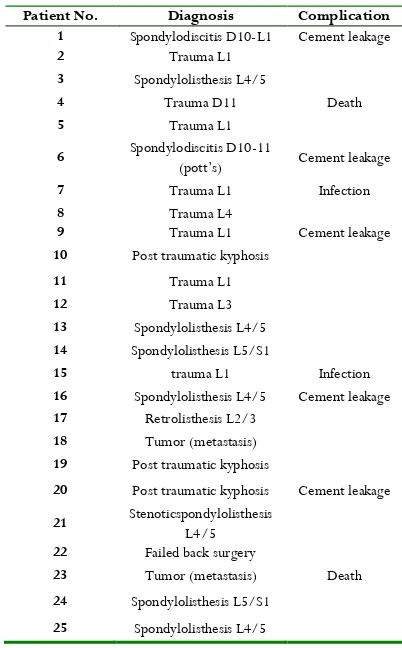
Table 3: Showing diagnoses and complications
Patient No. Diagnosis Complication
1 Spondylodiscitis D10-L1 Cement leakage
2 Trauma L1
3 Spondylolisthesis L4/5
4 Trauma D11 Death
5 Trauma L1
6 Spondylodiscitis D10-11
(pott’s) Cement leakage
7 Trauma L1 Infection
8 Trauma L4
9 Trauma L1 Cement leakage
10 Post traumatic kyphosis
11 Trauma L1
12 Trauma L3
13 Spondylolisthesis L4/5 14 Spondylolisthesis L5/S1
15 trauma L1 Infection
16 Spondylolisthesis L4/5 Cement leakage 17 Retrolisthesis L2/3
18 Tumor (metastasis) 19 Post traumatic kyphosis
20 Post traumatic kyphosis Cement leakage
21 Stenoticspondylolisthesis L4/5 22 Failed back surgery
23 Tumor (metastasis) Death 24 Spondylolisthesis L5/S1
Journal of Advanced Pharmacy Education & Research | Oct-Dec 2017 | Vol 7 | Issue4 408
Discussion
The bone–screw interface is greatly affected by bone density which decreases by age. The special configuration of the trabecular meshwork affects the mechanical grip of the screws and the integration at the interface between bone and metal. That is why surgery of the osteoporotic spine is burdened with a high incidence of implant failure as a result of pedicle screws loosening and backing out[8]. This can also be found during
revision surgeries or whenever a disease causes deterioration in bone quality. Many solutions for enhancing the grip of the screw have been described in literature[4, 5, 9]. Using screws with
a larger diameter than those previously used proved to be effective in revision surgery[10]. However, it is not always
possible to use bigger screws as they carry the risk of fracture of the pedicle[5, 11]
. Using longer screws, reaching the anterior cortex of the vertebral body, has also been proposed. Zindrick et al. found that the force increased significantly but could not ignore the risk of vascular injuries[12]
. Expansion screws in which the anterior part expands in diameter once the screw has passed through the pedicle have been tried. Results have shown that such screws are more resistant to pull-out in osteoporotic spine[5]. Hydroxyapatite coated pedicle screws can also improve
implant stability[4]. It was found that hydroxyapatite-coated
screws offered greater resistance to pull-out stresses than uncoated screws[9]. The use of cement has been a standard
procedure in orthopaedic surgery for decades. Many studies have proven that cement augmentation improves resistance to pull-out in osteoporotic and normal vertebrae[13-16]. In
osteoporotic bone, a gap is created between the threaded portion of the screw and the trabecular bone; cement strengthens the bone/metal interface at such gap. Cement augmented screws may increase both the primary stability and the fatigue resistance of the implants[6, 7]
, making them better able to withstand the axial stresses responsible for pull-out[13-15, 17]. Cement augmentation of pedicle screws can be carried out
by direct technique through injecting the cement then inserting the screw. But this technique carries the risk of increasing the pressure which may cause leakage of the cement, with possible venous embolism or cord damage[3]
. More recently, fenestrated screws, through which cement can be injected have been used[18]. In 2005, Yazu et al. compared the performance of
fenestrated screws with that of conventional screws[16]
.
Injection of cement can be done more accurately using fenestrated screws, reducing the risk of leakage into the canal[19]
.
Several pitfalls are to be mentioned. Cement leakage is the most common, can occur in the retroperitoneal space in case of defective anterior wall or in the canal in case of defective medial wall. Additionally, no movement should be done before the cement becomes solid, to avoid the breakage of cement bridges between the bone and the screw. Finally, because this technique carries the same risks as vertebroplasty, cement injection should be continuously controlled by fluoroscopy. In two of the patients reported in our study, such leakage occurred, this complication was probably due to our limited experience with this technique, as we were at the beginning of the learning curve. In one case, cement was removed intraoperative, while in the other case it was already solid and couldn't be removed.
Fransen et al. in 2007[18]
reported no cases of cement leakage, while Pare’ et al. in 2011[19] reported three cases of leakage
anterior and lateral. Becker et al. in 2008[20] had 20% leakage
which contributed to 4 cases, Frankel et al. in 2007[17]
also had 4 cases of anterior and posterior leakage. In 2011, Amendola et al. [3] had 5 cases of leakage, 2 discovered intraoperatively.
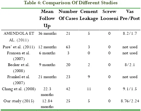
Comparison of different studies are shown in table 4.
To avoid this complication, CT scan is done for evaluation of the base of the pedicle and this technique is contraindicated in case of defective posterior wall or pedicle[18]
. Injection should be done under fluoroscopic monitoring, and should be stopped if any leakage is observed. The optimum amount to be injected is no more than 2 cc. Insertion of the screws should be done carefully to avoid breaching the medial cortex leading to cement leakage in the canal.
In our study, cement augmentation improved pedicle screw fixation in osteoporotic bone. The concentration of cement around distal part of the screw gives higher force than its uniform distribution, thus less failure rates. Distal fenestrations allow also delivery of the whole volume of cement into the vertebral body, anterior to the neurocentral junction. This technique provides safer option for spine surgeons when using cement for screw augmentation by decreasing the risk of leakage into the epidural space through an unrecognized pedicle breach.
Incidence of leakage is higher in osteoporotic spines, thus this technique is preferred in such cases[20]. No screw loosening was
recorded after a mean follow-up of 24.84 months.
Table 4: Comparison Of Different Studies
Mean Follow Up Number Of Cases Cement Leakage Screw Looseni Vas Pre/Post AMENDOLA ET AL. (2011)
36 months 21 5 0 8.2/1.7
Pare’ et al. (2011) 12 months 63 3 0 not used Fransen et al.
(2007)
6 months 3 0 0 not used
Becker et al. (2008)
9 months 20 2 0 8/2.1
Frankel et al. (2007)
21 months 23 9 0 not used
Chang et al. (2008) 22.3 months
42 11 0 9.1/1.5
Our study (2015) 12.84 months
25 5 0 8.76/2.24
Conclusion
Based on our results, we think that the use of this technique in patients with bone softening caused by osteoporosis, infection or malignancy is recommended. This is due to being helpful for increasing the stabilization of fixation and preventing screw loosening. The main complication is cement leakage.
Limitations Of The Study
409
Journal of Advanced Pharmacy Education & Research | Oct-Dec 2017 | Vol 7 | Issue4
References
1. Andersson, G.B., et al., consensus summary on the diagnosis and treatment of osteoporosis. spine, 1997.22(24): p. 63s-65s.
2. Ettinger, B., et al., reduction of vertebral fracture risk in postmenopausal women with osteoporosis treated with raloxifene: results from a 3-year randomized clinical trial. jama, 1999.282(7): p. 637-645.
3. Amendola, L., et al., fenestrated pedicle screws for cement-augmented purchase in patients with bone softening: a review of 21 cases. journal of orthopaedics and traumatology, 2011.12(4): p. 193-199.
4. Nicolialdini, N., et al., pedicular fixation in the osteoporotic spine: a pilot in vivo study on long‐term ovariectomized sheep. journal of orthopaedic research, 2002.20(6): p. 1217-1224.
5. Cook, S.D., et al., lumbosacral fixation using expandable pedicle screws: an alternative in reoperation and osteoporosis. the spine journal, 2001.1(2): p. 109-114. 6. Wuisman, P., et al., augmentation of (pedicle) screws
with calcium apatite cement in patients with severe progressive osteoporotic spinal deformities: an innovative technique. european spine journal, 2000.9(6): p. 528-533.
7. Burval, D.J., et al., primary pedicle screw augmentation in osteoporotic lumbar vertebrae: biomechanical analysis of pedicle fixation strength. spine, 2007.32(10): p. 1077-1083.
8. Cook, S.D., Et Al., biomechanical study of pedicle screw fixation in severely osteoporotic bone. the spine journal, 2004.4(4): p. 402-408.
9. Hasegawa, T., et al., hydroxyapatite-coating of pedicle screws improves resistance against pull-out force in the osteoporotic canine lumbar spine model: a pilot study. the spine journal, 2005.5(3): p. 239-243.
10. Polly Jr, D.W., j.r. orchowski, and r.g. ellenbogen, revision pedicle screws: bigger, longer shims‐what is best? spine, 1998.23(12): p. 1374-1379.
11. Hirano, T., et al., fracture risk during pedicle screw insertion in osteoporotic spine. journal of spinal disorders & techniques, 1998.11(6): p. 493-497.
12. Zindrick, M.R., et al., a biomechanical study of intrapeduncular screw fixation in the lumbosacral spine. clinical orthopaedics and related research, 1986.203: p. 99-112.
13. Rohmiller, M.T., et al., evaluation of calcium sulfate paste for augmentation of lumbar pedicle screw pullout strength. the spine journal, 2002.2(4): p. 255-260. 14. Turner, A.W., et al., hydroxyapatite composite resin
cement augmentation of pedicle screw fixation. clinical orthopaedics and related research, 2003.406(1): p. 253-261.
15. Renner, S.M., et al., augmentation of pedicle screw fixation strength using an injectable calcium phosphate cement as a function of injection timing and method. spine, 2004.29(11): p. e212-e216.
16. Yazu, M., Et Al., efficacy of novel-concept pedicle screw fixation augmented with calcium phosphate cement in the osteoporotic spine. journal of orthopaedic science, 2005.10(1): p. 56-61.
17. Frankel, B.M., S. D'agostino, And C. Wang, a biomechanical cadaveric analysis of polymethylmethacrylate-augmented pedicle screw fixation. 2007.
18. Fransen, P., increasing pedicle screw anchoring in the osteoporotic spine by cement injection through the implant. 2007.