-500-
ANTI- ANNEXIN 1 ANTIBODIES CORRELATION BETWEEN ANTIBODIES LEVEL AND
DISEASE ACTIVITY IN SOME AUTOIMMUNE CONNECTIVE TISSUE DISEASES
Khashaba S A, Metwalli M I, Ghanem A H and Zedan A A*.
Dermatology and Venereology Department, Faculty of Medicine, Zagazig University *Clinical Pathology Department, Faculty of Medicine, Zagazig University
ABSTRACT
Introduction: Annexin 1 is an anti-inflammatory protein, exerts a regulatory effect in both innate and adaptive immune responses. Autoantibodies against annexin 1 have been recognized in some patients with autoimmune connective tissue diseases. Aim: to detect presence of anti-annexin 1 antibodies in patients with rheumatoid arthritis, scleroderma, systemic and discoid lupus erythematosus and to evaluate whether there is a correlation between the antibodies levels and disease activity. Methods: determining the levels of IgM antibodies in serum samples by ELISA and correlating them with the disease activity, which is evaluated using specific activity index for each disease. Results: levels of anti- annexin 1 antibodies were found to be significantly higher in diseased groups when compared to normal healthy controls. When correlating antibodies levels with disease activity, rheumatoid arthritis and discoid lupus patients showed no significant correlation, however systemic lupus and scleroderma patients showed significant correlation between antibodies levels and disease activity. Conclusion: anti- annexin 1 antibodies might be a promising aid in diagnosis of these diseases and could have a prognostic capacity in some autoimmune connective tissue diseases.
Key words: Annexin 1- autoantibodies- autoimmune connective tissue diseases.
1- INTRODUCTION
utoimmune connective tissue diseases (ACTDs) are characterized by the spontaneous stimulation of the immune system with the production of
autoantibodies, which are specific for
self-components in the nucleus and cytoplasm, often macromolecular complexes of proteins and nucleic acids. ACTDs can affect any connective tissue of the human body via inflammation or destruction. The classic ACTDs include systemic lupus erythematosus (SLE), rheumatoid arthritis (RA), systemic sclerosis (SSc), Sjögren's syndrome (SS) and mixed connective
tissue disease (MCTD). [1]
Annexin 1(Anx 1) is the prototype of a new class of mediators able to exert a regulatory effect in innate and adaptive immune responses. It is widely distributed in the body in different cells and biological fluids. Anx 1 has been shown to have anti-inflammatory properties by suppressing the generation of inflammatory mediators, including prostaglandins, thromboxanes, and leukotrienes, by inhibiting neutrophil traffic, mast-cell degranulation and activation, and by stimulating neutrophil apoptosis and phagocytosis of apoptotic cells by macrophages. It has also been known to modulate Th1/ Th2 balance and T cell signaling. [2]
The idea that Anx1 could be an autoantigen in inflammatory and autoimmune diseases is a fascinating hypothesis that has been largely neglected it would be interesting to investigate the presence of Anx1 autoantibodies in autoimmune patients including rheumatoid arthritis (RA), scleroderma(SS), systemic lupus erythematosus (SLE) and discoid lupus erythematosus (DLE). [3]
2- PATIENTS AND METHODS
2.1. Patients
Eighty patients were collected from both Dermatology and Venereology Department and Rheumatology Department at Zagazig University Hospitals after being consented to participate in the study. Patients were divided into five groups, fifteen patients at each disease group RA, SLE, SS and DLE, in addition to twenty normal matched controls. All the patients have fulfilled the American College of Rheumatology (ACR) criteria specific for each disease. We recorded demographic data for all patients and controls.
The patients’ disease activities were evaluated using Disease Activity Score (DAS28) [4] for RA, SLE Disease Activity Index (SLEDAI-2K) [5] for SLE,Valentini Disease Activity Index [6] for SS and
The Cutaneous Lupus Erythematosus Disease Area
and Severity Index (CLASI) [7] for DLE.
Serum samples from patients and healthy individuals were collected and kept at−80 °C.
2.2. Detection of anti-anxexin 1 antibodies
To detect anti-annexin 1 antibodies in human sera, an ELISA was established. Wells were coated for two hours with 100µl of carbonate- bicarbonate buffer PH 9.6(provided by Biochemistry Department, Zagazig University) containing 1 µg purified recombinant human annexin 1 (USCN Life science INC., Houston, USA) at room temperature.
-501-
wells were incubated with AP-conjugated polyclonal goat anti-human IgM (Bioss INC, USA) diluted 1:1000 for 1hr at 37°C.After several washing steps, development was carried out with 100 µl phosphatase substrate for two to three minutes and then a stop solution was added. Absorbance values at 405 nm (A 405) were obtained with ELISA reader, and results were expressed in ELISA units. ELISA units up to 64 are considered normal. An ELISA unit was defined by the following equation proposed by Goulding [8]
ELISA unit = A 405 (sample) x sample dilution / reaction volume (ml)
2.3. Statistical analysis
For data analysis SPSS version 20 was used and the following tests were use; chi- square test (χ²), one way ANOVA (F) and Pearson’s correlation
coefficient(r). Results were expressed as mean ( ± standard deviation (SD), ranges, numbers or percentages. P-value ˂ 0.05 was considered statistically significant and P-value ˂ 0.001 was considered statistically highly significant
3- RESULTS
3.1. Characteristics of the study population
This study included 60 patients, 15 in each disease group in addition to 20 healthy controls. Descriptive and
comparative statistics of the demographic data of the studied patients are demonstrated in (table 1). The activity of the patients in the first four groups was estimated at the time of examination using specific activity index for each disease. The descriptive statistics of the activity index score of the studied groups are shown in (table 2).
3.2. Anti-annexin 1 antibodies
There is highly statistical significant difference (P< 0.001) when compare antibodies levels of the control group with the other four diseased groups and also highly statistical significant difference (P< 0.001) in the prevalence of the positivity between control and cases. Descriptive and comparative statistics of the antibody levels and prevalence of positivity among studied groups are shown in (tables 3and 4)
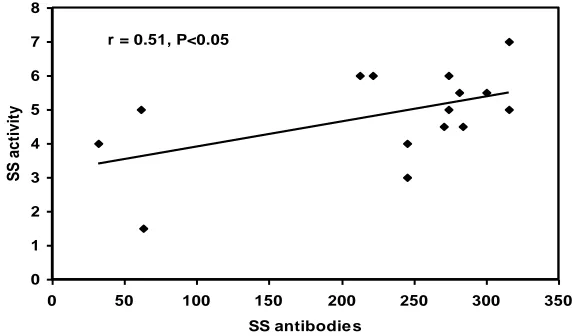
3.3. Correlation between anti-annexin 1 antibodies and diseases activity
There was no statistical significant correlation between antibodies in the serum and the disease activity in both group (1) RA and group (4) DLE. On the other hand, there was statistical significant correlation between antibodies levels in serum and disease activity in group (2) SLE and group (3) SS. (table 5) the positive correlations are represented in (Figure1 and 2)
Table (1):Descriptive and comparative statistics of the demographic data of the studied groups.
Group (1) RA N=15
Group (2) SLE N=15
Group (3) SS N =15
Group (4) DLE N =15
Group (5) CONTROL N = 20
Age(years)
± SD Range
44.5 ± 8.2 30 - 55
34.3 ± 10.6 15 – 50
41.2 ± 12.8 17 - 60
37.3 ± 8.3 14 - 48
38.8 ± 10.2 23 - 55
F = 2.2
P = 0.07 NS
Gender
(N. %) Male Female
1 6.7% 14 93.3%
2 13.3% 13 86.7%
5 33.3% 10 66.7%
7 46.7% 8 53.3%
4 20.0% 16 80.0%
χ² = 8.58
-502-
Table (2):Descriptive statistics of the activity index score of the studied groups.
Diseases Index score
± SD (Range)
Group (1) RA 5.7 ± 1.4 (3.48 – 7.61)
Group (2) SLE 27.5 ± 8.2 (16 – 44)
Group (3) SS 4.8 ± 1.36 (1.5 – 7)
Group (4) DLE 10.87 ± 6.5 (5 – 25)
Table (3):Descriptive and comparative statistics of the antibody levels among studied groups.
Groups Antibodies
± SD (Range)
F P
(1) RA 249.3 ± 103.3 (35 – 445)
16.7 0.001
Highly significant (2) SLE 256.2 ± 135.0 (35 – 497)
(3) SS 226.5 ± 95.2 (32 – 316)
(4) DLE 193.7 ± 133.8 (21 – 490)
(5) Control 20.9 ± 9.2 (4 – 31)
Table (4): Prevalence of positivity
+/-
N %
RA
SLE
SS
DLE
Control
χ² P
-ve 2 13.3 % 3 20 % 3 20 % 5 33.3 % 20 100 % 39.29 0.001 Highly significant +ve 13 86.7 % 12 80 % 12 80 % 10 66.7 % 0 0 %
Table (5): Correlation between antibodies and activity index
groups R P Significance
Group (1) RA 0.4 > 0.05 NS
Group (2) SLE 0.57 < 0.05 Sig
Group (3) SS 0.51 < 0.05 Sig
-503-
r = 0.57, P<0.050 5 10 15 20 25 30 35 40 45 50
0 100 200 300 400 500 600
SLE antibodie s
S
L
E
a
c
ti
v
it
y
Figure (1) showing the correlation between SLE antibodies and the disease activity.
r = 0.51, P<0.05
0 1 2 3 4 5 6 7 8
0 50 100 150 200 250 300 350
SS antibodie s
S
S
a
c
ti
v
it
y
Figure (2) showing the correlation between SS antibodies and the disease activity.
4- DISCUSSION
Annexin1 is an important member of the phospholipid-binding proteins named annexins. Functionally, it is an endogenous anti-inflammatory protein and plays a critical role in diverse cellular functions. Multiple researches have suggested that annexin1 was involved in certain autoimmune disorders as a possible antigen. [9]
Some previous researches have shown the presence of anti- annexin 1 antibodies in the sera of some patients with autoimmune diseases. In our study the levels of anti- annexin 1 antibodies have been estimated by ELISA and our results revealed in RA and SLE patients the prevalence of positivity was 86.7 % and 80 % respectively. The first study done to estimate anti -annnexin 1 antibodies by Hirata et al. [10] has detected the presence of the
antibodies in 53.8 % of his RA patients and 61.5 % of SLE patients; their method was bioassays to detect the presence or absence of antibodies and not a quantitative assessment of the antibody levels. On the other hand Goulding et al. [8] in their study, in which they have proposed the ELISA unit equation and the cut off level that we followed, the percentage of positivity in RA and SLE patients were 31.7 % and 47.2% respectively. The variability of the results may be attributed to the misdiagnosis of the patients as in RA they diagnosed the cases clinically without following the ACR criteria and in SLE only 16 patients out of 36 were fulfilling the ACR criteria.
-504-
technique, and then in these positive samples they determined the levels by ELISA and represent the results in absorbance units. Their positivity prevalence was 37.8 % in SLE patients.In SS, the prevalence of positivity was 80 %. Our study is the first study to estimate the level of anti- annexin 1 antibodies in the sera of scleroderma patients. As for DLE, the prevalence of positivity was 66.7 %. Only one previous study done by Kretz et al. [11], showed 32% positivity in DLE patients. They followed the same method but the antibody titre was expressed in arbitrary units relative to the reference serum.
In the group representing the controls, none of our patients have showed levels of antibodies above the cut off level of positivity. Similarly, Hirata et al. [10]
and Goulding et al. [8] both have no positive sera. On the other hand, Meng et al. [9] have 45 % prevalence of positivity among the control group; this might be due to the different method they followed that we mentioned before.
This highly statistical significant difference (P< 0.001) seen in the prevalence of the positivity when comparing between our controls and cases supports the value of considering anti- annexin 1 antibodies a candidate biomarker in the diagnosis of these diseases.
Considering the other part of our study, that focused on determining if there is a correlation between the antibodies levels and the diseases activity, our study revealed that both RA and DLE patients have no statistical significant correlation between antibodies in the serum and the disease activity (p> 0.05), while SLE patients have statistical significant correlation between antibodies in the serum and the disease activity (p<0.05). These results are consistent with the results of Hirata et al. [10], Goulding et al. [8] andKretz et al. [11].
In SS patients, there was a statistical significant correlation between antibodies in the serum and the disease activity (p<0.05). To our knowledge no previous studies have discussed this correlation in SS patients.
In conclusion, anti- annexin 1 antibodies are a promising biomarker for some autoimmune connective tissue diseases and further studies are required to determine their pathogenic role.
5- REFERENCES
1) Li J, Wang X, Zhang F, et al. (2013).Toll-like receptors as therapeutic targets for autoimmune connective tissue diseases. Pharmacol Ther; 138: 441–451.
2) Iaccarino L et al. (2011). Anti-annexins autoantibodies: Their role as biomarkers of autoimmune diseases, Autoimmun Rev; 10(9):553-8.
3) D’Acquisto F, Piras G, Rattazzi L (2013). Pro-inflammatory and pathogenic properties of Annexin-A1: The whole is greater than the sum of its parts. Biochem Pharmacol: 85(9):1213-8. 4) Aletaha D and Smolen J S (2005). The simplified
disease activity index (SDAI) and the clinical disease activity index (CDAI): a review of their usefulness and validity in rheumatoid arthritis. Clin Exp Rheumatol; 23(5 Suppl 39):S100- S108. 5) Gladmann D D, Ibanez D and Urowitz M B (2002).
Systemic lupus erythematosus disease activity index 2000. J Rheumatol; 29(2): 288- 91
6) Valentini G, Bencivelli W, Bombardieri S et al. (2003). European Scleroderma Study Group to define disease activity criteria for systemic sclerosis. III. Assessment of the construct validity of the preliminary activity criteria. Ann Rheum Dis; 62:901–903
7) Zuleika L, Joerg A, Andrea BT et al. (2008). The
Cutaneous Lupus Erythematosus Disease Area and Severity Index. A Responsive Instrument to Measure Activity and Damage in Patients With Cutaneous Lupus Erythematosus. Arch Dermatol; 144(2):173-180.
8) Goulding NJ, Podgorski MR, Hall ND et al. (1989). Autoantibodies to recombinant lipocortin-1 in rheumatoid arthritis and systemic lupus erythematosus. Ann Rheum Dis; 48: 843-850. 9) Meng Z, Shi Z-R, Tan G-Z et al. (2014) .The
association of anti-annexin1 antibodies with the occurrence of skin lesions in systemic lupus erythematosus. Lupus; 23: 183- 187.
10) Hirata F, Del Carmine R, Nelson CA et al. (1981). Presence of autoantibody for phospholipase inhibitory protein, lipomodulin, in patients with rheumatic diseases. Proc Natl Acad Sci USA; 78: 3190-3194.