K
RZYSZTOFW
IDERKIEWICZ1, D
OROTAD
ZIANOTT−P
ABIJAN2, B
ARBARAM
ASŁOWSKA2,
M
ARIAK
OTSCHY3Thrombomodulin in the Blood of Patients
with Acute Cerebral Ischaemia
Trombomodulina we krwi chorych na ostre niedokrwienie mózgu
1 Merck Sharp & Dohme Idea Inc., Poland
2 Department of Neurology Dr J. Biziel Memorial Voivodship Hospital, Bydgoszcz, Poland
3 Chair and Division of Pathophysiology, Collegium Medicum in Bydgoszcz, Nicolaus Copernicus University,
Poland
Adv Clin Exp Med 2006, 15, 2, 271–277 ISSN 1230−025X
ORIGINAL PAPERS
Abstract
Background.Thrombomodulin (TM) is a membrane−bound receptor for thrombin expressed by vascular endothe− lial cells.
Objectives. Determination of the blood levels of soluble TM (sTM) in patients with acute cerebral ischaemia.
Material and Methods.The sample consisted of 73 patients (36 female, 37 male) aged 42–90 (68.1 ±11.6) with acute cerebral ischaemia. sTM concentration was determined using the Imubind® Thrombomodulin ELISA Kit
assay from American Diagnostica Inc.
Results. sTM concentration was found to be significantly higher in all patients with acute cerebral ischaemia enrolled in the study compared to the control group. There were no significant differences in sTM concentration among patients with acute cerebral ischaemia (ACI) relative to the presence of selected risk factors for ACI. A com− parison of sTM concentration in patients with ACI relative to gender, age (below or above 70) and the presence or absence of a CT−confirmed ischaemic lesion did not reveal any statistically significant differences. Patients with ACI but without co−existing brain stem failure (ICT) had higher sTM levels. There were no differences in sTM concentration between patients with atherosclerotic and lacunar strokes or between patients with embolic strokes and transient ischaemic attacks. However, significantly lower sTM concentrations were seen in patients with embolic strokes or transient ischaemic attacks compared to patients with atherosclerotic or lacunar strokes.
Conclusions.Elevated sTM concentration is the result of vascular endothelial injury in the course of atheroscle− rosis in patients with acute cerebral ischaemia (Adv Clin Exp Med 2006, 15, 2, 271–277).
Key words:soluble thrombomodulin, ischaemic stroke.
Streszczenie
Wprowadzenie. Trombomodulina (TM) jest receptorem błonowym dla trombiny obecnym na powierzchni ko− mórek śródbłonka naczyń.
Cel pracy.Oznaczenie stężenia rozpuszczalnej TM (sTM) we krwi chorych z udarem niedokrwiennym mózgu.
Materiał i metody.Do badań włączono 73 chorych (36 kobiet i 37 mężczyzn) w wieku 42–90 lat (68,1 ± 11,6) z ostrym niedokrwieniem mózgu. Stężenie sTM oznaczano za pomocą testu Imubind® Thrombomodulin ELISA
Kit firmy American Diagnostica Inc.
Wyniki.W całej analizowanej grupie chorych na ostre niedokrwienie mózgu stężenie sTM było istotnie większe niż w grupie kontrolnej. Nie stwierdzono istotnych statystycznie różnic stężenia sTM między chorymi na ostre nie− dokrwienie mózgu w zależności od występowania wybranych czynników ryzyka tej choroby. Porównując stężenie sTM u chorych na ostre niedokrwienie mózgu w zależności od płci, wieku > 70. lub < 70. r.ż. oraz od stwierdza− nego w tomografii komputerowej ogniska niedokrwiennego, nie stwierdzono istotnych statystycznie różnic. U cho− rych na ostre niedokrwienie mózgu bez współistniejącej niedomogi pnia mózgu stwierdzono większe stężenia sTM. Nie stwierdzono różnic stężenia sTM między chorymi z udarem o etiologii miażdżycowej a lakunarnej oraz między chorymi z udarem o etiologii zatorowej a przemijającym niedokrwieniem mózgu. Zaobserwowano nato− miast istotnie mniejsze stężenia sTM u chorych z udarem mózgu o etiologii zatorowej lub TIA w porównaniu z chorymi z udarem o etiologii miażdżycowej lub lakunarnej.
Wnioski. Zwiększone stężenie sTM jest wynikiem uszkodzenia śródbłonka naczyniowego przez proces miażdży− cowy u chorych na ostre niedokrwienie mózgu (Adv Clin Exp Med 2006, 15, 2, 271–277).
Human thrombomodulin is a integral mem− brane protein made up of 557 amino acids that bears some structural resemblance to the LDL re− ceptor. Expressed by vascular endothelial cells and functioning as a thrombin receptor, thrombomod− ulin is one of the factors responsible for the anti− coagulant properties of the vascular endothelium [1, 2].
There are 30 000–50 000 thrombomodulin mo− lecules expressed on an endothelial cell, represent− ing 50–60% of all thrombin binding sites. Throm− bomodulin is present on all endothelial cells ex− cept the sinusoidal hepatic lining cells and postcapillary endothelial cells in lymph node veins. It is also found in the mucosal mesothelium and lining of body cavities, blood plasma, plate− lets, monocytes, neutrophils, urine and placenta [2–6].
Thrombomodulin antigen (TM Ag) has been found in all blood vessels in the CNS: capillaries supporting the spinal cord, the white and grey mat− ter of the medulla oblongata, pons, mesen− cephalon, cerebellum, diencephalon and telen− cephalon. Membranes and large arteries responded to exposed TM much more strongly than veins and capillaries. A strongly positive response in blood vessels did not depend on the organ investigated, but was related to the quality of blood flow [4, 7]. Thrombomodulin complexed with thrombin is a co−factor in protein C activation, accelerating this reaction more than 1000−fold. As an enzyme responsible for the proteolytic degradation of fac− tors Va and VIIIa, activated protein C, aided by protein S, is a major inhibitor of coagulation [8–10]. Also, protein C inhibits the formation of new thrombin molecules by binding factor Xa [9] and has profibrinolytic properties since it inhibits the tissue plasminogen activator inhibitor (PAI−1). Thus, owing to its ability to catalyse protein C activation, TM plays a major role in maintaining blood in a liquid state and preventing intravascular coagulation. Moreover, thrombin complexed with thrombomodulin loses its pro−coagulation proper− ties (the proteolytic effect) in the conversion of fibrinogen into fibrin, activation of factors V, VIII and XIII, inactivation of protein S and induction of platelet aggregation. Thus, TM also acts to inhibit intravascular coagulation [11]. TM is also known to bind factor Xa, thus inhibiting the activation and conversion of prothrombin into thrombin. It also speeds up thrombin−mediated activation of factor XI and inactivation of urokinase. TM also acceler− ates thrombin inactivation by antithrombin III [6–8]. The thrombomodulin−thrombin complex has also both profibrinolytic (PAI inactivation) and antifibrinolytic (activation of the thrombin− activable fibrinolysis inhibitor TAFI) properties
[12]. TM is metabolised in the liver and eliminat− ed via renal route [6].
Clinical studies have shown elevated levels of soluble thrombomodulin in various pathological conditions, such as the diffuse intravascular coag− ulation syndrome, pulmonary embolism, lung fail− ure syndrome, chronic renal failure, acute liver failure, diabetic microangiopathy, systemic lupus erythematosus, peripheral and coronary athero− sclerosis [6, 13–16], thrombotic thrombopenic po− lycythaemia, Schönlein−Henoch purpura, vasculi− tis [17], in malignancies such as lymphomas, leukaemias, carcinomas and other conditions [14, 18, 19].
Even though the clinical significance of ele− vated TM levels has not been ascertained to date, in view of the nature of conditions in the course of which this abnormality is seen, it is assumed that TM elevation is mostly associated with damage to the vascular endothelium and endothelial activa− tion. In view of this, TM is regarded as a specific indicator of endothelial injury. The term soluble or circulating TM (sTM, cTM) refers to thrombo− modulin released into the blood as a result of endothelial damage or activation [6, 8, 11, 13].
Since the main underlying condition in ischae− mic stroke is atherosclerosis−associated endothe− lial injury, it may be supposed that TM is released into the blood in this condition.
In view of the paucity of data on changes in TM activity in ischaemic disease, the aim of our study was to evaluate the concentration of soluble TM in the blood of patients with ischaemic cere− bral stroke and assess the effect of risk factors on TM concentration.
Material and Methods
The study was carried out from September 1999 to February 2000. The sample consisted of 73 pa− tients (36 women, 37 men) aged 42–90 (68.1±11.6) with first−ever episodes of acute anterior cerebral ischaemia treated at the Department of Neurology of the J. Biziel Memorial Voivodship Hospital in Bydgoszcz.
All patients had given their consent to under− go the examinations.
A control group was formed consisting of 40 people (25 women, 15 men) aged 32–63, who were healthy and without the clinical indicators of arteriosclerosis.
The Regional Ethical Committee for Scientific Research in Bydgoszcz gave its consent to carry out the study.
Neurological examinations of the patients with acute cerebral ischaemia revealed paresis of various intensity in 59 patients (81%), paralysis in 14 patients (19%) and additional signs and symp− toms of brain stem failure in 7 patients (10%). The patients were divided on the basis of the clinical picture into those suffering from: atherosclerotic stoke (38–51%), lacunar stroke (15–21%), embol− ic stroke (15–21%) and transient ischaemic attack (5–7%). A CT scan performed within 24 hours of the onset of ischaemia revealed areas of infarction in 19 (26%) patients.
The number of patients with acute cerebral ischaemia and co−existing risk factors for arte− riosclerosis and embolus formation is presented in Table 1.
Blood samples for examination were obtained from the patients (within 24 hours of admission) and healthy controls in the morning (between 7 and 8 a.m.) into a 3.2% solution of sodium citrate at a ratio of 9 : 1. Platelet−poor plasma was obtained by centrifuging the citrated blood at 3000 rpm for 20 minutes at 4°C. Following centrifuga− tion, the citrated plasma was refrigerated at –20°C for no longer than 3 months.
The concentration of thrombomodulin (TM) was determined using the Imubind®Thrombomo−
dulin ELISA Kit assay from American Diagnos− tica Inc. This assay is able to recognize the intact and partially degraded forms of thrombomodulin (reference values: women – age−dependent: 2.73 ng/ml for those aged 21–30, then increases up to 4.79 ng/ml for those aged 61–70; men – age−inde− pendent: 4.00–5.35 ng/ml).
The results were then processed using Micro− soft® Excel 2000 and StatSoft® STATISTICA for WINDOWS 5.0. The significance level was deter− mined at p < 0.05. The Kolmogorov−Smirnov test was used to test for goodness of fit to the normal distribution. TM concentrations following a non− normal distribution were tabulated as medians and the upper and lower quartiles. The significance of differences between parameters was determined using the Mann−Whitney U test for independent groups. The incidence of risk factors for athero− sclerosis in the subgroups was compared using the chi−squared distribution goodness of fit test. Spearman’s method was used to determine corre−
lations between parameters with non−normal dis− tributions. The level of significance of the defined coefficient of correlation was also tested.
Results
Table 2 presents TM concentrations in the patients with acute cerebral ischaemia and in the controls. TM concentration was consistently and significantly higher in all patients compared to the control group.
There were no statistically significant differ− ences in TM concentration in the patient group rela− tive to the presence of risk factors for acute cerebral ischaemia, such as arterial hypertension, hypercho− lesterolaemia, cigarette smoking, diabetes mellitus, ischaemic heart disease and atrial fibrillation.
A comparison of TM concentration in patients with acute cerebral ischaemia relative to gender,
Table 1. Incidence of risk factors for atherosclerosis and embolus formation
Tabela 1. Występowanie czynników ryzyka miażdżycy tętnic oraz powstawania materiału zatorowego
Risk factor Number of patients
(Czynnik ryzyka) (Liczba chorych) n (%)
Arterial hypertension 53 (73) (Nadciśnienie tętnicze)
Hypercholesterolaemia 53 (73)
(Hipercholesterolemia)
Cigarette smoking 16 (22)
(Palenie papierosów)
Type 2 diabetes mellitus 19 (26) (Cukrzyca typu 2)
Ischaemic heart disease (Choroba niedokrwienna serca)
– without atrial fibrillation 19 (26) (bez migotania przedsionków)
– with atrial fibrillation 10 (14) (z migotaniem przedsionków)
Table 2. TM levels in patients with acute cerebral ischaemia and normal controls
Tabela 2. Stężenia TM u chorych na ostre niedokrwienie mózgu i u osób zdrowych
Study group Control group p (Grupa badana) (Grupa kontrolna)
(n = 73)* (n = 40)*
TM ng/ml TM ng/ml
4.72 (4.0; 5.48) 4.32 (3.64; 4.68) 0.0349 * – median (1stquartile; 3rdquartile)
age (below or above 70 years) and the presence or absence of an ischaemic lesion in a CT scan did not reveal any statistically significant differences. Higher TM concentrations were seen in patients with acute cerebral ischaemia but without co− existing brain stem failure.
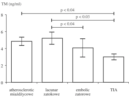
The differences in TM concentration in patients with acute cerebral ischaemia of different aetiology are charted in Figure 1.
There were no differences in TM concentra− tion between patients with atherosclerotic and la− cunar strokes or between patients with embolic strokes and transient ischaemic attacks. However, significantly lower TM concentrations were seen in patients with embolic strokes or transient ischaemic attacks compared to patients with ather− osclerotic or lacunar strokes.
Discussion
Pathological processes in the vascular endo− thelium disturb the coagulation system and may thus be a major factor underlying acute cerebral ischaemia. These disturbances are associated with the response of endothelial cells to pathological processes in vessel walls e.g. in the course of ath− erosclerosis.
Thrombomodulin is chiefly present in the vas− cular endothelium and contributes considerably to its anticoagulant properties. TM circulating in the blood is in a soluble form. Soluble TM probably forms as a result of cell−bound TM having been split by one (or more) proteolytic enzymes present in the blood or endothelial cells. It is assumed that this process is intensified under pathological con− ditions following damage to the vascular endothe− lium [13, 20]. Consequently, elevated TM con−
centration is regarded as a sign of endothelial damage [18].
Our study demonstrated that TM levels are elevated in patients with acute cerebral ischaemia. A search through available data bases did not reveal any studies specifically designed to deter− mine TM levels in the blood of patients with acute cerebral ischaemia. Still, our findings concur with reports by other authors, who found elevated TM in disorders associated with vessel wall injury and increased coagulation. This finding suggests that the release of soluble TM into circulating blood is intensified by proteolytic action on the surface of damaged endothelium [6, 13, 14, 17].
Elevated TM levels have been observed main− ly in conditions associated with small vessel injury. Since it is not clear whether damage limit− ed to major vessels would also result in increased production of sTM, some authors are not inclined to look at TM as an independent risk factor in all vascular disorders [6, 21]. Our study apparently supports their reservations as we found higher TM
Table 3. Effect of risk factors on TM concentration in pa− tients with acute cerebral ischaemia
Tabela 3. Wpływ czynników ryzyka na stężenia TM u chorych na ostre niedokrwienie mózgu
Risk factor n TM ng/ml *
(Czynnik ryzyka)
Arterial hypertension 53 4.96 (4.08; 5.48) (Nadciśnienie tętnicze)
Without arterial hypertension 20 4.28 (3.76; 5.22) (Bez nadciśnienia tętniczego)
Cholesterol level > 200 mg/dl 53 4.76 (4.08; 5.56) (Poziom cholesterolu)
Cholesterol level ≤200 mg/dl 20 4.48 (3.76; 5.4) (Poziom cholesterolu)
Smokers 16 4.48 (4.16; 5.04)
(Palący papierosy)
Non−smokers 57 4.88 (3.88; 5.56)
(Niepalący papierosów)
Diabetes mellitus 19 4.72 (3.84; 5.96) (Cukrzyca)
Without diabetes mellitus 55 4.8 (4.0; 5.4) (Bez cukrzycy)
Ischaemic heart disease 19 4.24 (3.84; 5.96) (Choroba niedokrwienna serca)
Without ischaemic heart disease 54 4.96 (4.08; 5.48) (Bez choroby niedokrwiennej serca)
Atrial fibrillation 10 5.2 (3.44; 6.2) (Migotanie przedsionków)
Without atrial fibrillation 63 4.64 (4.0; 5.4) (Bez migotania przedsionków)
* – median (1stquartile; 3rdquartile)
* – mediana (kw I; kw III)
Fig. 1. TM levels in patients with acute cerebral ischaemia relative to type of ischaemia
Ryc. 1. Stężenia TM u chorych na ostre niedokrwienie mózgu w zależności od typu niedokrwienia
p < 0.04
p < 0.03
p < 0.04 TM (ng/ml)
atherosclerotic miażdżycowe
lacunar zatokowe
embolic zatorowe
TIA 0
concentrations in patients with atheromatous and lacunar strokes than in those with embolic strokes and TIAs.
In last years some studies appeared about pres− ence of plasma haemostatic factors, among them TM, in human atherosclerotic carotid plaques.
Table 4. TM concentration in patients with acute cerebral ischaemia relative to gender, age (below or above 70 years), pre− sence of an ischaemic lesion on CT scan and presence of signs of brain stem failure (ITC)
Tabela 4. Stężenia TM u chorych na ostre niedokrwienie mózgu w zależności od płci, wieku > 70. lub < 70. r. ż., obecności ogniska niedokrwiennego wykazanego w tomografii komputerowej i występowania objawów niewydolności pnia mózgu (ITC)
n TM ng/ml * p
Gender male 37 4.52 (3.92; 5.48) ns.
(Płeć) (męska)
female 36 4.96 (4.08; 5.48)
(żeńska)
Age < 70 years 35 4.64 (4.16; 5.32) ns.
(Wiek) < 70 (lat)
> 70 years 38 4.96 (4.08; 5.48)
> 70 (lat)
CT ischaemic lesion present 19 5.22 (4.32; 5.56) ns.
(KT) (ognisko niedokrwienne)
ischaemic lesion absent 54 4.56 (3.84; 5.32) (bez ogniska niedokrwiennego)
ITC ITC signs present 7 3.92 (3.44; 4.32) 0.0344
(z objawami ITC)
ITC signs absent 66 4.96 (4.08; 5.52)
(bez objawów ITC) * – median (1stquartile; 3rdquartile)
* – mediana (kw I; kw III).
Table 5. TM levels in patients with acute cerebral ischaemia relative to type of ischaemia
Tabela 5. Stężenia TM u chorych na ostre niedokrwienie mózgu w zależności od typu niedokrwienia
Defined types of ischaemia n TM ng/ml* p
(Porównywane typy niedokrwienia)
Atherosclerotic 38 4.84 (4.12; 5.36) ns.
(Miażdżycowy)
Lacunar 15 5.24 (4.48; 5.96)
(Lakunarny)
Atherosclerotic 38 4.84 (4.12; 5.36) ns.
(Miażdżycowy)
Embolic 15 4.08 (3.44; 5.2)
(Zatorowy)
Atherosclerotic 38 4.84 (4.12; 5.36) 0.0307
(Miażdżycowy)
TIA 5 3.02 (2.68; 3.36)
Lacunar 15 5.24 (4.48; 5.96) 0.0359
(Lakunarny)
Embolic 15 4.08 (3.44; 5.2)
(Zatorowy)
Lacunar 15 5.24 (4.48; 5.96) 0.0254
(Lakunarny)
TIA 5 3.02 (2.68; 3.36)
Embolic 15 4.08 (3.44; 5.2) ns.
(Zatorowy)
TIA 5 3.02 (2.68; 3.36)
* – median (1stquartile; 3rdquartile)
These observations indicate that concentration of examined parameters in carotid plaques were much higher as in plasma, but no correlation between their concentration in plaques and plasma were observed [22–24].
The authors conclude that patients with acute cerebral ischaemia had elevated levels of TM, which may be the result of damage to the vascu− lar endothelium. Higher TM concentrations were
observed in patients with atherosclerotic and lacu− nar strokes or between patients with embolic strokes and transient ischaemic attacks. In patients without generalised signs of CNS dam− age (brain stem failure – ITC), TM levels were higher than in patients with ITC. Exposure to risk factors for cerebral ischaemia does not influence TM concentration in patients with acute cerebral ischaemia.
Referecnes
[1] Coolman RW: Review of normal hemostasis. In: Thrombolytic therapy for peripheral vascular disease. Eds.: Comerota AJ, J.B. Lippincott Company, 1995, 266–269.
[2] Jaffe AE:Vascular function in hemostasis. In: Hematology. Eds.: Beutler E, Lichtman MA, Coller BS, Kipps TJ, Williams McGraw−Hill, Inc. 1995, 125, 1261–1276.
[3] Bajaj MS, Kuppuswamy MN, Manepalli AN, Bajaj SP:Transcriptional expression of tissue factor pathway inhibitor, thrombomodulin and von Willebrand factor in normal human tissues. Thromb Haemost 1999, 82, (3), 1047–1052.
[4] Boffa MC, Burke B, Haudenschild CC:Preservation of thrombomodulin antigen on vascular and extravascular surfaces. J Histochem Cytochem 1987, 35, (11), 1267–1276.
[5] Ishii H, Majerus PW:Trombomodulin is present in plasma and urine. J Clin Invest 1985, 76, 2178–2181.
[6] Seigneur M, Dufourcq P, Conri C, Constans J, Mercié P, Pruvost A, Amiral J, Midy D, Baste JC, Boisseau MR:Levels of plasma thrombomodulin are increased in atheromatous arterial disease. Thromb Res 1993, 71, 423–31.
[7] Boffa MC, Jackman RW, Peyri N, Boffa JF, George B:Thrombomodulin in the central nervous system. Nouv Rev Fr Hematol 1991, 33(6), 423–429.
[8] Wang L, Tran ND, Kittaka M, Fisher MJ, Schreiber SS, Zlotowic BV:Thrombomodulin expression in bovine brain capillaries: Anticoagulant function of the blood−brain barrier, regional differences, and regulatory mecha− nisms. Arteriosc Thromb Vasc Biol 1997, 17, (11), 3139–3156.
[9] Valen G, Sigurdardottir O, Vaage J:Systemic release of thrombomodulin, but not from the cardioplegic, reper− fused heart during open heart surgery. Thromb Res 1996, 83(4), 321–328.
[10] Grau GE, De Moerloose P, Bulla O, Lou J, Lei Z, Reber G, Mili N, Ricou B, Morel DR, Suter PM:
Haemostatic properties of human pulmonary and cerebral microvascular endothelial cells. Thromb Haemost 1997, 77, (3), 585–590.
[11] Ikeda E, Maruyama I, Hosoda Y:Expression of thrombomodulin in patients with spontaneous occlusion of the circle of Willis. Stroke 1993, 24, (5), 657–660.
[12] Plow EF, Herre T, Redlitz Miles LA, Hoover−Plow JL:The cell biology of the plasminogen system. FASEB J 1995, 9, 939–945.
[13] Blann AD, McCollum CN:Von Willebrand factor and soluble thrombomodulin as predictors of adverse events among subjects with peripheral or coronary atherosclerosis. Blood Coagul Fibrinolysis 1999, 10, (6), 375–380.
[14] Takano S, Kimura S, Ohdama S, Aoki N:Plasma Thrombomodulin in health and diseases. Blood 1990, 76, (10), 2024–2029.
[15] Wiśniewska E, Wodyńska T, Kulwas A, Kotschy M, Nartowicz E, Paczuski R, Michalski A:
Thrombomodulin – endothelial thrombin receptor in blood of patients with unstable angina pectoris. Med Sc Mon 2001, 7, 256–259.
[16] Kotschy M, Polaszewska−Muszyńska M, Moskal S, Będowska−Gontarz W, Sikorska Z, Paczuski R:Some vascular endothelial cell markers in diabetes type 2 with proliferative retinopathy. Thromb Haemost 2001, Suppl. Abstract: CD3506.
[17] Besbas N, Erbay A, Saatçi Ü, Özdemir S, Bakkaloglu A, Ozen S, Topaloglu R:Thrombomodulin, tissue plas− minogen activator and tissue plasminogen activator inhibitor−1 in Henoch−Schönlein purpura. Clin Exp Rheumatol 1998, 16, 95–98.
[18] Jansson JH, Boman K, Brännström M, Nilsson K:Increased levels of plasma thrombomodulin are associated with vascular and all−cause mortality in patients on long−term anticoagulant treatment. Eur Heart J 1996, 17, 1503–1505.
[19] Fareed J, Messmore HL:Plasma trombomodulin level as a predictor of hemorrhagic (and thrombotic) events in patients on long−term anticoagulant treatment. Circulation 1997, 96, 2765–2768.
[20] Shieh SD, Shiang JC, Lin YF, Shiao WY, Wang JY: Circulating angiotensin−converting enzyme, von Willebrand factor antigen and thrombomodulin in exertional heat stroke. Clin Sci Colch 1995, 89, (3), 261–265.
[22] Migdalski A, Jawień A, Kotschy M, Knapik−Bieniek A:Selected haemostatic factors in carotid bifurcation plaques of patients undergoing carotid endarterectomy. Eur J Vasc, Endovasc Surg 2004, 27, 172–179.
[23] Kotschy M, Knapik−Bieniek A, Migdalski A, Kotschy D, Jawień A:Selected endothelial markers in athero− sclerotic plaques and plasma of patients undergoing carotid endarterectomy. Acta Angiol 2004, 10, 173–180.
[24] Migdalski A, Kotschy M, Jawień A: tissue factor, tissue factor pathway inhibitor and vascular endothelial growth factor−A in carotid atherosclerotic plaques. Eur J Vasc Endovasc Surg 2005, 30, 41–47.
Address for correspondence:
Maria Kotschy Odon Bujwid 28/20 50−368 Wrocław Poland
Conflict of interest: None declared Received: 8.07.2005
Revised: 16.09.2005 Accepted: 28.11.2005
Praca wpłynęła do Redakcji: 8.07.2005 r. Po recenzji: 16.09.2005 r.