© 2014 Manal Mohamed Elhassan Taha et al. This is an open access article distributed under the terms of the Creative Commons Attribution License -NonCommercial-ShareAlikeUnported License (http://creativecommons.org/licenses/by-nc-sa/3.0/).
Journal of Applied Pharmaceutical Science Vol. 4 (11), pp. 042-046, November, 2014 Available online at http://www.japsonline.com
DOI: 10.7324/JAPS.2014.4118 ISSN 2231-3354
In vitro hepatotoxcity of Catha edulis Forsk. (khat) phenolic-rich
extract on human hepatocytes
Manal Mohamed Elhassan Taha, Siddig Ibrahim Abdelwahab
*, Rashad Al-sanousi
Medical Research Center, Substance Abuse Research Centre, Jazan University, Jazan,Kingdom of Saudi Arabia.
ARTICLE INFO ABSTRACT
Article history: Received on: 09/12/2013 Revised on: 18/01/2014 Accepted on: 03/03/2014
Available online: 27/11/2014
In our previous research, Khat has been shown to be associated with hepatic hypertrophy and hepatotoxicity in male and female SD-rats. However, no in vitro studies have been previously done to test on hepatotoxicity of khat on human experimental models. In vitro toxicological evaluation system plays an important role in the early phase of pharmaceutical development and drug safety. Therefore, the current study was designed to examine the in vitro hepatotoxic effect of phenolic-rich extract of khat(Catha edulis Forsk.). Khat was randomly collected from different places in Yemen [Dhamar (DMR), Ibb (IB), Taiz (TZ) cities] and Ethiopia (HAR). Phenolic-rich extraction was performed using 60% methanol. The effects of khat on HepG2 human hepatocyte cell line were measured using the MTT assay. HepG2 cells are a suitable in vitro model system for the study of human hepatocytes. The potency of cell growth inhibition for khat was expressed as an IC50 value. Cellular proliferation
following 24h of exposure to khat samples showed considerable inhibition in khat-treated cells compared to non-treated cells (controls). The IC50 values of DMR, IB, TZ and HAR were 5.1±0.03, 15.3±0.12, 10.2±0.20 and
8.7±0.47 μg/mL, following 24 h of treatment, respectively. The proliferation of khat-treated cells decreased as the khat concentration increased. The method used appears to be a useful and reproducible technique for the in vitro assessment of the khat-induced cytotoxicity in a human liver cell line. Further studies are recommended to understand the molecular mechanism of Khat induced HepG2 cytotoxicity.
Key words:
Khat (Catha edulis F.), phenolic compounds, Antioxidative activity, HepG2 cells.
INTRODUCTION
Catha edulis Forsk. (Khat) is an undeciduous tree
growing East Africa and the Arabian Peninsula. There is an argument as to the exact origin where khat cultivation and chewing habituation originated, with some authors suggesting Ethiopia and others suggesting Yemen (Habori and Mamary, 2004; Al-Mamary et al., 2006; Al-Zubairi et al., 2008; Brostoff et al., 2006). Chewing the leaves of khat has become a widespread practice in several countries due to its pyschostimulant properties. This effect is attributed to its major constituents (cathine and cathinone). Beside this daily habitual use, khat has also some ethno-pharmacological uses (Krizevski et al., 2007; Nyongesa et al.,
2008; Pennings et al., 2008). Cathine and cathinone, as alkaloids, were found to have a negligible contribution to the antioxidative activity. Khat has a potential to affect the systemic capacity to handle free radicals (Al-Habori and Al-Mamary, 2004; Dudai et al., 2006) but may also have some antioxidant effects owing to some of its antioxidant constituents such as flavonoids (Al-Zubairi
et al., 2003).
.
* Corresponding Author
Email: [email protected]; TEL +966173233317
Flavonoids compose the major and important studied compounds of plant polyphenols (Santos-Buelga et al., 2010).
On the other hand, the growth retardation of animals treated with Khat extract might be attributed to the presence of tannins and other polyphenolic compounds, since tannins and polyphenolic compounds could inhibit the digestive enzymes (Abdul Ghani et al., 1987). Khat has a potential to affect the systemic capacity to handle free radicals (Habori and Al-Mamary, 2004; Dudai et al., 2006) but may also have some antioxidant effects owing to some of its antioxidant constituents such as flavonoids (Al-Zubairi et al., 2003). The balance between oxidation and antioxidation (redox balance) is critical in maintaining a healthy biological system. In cellular redox state, the double-edged effect does not only concern ROS, but also antioxidants. Physiologic doses of exogenous antioxidants are required to maintain or re-establish redox homeostasis (Bouayed and Bohn, 2010; Martin and Barrett, 2002). However, high doses of exogenous antioxidants may disrupt redox balance. At high concentrations, ROS are toxic compounds leading to lipid peroxidation and the oxidation
When this situation occurs, cells enter an oxidative stress state, characterized by the disequilibrium between oxidant production and antioxidant protection in favor of the former (Bouayed and Bohn, 2010). In our previous research we have shown that Khat could be associated with hepatic hypertrophy and hepatotoxicity in male and female SD-rats. However, no in vitro studies have been previously done to test on hepatotoxicity of khat on human experimental models. In vitro toxicological evaluation system plays an important role in the early phase of pharmaceutical development and drug safety. This study aims to examine the in vitro hepatotoxic effect of phenolic-rich extract of khat (Catha edulis Forsk.). Khat was randomly collected from different places in Yemen [Dhamar (DMR), Ibb (IB), Taiz (TZ) cities] and Ethiopia (HAR).
MATERIALS AND METHODS Plant material
Three plant materials were randomly collected from different regions in Yemen (Dhamar, Ibb and Ta’az) and Ethiopian. Samples are a generous gift from Dr. Rasheed Almagtary, Faculty of Dentistry, Dhamar University, Yemen.
Phenolic rich extraction
Extraction was performed according to the method described previously (Mariod et al., 2009). Dried ground leaves of Khat were extracted successively with 60% methanol (3x300 ml) for two days at room temperature to obtain crude methanolic extract; the extracts were pooled and the solvent was evaporated using rotary evaporator (Buchi, Flawil, Switzerland). The yield of these phenolic-richextracts was determined and kept at 4 °C for further analysis.
Chemicals and Reagents
Cispatin (>99% pure) was purchased from Sigma (St. Louis, MO, USA). HepG2 liver cell lines used in this study were obtained from ATTC, USA. RPMI 1460, fetal bovine serum (FBS) and penicillin-streptomycin were obtained from Bioscience Ltd. (Wokingham, UK). Phosphate buffered saline (PBS), ethanol (95%), 3-(4,5-dimethylthiazol-2yl)-2,5-diphenyltetrazolium bromide (MTT) was from Santa Cruz Biotechnology (Santa Cruz, CA USA).
Cell Culture and Viability Assay
Cell cultures were maintained in a humidified atmosphere with 5% CO2 at 37 °C. To study proliferation and determine the survival of cells after treatment, the MTT assay was used. Briefly, the cells were plated on a 96-well plate at 2 × 10⁵
cell/mL in 100 μL culture medium. They were plated in triplicate.
Different concentrations of khat samples were prepared by serial dilution. All serial dilutions were transferred to the cells in the 96-well plate; the plate included untreated cells as control and was incubated for 24 h. After incubation, the viability of the cells was assessed using 3-[4,5-dimethylthiazol-2-yl]-2,5-diphenyl
tetrazolium bromide (MTT, 5 mg/mL); 20 μL were added to the
cells in a dark place, and the cells were then covered with aluminium foil and incubated for 4 h. After incubation, all of the
media were removed and 100 μL of DMSO were added to the cells
in order to solubilise the formazan crystals. Subsequently, the absorbance was read at a wavelength of 570 nm using a micro plate reader. The potency of cell growth inhibition for the test agent was expressed as an IC50 value.
Statistical Analysis
Results were reported as means±SEM of at least three analyses for each sample. Normality and Homogeneity of variance assumptions were checked. Statistical analysis was performed according to the SPSS-16.0 package.
RESULTS AND DISCUSSION
Khat becomes a serious public health issue in Saudi Arabia due to its widespread use. Many unfavorable sideffects have been associated with khat chewing. Accordingly, the prolonged use of Khat could produce in psychoneurological disturbances such as neurosis. In addition, increased diastolic blood pressure and vasoconstriction of coronary vasculature were also reported (Alsalahi et al., 2012; Nyachieo et al., 2012). Furthermore, Luqman and Danowski reported that liver cirrhosis that was observed among Yemeni Khat chewers might be due to Khat consumption, but at that time it was not further investigated, and hepatotoxicity of Khat chewing is still debated in humans. In animals, the administration of crude extract of Khat to New Zealander white rabbits for three months suggested toxic hepatocellular jaundice as well as histopathological abnormalities in livers of such animals. On the other hand, the growth retardation of animals treated with Khat extract might be attributed to the presence of tannins and other polyphenolic compounds, since tannins and polyphenolic compounds could inhibit the digestive enzymes (al'Absi et al., 2013; Al-Hashem and Shatoor, 2012).
Growth and cell viability assays are an alternative technique of measuring cellular response to harmful agent and newly developed drugs (Kepp et al., 2011). These assays depend on quantifying growth of cells in short term cell culture. Different methods of measuring the number of living cells have been used, e.g., dye exclusion, isotope uptake, staining with crystal violet, and quantifying with computerized image analysis, and more recently staining with a fluorescent DNA-specific dye, Hoechst 33258. The MTT assay quantifies metabolically viable cells by their ability to reduce MTT. The advantages of the MTT assay include rapid semi-automated reading, objective assessment, comparative low cost, high reproducibility, low number of cells required, and the facility to quantify cells grown in suspension, on monolayer or in spheroids or colonies (Fotakis and Timbrell, 2006).
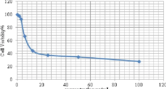
The potency of cell growth inhibition for khat was expressed as an IC50 value. Cellular proliferation following 24h of exposure to khat samples showed considerable inhibition in khat-treated cells compared to non-treated cells (controls). The IC50 values of DMR, IB, TZ and HAR were 5.1±0.03, 15.3±0.12, 10.2±0.20 and
8.7±0.47 μg/mL, following 24 h of treatment, respectively (Table
1). The proliferation of khat-treated cells decreased as the khat concentration increased. The method used appears to be a useful and reproducible technique for the in vitro assessment of the khat-induced cytotoxicity in a human liver cell line as shown in Figure 1, 2, 3 and 4, respectively. Khat has a potential to affect the systemic capacity to handle free radicals (Habori and Al-Mamary, 2004; Dudai et al., 2006) but may also have some antioxidant effects owing to some of its antioxidant constituents such as flavonoids (Al-Zubairi et al., 2003). The balance between oxidation and antioxidation (redox balance) is critical in
maintaining a healthy biological system. In cellular redox state, the double-edged effect does not only concern ROS, but also antioxidants. Physiologic doses of exogenous antioxidants are required to maintain or re-establish redox homeostasis (Bouayed and Bohn, 2010; Martin and Barrett, 2002). However, high doses of exogenous antioxidants may disrupt redox balance. At high concentrations, ROS are toxic compounds leading to lipid peroxidation and the oxidation of other sensitive biomolecules such as proteins and DNA. When this situation occurs, cells enter an oxidative stress state, characterized by the disequilibrium between oxidant production and antioxidant protection in favor of the former (Bouayed and Bohn, 2010). It could be concluded that
the in vitro hepatotoxcity of Catha edulis (khat) phenolic-rich
extract on human hepatocytes could be attributed to the disturbance of intracellular redox status of HepG2. Further studies are recommended to support such claim.
Table. 1: The 50% inhibitory concentrations of khat phenolic-rich extracts (Dhamar (DMR), Ibb (IB), Taiz (TZ) and Ethiopia (HAR) varieties) on the HepG2
liver cells.
Treatment IC50 (µg/mL)
DMR 5.1±0.03a
IB 15.3±0.12b
TZ 10.2±0.20c
HAR 8.7±0.47c
Cisplatin 3.2±0.08d
Results were reported as means ± SEM of at least three analyses for each sample.
Fig. 1: MTT (3-(4,5-dimethylthiazol-2-yl)-2,5-diphenyltetrazolium bromide) cell viability assay on human hepatic cells ( HepG2) for Dhamar (Yemen) sample.
ACKNOWLEDGEMENTS
The present project is supported by the Research Fund from Substance Abuse Research Centre, Jazan University, Jazan, Saudi Arabia.
REFERENCES
AbdulGhani N, Eriksson M, Kristiansson B, Qirbi A. The influence of khat-chewing on birth-weight in full-term infants. Soc Sci Med, 1987; 24, 625-627.
al'Absi M, Khalil NS, Al Habori M, Hoffman R, Fujiwara K, Wittmers L. Effects of chronic khat use on cardiovascular, adrenocortical, and psychological responses to stress in men and women. Amer Addi. 2013; 12:125-29.
Al-Habori M, Al-Mamary M. Long-term feeding effects of Catha edulis leaves on blood constituents in animals. Phytomed. 2004; 11:639-44.
Al-Hashem FH, Shatoor AS. Acute oral administration of Khat (Catha edulis) aqueous extract elevates blood pressure and prolongs QT and QTC intervals in Wistar albino rats. Saudi Med J. 2012; 33:1065-72.
Al-Mamary M, Al-Habori M, Al-Shoaibi Z, Shamsan B. Nitrosamine formation from different Catha edulis leaves extracts under simulated gastric condition. Food Chem. 2006; 97:586-90.
Al-Zubairi A, Al-Habori M, Al-Geiry A. Effect of Catha edulis (khat) chewing on plasma lipid peroxidation. J of Ethnopharmaco. 2003; 87:3-9.
Al-Zubairi A, Ismail P, Pei Pei C, Rahmat A. Genotoxic effect of Catha edulis (khat) crude extract after sub-chronic administration in rats. Envir Toxicol Pharmacol. 2008; 25:298-303.
Alsalahi A, Abdulla MA, Al-Mamary M, Noordin MI, Abdelwahab SI, Alabsi AM, Shwter A, Alshawsh MA. Toxicological features of catha edulis (Khat) on livers and kidneys of male and female sprague-dawley rats: a subchronic study. ECAM; 2012. 2012:Article ID 829401, 11 pages.
Bouayed J, Bohn T. Exogenous antioxidants—double-edged swords in cellular redox state: health beneficial effects at physiologic doses versus deleterious effects at high doses. Oxid Med Cell Longevity. 2010; 3:228-37.
Brostoff JM, Plymen C, Birns J,. Khat -- a novel cause of drug-induced hepatitis. Euro J Inter Med. 2006; 17:383-83.
Dudai N, Fischer R, Segev D, Chaimovitsh D, Rosenzweig N, Shimoni, M. 2006. Antioxidative Activity of Khat (Catha edulis Forsk.). : II International Symposium on Natural Preservatives in Food, Feed, and Cosmetics.
85-92.
Fotakis G, Timbrell JA. In vitro cytotoxicity assays: comparison of LDH, neutral red, MTT and protein assay in hepatoma cell lines following exposure to cadmium chloride. Toxicol lett. 2006; 160: 171-177.
Fig. 3: MTT (3-(4,5-dimethylthiazol-2-yl)-2,5-diphenyltetrazolium bromide) cell viability assay on human hepatic cells ( HepG2) for IBB (Yemen) sample.
Kepp O, Galluzzi L, Lipinski M, Yuan J, Kroemer G. Cell death assays for drug discovery. 2011; Nat Rev Drug Disc. 2011; 10:221-37.
Krizevski R, Dudai N, Bar E, Lewinsohn E. Developmental patterns of phenylpropylamino alkaloids accumulation in khat (Catha
edulis, Forsk.). J Ethnopharmacol. 2007; 114:432-38.
Mariod AA, Ibrahim RM, Ismail M, Ismail N, Antioxidant activity and phenolic content of phenolic rich fractions obtained from black cumin (Nigella sativa) seedcake. Food Chem. 2009: 116: 306-312.
Martin K, Barrett J. Reactive oxygen species as double-edged swords in cellular processes: low-dose cell signaling versus high-dose toxicity. Hum Exper Toxicol. 2002: 21;71-75.
Nyachieo A, Kiraithe MM, Spiessens C, Chai DC, Kiulia NM, D'Hooghe TM, Mwenda JM. Short-Term Effects of High-Dose Khat on Sperm Parameters and Reproductive Hormonal Levels in Olive Baboons (Papio anubis). Gynecol Obs Invest. 2012; 75: 109-114.
Nyongesa AW, Patel NB, Onyango DW, Odongo HO, Wango EO. Khat (Catha edulis) lowers plasma luteinizing hormone (LH) and
testosterone secretion, but increases cortisol levels in male rabbits. J Ethnopharmacol. 2008; 116:245-50.
Pennings EJM, Opperhuizen A, van Amsterdam JGC. Risk assessment of khat use in the Netherlands: A review based on adverse health effects, prevalence, criminal involvement and public order. Regul Toxicolo and Pharmacol. 2008: 52;199-207.
Santos-Buelga C, Escribano-Bailon MT, Lattanzio V. 2010. Recent Advances in Polyphenol Research. April 2012, Hardcover, Wiley-Blackwell.
How to cite this article: