Isil Fidan
A, C, D, F, Ayse Kalkanci
B, C, E, Emine Yesilyurt
B, C, Berna Erdal
B, CIn Vitro
Effects of
Candida albicans
and
Aspergillus fumigatus
on
Dendritic Cells
and the Role of Beta Glucan in this Effect
Gazi University Faculty of Medicine, Department of Medical Microbiology, Ankara, Turkey
A – research concept and design; B – collection and/or assembly of data; C – data analysis and interpretation;
D – writing the article; E – critical revision of the article; F – final approval of article; G – other
Abstract
Background. Dendritic cells (DCs) are able to initiate and regulate the immune response to fungal infections. β-glucan stimulates the immune system, modulating cellular and humoral immunity. It has a beneficial effect in fighting fungal infections.
Objectives. We investigated the in vitro effect of C.albicans and A.fumigatus infection on human DCs. The cytokine levels were determined by ELISA.
Material and Methods. Human PBMCs isolation was performed by Ficoll-hypaque density gradient centrifugation method. DCs maturation was analysed by using flow cytometry. The cytokine levels were determined by ELISA.
Results. DCs stimulated by C. albicans and A. fumigatus induced DC maturation by increasing CD80 and CD86 co-stimulatory molecules. DCs stimulated by fungi produced IL-8 and IL-12p70. Whereas IL-10 production from the stimulated DCs did not differ from uninfected DCs. Also, the addition of β-glucan to the DCs stimulated by fungi promoted the activation and maturation of DCs.
Conclusions. Our results suggest that DCs are capable of initiating an innate and adaptive immune response against fungal infections. In addition, β-glucan can be used as a novel stimulator to DC-based vaccination against fungal infections (Adv Clin Exp Med 2014, 23, 1, 17–24).
Key words: dendritic cells, fungal infection, β-glucan.
Adv Clin Exp Med 2014, 23, 1, 17–24 ISSN 1899–5276
ORIGINAL PAPERS
© Copyright by Wroclaw Medical University
Dendritic cells (DCs) are antigen presenting cells that reside in the peripheral regions such as skin, nose mucosa, the respiratory system and the digestive system and in all tissues apart from the brain, testis and eyes, and they play key roles in the initiation of adaptive immunity. These cells, recog-nized as professional antigen presenting cells, in-duce the immune response by stimulating the
un-differentiated T cells [1]. When activated, dendritic
cells mature and migrate to regional lymph nodes
in order to activate antigen specific T cells [2].
Moreover, DCs play an effective role in the inter-action between innate and adaptive immunity [3]. DCs also exist in nonlymphoid tissues consisting of antigen entrance sites such as the skin while they are widespread in lymphoid tissues [4].
DCs express great amounts of CD11c and MHC class II molecules. During their migration, DCs have some phenotypic and functional chang-es. When they mature, MHC molecules,
co-stim-ulatory moleculessuch as CD86, CD80 and CD40
are activated. These molecules are essential for the
activation of T cells. During maturation, Th cells
(T-helper cells) are differentiated as Th1 and Th2
according to the cytokine type secreted. IL-12 se-creted from DCs causes Th1 differentiation and it is important in developing the immune response against bacterial infections. At the same time, IL-10 secreted from DCs causes Th2 differentiation and this carries some importance for the immune re-sponse against extracellular parasites [5].
DCs are the immune cells that have been the focus of many studies recently. There have been some findings that many pathogens at-tempt to affect the host immune response by in-teracting with DC functions [6]. They have cru-cial roles in infections, host-microbe interactions and the balance between protective immunity and immunopathology [6].
Opportunistic fungal infections such as
Can-dida albicans (C.albicans) and Aspergillus fumiga-tus (A.fumigafumiga-tus) cause some life threatening in-fections, especially during immunosuppressive conditions such as AIDS or organ transplants, and they have an increasing incidence. Some different host defense mechanisms play effective roles dur-ing infections against fungi. The innate immune response is basically effective during the early
stag-es of the infection[6].
DCs have important roles in the initiation of cellular response against fungi. It has been
discov-ered that DCs have direct effects on both in vitro
and in vivo anti-fungal immunity. DCs
phagocy-tose bothyeast and hyphal forms of the fungus [6].
Phagocytosis of A.fumigatus conidia by DCs leads
to the secretion of some cytokines. So, it is con-sidered that DCs initiate the adaptive immune
re-sponses to Aspergillus species and directly influence
the outcome of an Aspergillus infection [3, 7].
The changes occurring in dendritic cell struc-tures and functions because of fungal infections play important roles in the development of anti-fungal immunity [3]. Therefore, the use of ther-apeutic approaches in fungal infections targeting DCs carry a great importance in the prevention of the agents that cause mortality especially in immu-nosuppressive patients and the treatment of these infections.
β-glucan (beta-glucan), a polysaccharides, is
a component of the cell wall structure in various microorganisms such as pathogen bacteria and
fungus. β-glucan is one of the strongest immune
system stimulators known so far and plays an ef-fective role in the protection against several
infec-tion agents. β-glucan has some strong effects in the
immunotherapy of some diseases such as cancer.
Recently, β-glucan has been accepted as an
impor-tant pharmacological agent in human beings since its several immunomodulatory effects such as an-ti-cytotoxic, anti-mutagenic and anti-tumorigenic effects have been identified [8].
In our study, we aimed to identify the changes in the structure and function of DCs due to some
fungi and the effect of β-glucan administered
ex-ternally on those changes.
Experiment
Isolation of Human Peripheral
Blood Mononuclear Cells
(PBMC)
Heparinized blood was collected from 20 heal-thy human donors. PBMCs were separated from the blood by Ficoll-hypaque density gradient cen-trifugation (Sigma, UK). After cencen-trifugation, buffy coats were collected and washed in Phosphate Buff-ered Saline (PBS, Gibco, Germany) 3 times and
re-suspended at a concentration of 2 × 10⁶ cells/mL
in a complete RPMI 1640 medium containing
2 mM L-glutamine, 100 U/mL penicillin, 100 µg/mL
streptomycin, 0.05 mM 2ME and supplement-ed with 10% fetal calf serum (Gibco, Germany). Cell viability was 95% by the trypan blue exclusion test [2]. PBMCs were allowed to adhere to 6-well
plates. After 2 h at 37°C the nonadherent cells were
removed and the adherent cells were separated by
incubated with Mg2+ and Ca2+ free PBS containing
0.5 mM EDTA at 37°C [2].
Preparation of Dendritic
Cell Culture
The cell culture medium (Complet RPMI) was RPMI-1640 (Sigma, Germany) supplemented with Penicillin (100 U/mL, Biochrom AG, Germany), Streptomycin (100 µg/mL, Biochrom AG, ny), L-glutamin (2 mM, Biochrom AG, Germa-ny), 2-mercaptoethanol (50 µM, Fluka, Switzer-land), and 10% heat-inactivated FCS (Biochrom AG, Germany). At day 0, the cells were seeded at
2 × 10⁶ per 100 mm dish in 10 mL complete RP-
-MI containing 50 ng/mL rhGM-CSF (Biosource, USA) and 1.000 U/mL IL-4. The culture medium was partially replaced every 3 days and fresh cy-tokines (rmGM-CSF and IL-4) were added. For complete maturation, on day 10, non-adherent cells were collected by gentle pipetting, centrifuged at 300 × g for 5 min at RT, and resuspended with 10 mL fresh complete RPMI in a 100 mm tissue culture plastic dish containing 100 U/mL rmGM- -CSF and lipopolysaccharide (LPS, Sigma, Germa-ny) at 1 µg/mL. The cells were then cultured for 1 or 2 more days. The cultured cells were washed once and an aliquot volume mixed 1 : 1 in Trypan Blue solution (Sigma, Germany). DCs were count-ed as viable under the microscope in a Neubauer
chamber and the number was adjusted to 5 × 10⁶
Cell Separation
and Flow Cytometry
CD11c+ DCs were purified by magnetic cell
sorting (MACS) by using positive selection,
ac-cording to the manufacturer’sprotocol
(Milte-nyi, USA). Briefly,the cells were incubated with
magnetic microbeads conjugatedwith
monoclo-nal anti-human CD11c antibodies in MACS
buf-ferfor 15 min at 4°C. After this, the cells were run
through a MACS column(Miltenyi) in a
magnet-ic field. The column was then removedfrom the
magnet, and the positive cells were flushed out.
Then CD11c+ DCs were analyzed further by flow
cytometer (Coulter, USA).
Introduction of Fungi and DCs
One mL of BM-DCs (106) were delivered in
per well. One mL of A. fumigatus (106 conidia/mL)
and C. albicans (106 yeast/mL) were added to the wells, respectively. Thusly, the DCs were
stimulat-ed by the fungi. Amphotericin B was used to
pre-vent fungal overgrowth in the wells. Control cul-ture flasks did not contain fungi, only the medium and DCs. Each sample was studied in triplicate. β-glucan (Immunex) was added to the control cul-ture plates and culcul-ture plates containing fungi. All
plates were incubated for 24 h in 5% CO2 at 37οC.
At the end of the time period, the contents of the culture plates were transferred into tubes and cen-trifuged. The culture supernatants were removed
and stored at –70°C until used in ELISA.
Flow Cytometric Analysis
of PBMCs
The pelleted cells were washed in PBS and
stained with antibodies for 30 min at 4οC. The DCs
(2 × 10⁶ cells/mL) were incubated with
monoclo-nal antibodies specific to human CD antigens. The antibodies used were anti-CD80, and anti-CD68 (e-bioscience, USA). IgG1-FITC/IgG2-PE were used as isotypic control. Flow cytometric analyses were done by using a Coulter FC500 flow cytome-ter (Coulcytome-ter, USA).
Cytokine Secretion
The levels of cytokines such as IL-8
(İnterleukin-8), IL-10 and IL-12p70 were
deter-mined by specific enzyme-linked immunosorbent assay (ELISA) techniques according to the man-ufacturer’s instructions (Biosource, USA). The concentration of cytokines was determined spec-trophotometrically. The absorbance was read at
450 nm (BioTek, USA). We constructed a stan-dard curve using cytokine stanstan-dards. The cytokine concentrations for unknown samples were calcu-lated according to the standard curve.
Statistical Analysis
The data was analyzed with the SPSS statisti-cal package (15.0 Version). Results are expressed as mean + standard deviation (SD). The Kolmogo-rov-Smirnov test was used to analyze the normal distribution of the variables. The Levene’s test was used to assess variance homogeneity. Data with a normal distribution was analyzed with one-way analysis of variance (ANOVA). The
Bonferro-ni test was used as post hoc analysis. P < 0.05 was
considered to be significant.
Results
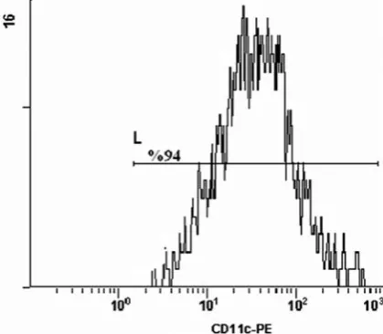
Analysis of DCs
The analysis of cells by flow cytometry on
a plot of CD11c vs side scatter showed that more
than 94% of the purifiedcells expressed CD11c+
(Fig. 1).
DCs Maturation
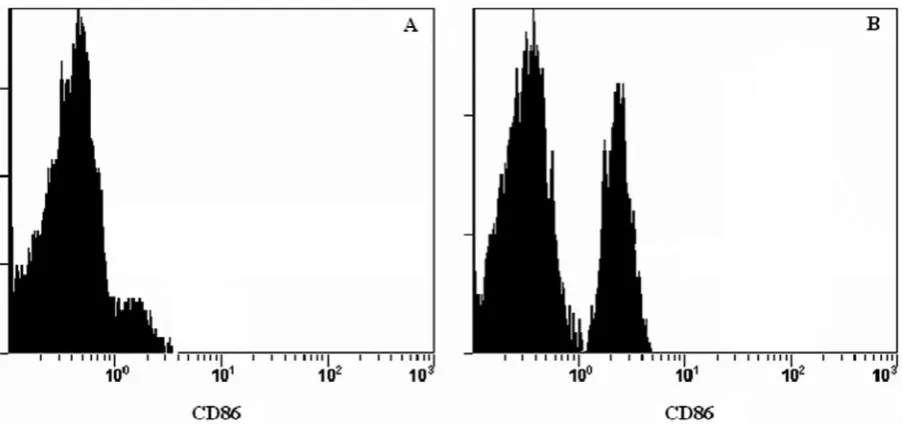
In the analysis of the effectiveness of C.
albi-cans and A. fumigatus on DC maturation, it was
found that the expression of DC co-stimulatory molecules such as CD80 and CD86 remarkably in-creased after the stimulation of DCs with both of the fungi. In addition, the expression of these co-stimulatory molecules increased in the wells con-taining DCs infected by fungi with the addition of
β-glucan after fungal infection; however, this crease was not significantly different from the in-crease in the wells infected by fungus without
β-glucan (p > 0.05).
The increase in the expression of these co-stim-ulatory molecules after the addition of β-glucan to the DCs stimulated by fungus shows that DCs have effective antigen-presenting cell function after fun-gal infection and in the presence of β-glucan.
The Levels of Cytokines
In our study, we observed that the IL-8 and IL-12p70 cytokine levels increased in DCs
stimu-lated by C. albicans and A. fumigatus compared to
the wells without any infection. However, these in-creases were not statistically significant (p > 0.05). The stimulation of DCs with fungus did not lead to any significant changes in IL-10 levels compared to
the wells with only DCs. Nevertheless, the addition of β-glucan to DCs stimulated with fungus caused statistically significant changes in the levels of IL-8, IL-10 and IL-12p70 (p < 0.05).
The changes observed in IL-8 cytokine levels after 24, 48 and 72 h are shown in Fig. 3. According to the figure, IL-8 cytokine levels after 24, 48 and 72 h in DCs stimulated with fungus increased com-pared to the wells with only DCs, but the changes were not statistically significant (p = 0.291, 0.445, 1, respectively). The addition of β-glucan to the
DCs stimulated with C. albicans led to statistically
significant changes after 24, 48 and 72 h (p < 0.05)
(Fig. 3).
The changes observed in IL-10 cytokine levels
in the wells containing DCs stimulated by C.
al-bicans after 24, 48 and 72 h are shown in Fig. 4. The stimulation of DCs by fungus did not lead to significant changes in IL-10 secretion after 24, 48
Fig. 2. The expression of CD86 of DCs (A. Dendritic cell, B. Dendritic cell infected with C. albicans)
and 72 h in the wells containing only DCs (p = 1). While the addition of β-glucan to DCs stimulated by C. albicans did not cause any statistically sig-nificant increases in IL-10 levels after 24 and 48 h compared to the wells without β-glucan, there was a statistically significant increase in IL-10 levels
af-ter 72 h (p < 0.05) (Fig. 4).
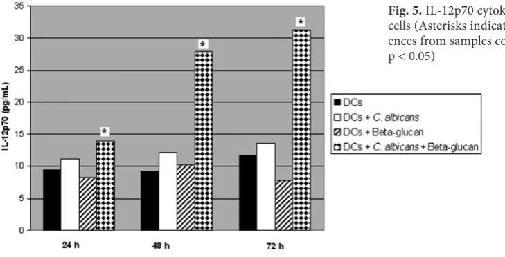
The changes in IL-12p70 cytokine levels in the
wells containing DCs stimulated with C. albicans
after 24, 48 and 72 h are shown in Fig. 5. When DCs were stimulated with fungus, there was an in-crease in the secretion of IL-12p70 compared to the wells containing DCs non-stimulated by fungus. However, the changes after 24, 48 and 72 h were not found statistically significant (p = 0.292, 0.089, 0.477, respectively). Still, the addition of β-glucan
to DCs stimulated with C.albicans led to
statis-tically significant changes after 24, 48 and 72 h (p < 0.05) (Fig. 5).
The same results were obtained with DCs
stim-ulated by A. fumigatus (data not shown).
Discussion
DCs, professional antigen presenting cells, play important roles in the initiation of the cel-lular response against foreign antigens. They are also effective in providing innate defense against microbial antigens. DCs are activated by different
microorganisms or their products[9]. Having been
activated when the infecting agent is recognized by the DC, DCs play some roles in the activation and control of the adaptive immunity through the
cy-tokines they secrete[10].
DCs are also important in the defense against
fungal infections. DCs are effective against C.
albi-cans infections as antigen presenting cells. They al-so phagocytose both yeast and hyphal forms of the fungus. It has been found that DCs not only phago-cytose, process and present fungal to T cells, but
al-so kill fungi as efficiently as macrophages do[11].
DCs also have an important role in the initiation of the cellular immune response against fungi. Fungi
Fig. 4. IL-10 cytokine levels in dendritic cells (Asterisks indicate significant differ-ences from samples containing only DCs, p < 0.05)
and its products cause a strong activation stimulus in DCs, leading to DC maturation with upregula-tion of co-stimulatory molecules and producupregula-tion
of cytokines leading to different T cell
respons-es. DCs are important for further treatment ap-proaches since they are effective in enhancing the
anti-fungal immune response[12]. Perruccio et al.
reported that the adoptive transfer of DCs activate Th cells and effect the outcome of fungal infections
in hematopoietic stem cell transplantation[6].
In our study, aimed to determine DC activa-tion signals and the release of cytokines such as IL-8, IL-10 and IL-12p70 following DCs stimulated
with A. fumigatus and C. albicans strains, we
ob-served some changes in the cell surface co-stimu-latory molecules and DC functions after DCs were
infected by fungi such as A. fumigatus and C.
albi-cans. Furthermore, the presence of β-glucan
affect-ed the activation of DCs infectaffect-ed by fungi.
There have been a lot of studies focusing on the immunomodulatory effects of β-glucan. How-ever, there have been few dealing with the effects of β-glucan on the maturation and activation of DCs after fungal infections. For this reason, we aimed to determine the effects of β-glucan administration on DCs during fungal infections.
The DC maturation process is one of the portant phases in the initiation of the adaptive im-mune response. The maturation signals for DCs may derive from various sources. This process is regulated by extracellular stimulators such as mi-crobial products or membrane associated ligands. DC maturation occurs due to the changes in mor-phological, phenotypical and functional features of DCs. DC activation is characterized by cyto-kine secretion and the expression of certain surface markers. These are essential for DCs to stimulate
effective T cell response. DCs affect immune
re-sponse by increasing co-stimulatory molecule ex-pression. CD80 and CD86 are the co-stimulatory
molecules crucial for both T cell response and Th
differentiation.
In our study, DCs stimulated by A. fumigatus
and C. albicans led to DC maturation. DC matura-tion is characterized by an increase in the expres-sion of co-stimulatory molecules on the dendritic cell surface and by the secretion of cytokines such
as IL-12 that provide strong stimulators for T cell
development and differentiation [13]. Our study showed that co-stimulatory molecules (CD80 and CD86) and proinflammatory cytokines (8,
IL-12p70) increased after DC was infected by A.
fu-migatus and C. albicans.
The increase in the expression of these co- -stimulatory molecules and cytokine secretion in
the DCs stimulated by A. fumigatus and C.
albi-cans indicate the importance of DC activation
during such fungal infections. However, the in-fection of DCs by fungus did not cause a remark-able change in IL-10 levels, an anti-inflammatory cytokine eliciting Th2 type response, compared to the DCs non-stimulated by fungus. The insignifi-cant change in IL-10 levels after fungal infections may be explained by the fact that Th1 cell-mediat-ed response plays an effective role in the protection
against fungal infections[14].
Romagnoll et al. have reported that different
forms of C. albicans enhance DC maturation and
stimulate Th1 type response[15].
Gafa et al. have also reported that in vitro
stim-ulation of DC by A. fumigatus increases the surface
expression of CD11b and CD18 activation signals and they have added that DC plays an important role in the regulation of the innate and adaptive
immune response against A. fumigatus by enabling
the production of various chemokines[3].
Netea et al. have indicated that although DC
stimulated by C. albicans produced a great amount
of TNF-α and IL-8, TNF and IL-8 release by DC
was only 5–10% of that released by monocytes or
macrophages[11].
IL-12 is a cytokine that plays an important role in the interaction between innate and adap-tive immunity. The expression of IL-12 is one of the most specific markers for DCs functionally
ac-tivated[15]. IL-12 stimulates natural killer cells and
T cells to secrete cytokines and to initiate lytic
ac-tivity. Moreover, IL-12p70 directs the immune
sys-tem towards Th1 response[16]. DCs stimulated by
C. albicans and A. fumigatus play important roles in Th1 cell differentiation by generating IL-12 secre-tion. Clinical and experimental studies have shown that Th1 cell reactivity plays an important role in the control of fungal infections such as invasive
aspergillosis[3].
β-glucan has several immunopharmacological
effects such as IL-6, TNF and nitrogen oxide se-cretion from macrophages, the adjuvant effect in
the antibody production and antitumor effect[17].
Since it enhances DC maturation, it can be used in DC-based vaccination and in combination
thera-pies to boost DC maturation[15].
In our study, the addition of β-glucan, an
im-portant immunomodulator, to DCs stimulated by fungus caused an increase in the expression of co- -stimulatory molecules and also in the proinflam-matory and antiinflamproinflam-matory cytokine response.
IL-8 and IL-12p70 cytokines in DCs stimulated by fungus indicates the importance of Th1 response during fungal infections. Moreover, the infection of DCs by fungus did not lead to significant chang-es in the secretion of IL-10, which is the indicator of Th2 response, and emphasizes again the impor-tance of Th1 response during fungal infections.
Avasti et al. have reported that in vitro
infec-tion of bone-marrow derived DC in mice with
dimorphic fungi such as Coccidioides posadasii
lead to a significant increase in the levels of IL-12. They also suggest that the differences in the activation status of DC in mice may be respon-sible for the discrepancy in their susceptibility to
C. posadasii[18].
The findings derived from our study show that β-glucan has strong effects on DC maturation and activation during fungal infections. It is con-sidered that by enabling DC maturation and acti-vation, β-glucan activates cytokine response, and
so it is considered to effect the regulation of
im-mune response[19]. Kikuchi et al. have also
re-ported that Candida β-glucan may augment DC
maturation [15].
Consequently, DC-based therapeutic ap-proaches have already been considered to be im-portant in the prevention of fungal infections. As was observed in our study, the changes in the mat-uration and activation of DC during fungal infec-tions becomes more remarkable in the presence of β-glucan and so this agent may significantly en-hance DC maturation and activation. The effect of β-glucan on DC endows it with an important role during the immune response. Since β-glucan is an important alternative for the therapeutic ap-proaches regulating host immune response, DC based immunotherapeutic approaches in the pres-ence of β-glucan for immunosuppressed patients with fungal infections are expected to be very ef-fective in future treatments.
References
[1] Faivre V, Lukaszewicz, Alves A, Charron D, Payen D, Haziot A: Accelerated in vitro differentiation of blood monocytes into dendritic cells in human sepsis. Clin Exp Immunol 2007, 147, 426–439.
[2] Sallusto F, Lanzavecchia A: Efficient presentation of soluble antigen by cultured human dendritic cells is main-tained by granulocyte/Macrophage colony-stimulating factor plus interleukin 4 and downregulated by tumor necrosis factor α. J Exp Med 1994, 179, 1109–1118.
[3] Gafa V, Remoli MR, Giacomini E, Gagliardi MC, Lande R, Severa M, Grillot R, Coccia EM:In vitro infection of human dendritic cells by Aspergillus fumigatus conidia triggers the secretion of chemokines for neutrophil and Th1 lymphocyte recruitment. Microb Infect 2007, 9, 971–980.
[4] Sundquist M, Rydström A, Wick MJ: Immunity to salmonella from a dendritic point of view. Cell Microbiol 2004, 6, 1–11.
[5] Flohe SB, Agrawal H, Schmitz D, Gertz M, Flohe S, Schade FU: Dendritic cells during polymicrobial sepsis rap-idly mature but fail to initiate a protective Th1-type immune response. J Leuko Biol 2006, 79, 473–481.
[6] Perruccio K, Boza S, Montagnoli C, Bellocchio S, Aversa F, Martelli M, Bistoni F, Velardi A, Romani L:
Prospects for dendritic cell vaccination against fungal infections in hematopoetic transplantation. Blood Cells, Mol Dis 2004, 33, 248–255.
[7] Brakhage AA, Bruns S, Thywissen A, Zipfel PF, Behnse J: Interaction of phagocytes with filamentous fungi. Curr Opin Microbiol 2010, 13, 409–415.
[8] Lull C, Wichers HJ, Savelkoul FJ: Antiinflammatory and immunomodulating properties of fungal metabolites. Mediators Inflamm 2005, 2, 63–80.
[9] Aiba S, Tagami H: Dendritic cell activation induced by various stimuli, e.g. exposure to microorganisms, their products, cytokines, and simple chemicals as well as adhesion to extracellular matrix. J Dermatol Sci 1999, 20, 1–13.
[10] Granucci F, Andrews DM, Degli-Esposti MA: Ricciardi-Castagnoli P. IL-2 mediates adjuvant effect of dendritic cells. Trends Immunol 2002, 23, 169–171.
[11] Netea MG, Gijzen K, Coolen N, Verschueren I, Figdor C, Van der Meer JWM, Torensma R, Kullberg BJ:
Human dendritic cells are less potent at killing Candida albicans than both monocytes and macrophages. Microb Infect 2004, 6, 985–989.
[12] Buentke E, Scheynius A: Dendritic cells and fungi. APMIS 2003, 111, 789–796.
[13] Lin YL, Lee SS, Hou SM, Chiang BL: Polysaccharide purified from Ganoderna lucidum induces gene expression changes in human dendritic cells and promotes T Helper 1 immune response in BALB/c mice. Mol Pharmacol 2006, 70, 637–644.
[14] Romagnoll G, Nisini R, Chlani P, Marlotti S, Teloni R, Cassone A, Torosantucci A: The interaction of human dendritic cells with yeast and germ-tube forms of Candida albicans leads to efficient fungal processing, dendritic cell maturation, and acquisition of a Th1 response-promoting function. J Leukol Biol 2004, 75, 117–126.
[15] Kikuchi T, Ohno N, Ohno T: Maturation of dendritic cells induced by Candida β-D-glucan. Int Immunopharma 2002, 2, 1503–1508.
[17] Akramiene D, Kondrotas A, Didziapetriene J, Kevelaitis E: Effects of β-glucans on the immune system. Medicina 2007, 43, 597–606.
[18] Awasthi S, Magee DM: Differences in expression of cell surface co-stimulatory molecules, Toll-like receptor genes and secretion of IL-12 by bone marrow-derived dendritic cells from susceptible and resistant Mouse strains in response to Coccidioides posadasii. Cell Immunol 2004, 231, 49–55.
[19] Carmona E, Vassallo R, Vuk-Pavlovic Z, Standing JE, Theodore TJ, Limper AH: Pnuemocystis cell wall β-Glucans induce dendtitic cell co-stimulatory molecule expression and inflammatoryactivation through a Fas-Fas ligand mechanism. J Immunol 2007, 177, 459–467.
Address for correspondence:
Isil Fidan Gazi University Faculty of Medicine
Department of Medical Microbiology Dekanlik Binasi 2.Kat
Beşevler/Ankara 06500 Turkey
Tel.:+90 312 202 46 26 E-mail: [email protected]
Conflict of interest: None declared