Grzegorz J. Lis
1, Ewa Jasek
1, Mariusz Gajda
1, Jan A. Litwin
1, Urszula Czubek
2,
Małgorzata Jasińska
1, Bogusław Kapelak
3, Jerzy Sadowski
3Participation of Tenascin C in Native and Homograft
Aortic Valve Degeneration*
Udział tenascyny C w degeneracji natywnych i homogennych
aortalnych zastawek serca
1 Chair and Department of Histology, Jagiellonian University Medical College, Cracow, Poland 2 Department of Coronary Disease, Jagiellonian University Medical College, Cracow, Poland
3 Department of Cardiac and Vascular Surgery and Transplantology, Jagiellonian University Medical College,
Cracow, Poland
Abstract
Background. Tenascin C (TnC) is a connective tissue matrix glycoprotein influencing cell proliferaton, migration and apoptosis. TnC participates in the development of pathological processes in the circulatory system including, as recently demonstrated, valvular degeneration leading to aortic valve stenosis. Homograft durability is limited by its degeneration, similar to changes appearing in cases of a typical aortic stenosis, which currently are regarded as an active remodeling process with expression of inflammatory modulators, as well as participation of cells and extracellular factors (including TnC), which under physiological conditions are involved in bone formation.
Objectives. The aim of the study was a comparison of TnC expression in native stenotic aortic valves and degen-erating aortic valve homografts.
Material and Methods. The material included two groups of aortic valves removed during routine surgery: native stenotic valves (n = 10) and dysfunctional aortic valve homografts (n = 10). Frozen valve sections were used for immunohistochemical detection of TnC, and also for identification of cells differentiated towards myofibroblasts (SMA), macrophages (CD68) and endothelial cells (CD34, vWF).
Results. TnC immunoreactivity was observed in both valve types, being significantly higher in native stenotic valves than in homografts (p = 0.007). The proportion of myofibroblasts in the total valvular cell population was insignificantly higher in native valves than in homografts. In homografts showing significantly lower total celullar-ity than native valves (p = 0.003), macrophages were more numerous (p = 0.005), and located mostly on the valve leaflet surfaces, while in native valves they were almost absent in this location.
Conclusions. The presence of TnC in the stroma of native stenotic aortic valves and in homografts indicates a par-ticipation of active remodeling mechanisms in the pathogenesis of degeneration in both types of valves. However, a significantly lower level of TnC and lower cellularity in homografts suggests that changes leading to their dysfunc-tion, to some extent, include passive degeneration (Adv Clin Exp Med 2011, 20, 2, 157–164).
Key words: tenascin, aortic valve, homograft, extracellular matrix proteins, immunofluorescence.
Streszczenie
Wprowadzenie. Tenascyna C (TnC) jest glikoproteiną macierzy tkanki łącznej biorącą udział w modyfikacji aktyw-ności proliferacyjnej, migracyjnej oraz różnicowaniu i apoptozie komórek. TnC uczestniczy w rozwoju procesów patologicznych w układzie naczyniowym, a ostatnio stwierdzono jej udział również w powstawaniu zastawkowych zmian degeneracyjnych prowadzących do stenozy aortalnej. Czas funkcjonowania homograftów aortalnych jest ograniczony ich degeneracją, przypominającą zmiany zachodzące w przypadku typowej stenozy aortalnej, które obecnie są uważane za proces aktywnej przebudowy (ekspresja modulatorów procesu zapalnego, udział komórek i czynników pozakomórkowych – w tym tenascyny C, fizjologicznie warunkujących osteogenezę).
Cel pracy. Porównanie ekspresji TnC w natywnych stenotycznych aortalnych zastawkach i degenerujących homo-graftach aortalnych.
Adv Clin Exp Med 2011, 20, 2, 157–164 ISSN 1230-025X
orIGINAL PAPErS
© Copyright by Wroclaw Medical University
Aortic valve degeneration is an age-related process leading to valvular sclerosis and stenosis affecting up to 5% of elderly patients [1]. In the course of the disease, dystrophic calcification is a common phenomenon. over the years it was regarded as a passive, purely destructive, non-modifiable process, leading to calcium salt ac-cumulation in defective valves. However, recent years have brought strong evidence supporting the concept of active mechanisms participating in the pathophysiology of valvular as well as vascular cal-cifications. The players involved include endothe-lial, inflammatory and activated interstitial cells, cytokines produced by these cells (TNF-alpha, TGF-beta1), extracellular lipids (apolipoproteins), various matrix proteins (metalloproteinases, bone morphogenetic proteins, bone matrix glycopro-teins) and renin-angiotensin systems [2].
Participation of the above mentioned active mechanisms in homograft pathology is still poorly understood. In calcified areas of explanted aortic valve homografts, Shetty et al. [3] found interstitial cells which expressed markers of osteoblastic dif-ferentiation and core binding factor α1 (rUNX2/ Cbfa1) as well as osteopontin, osteonectin, rANK and rANKL, indicating the osteogenic potential of the homograft valve tissue.
It is postulated that chondroblastic and/or os-teoblastic transdifferentiation of cells localized in the valvular stroma can lead to calcification and heterotopic bone formation [4, 5].
Dystrophic calcification is also observed in atherosclerosis, however, comparing to aortic valve stenosis (AVS), it seems to be a relatively late event. Studies have shown that coronary artery disease risk factors (hyperlipidemia, hypertension, diabetes) are also valid in cases of AVS. Many similarities between atherosclerosis and AVS ex-ist, including at pathophysiological and molecular levels: endothelial cell damage, lipid deposition, oxidation of lipoproteins and inflammatory infil-tration [6].
Tenascin C (TnC) is a member of a highly conserved family of large connective tissue matrix glycoproteins [7]. During embryogenesis, TnC participates in the modification of cellular activ-ity (proliferation, migration, differentiation and apoptosis). It plays also an important role in the development of vascular pathology, and the in-volvement of TnC in aortic valve degeneration leading to stenosis has been demonstrated recently [8]. However, its role in homograft pathology is not known.
The aim of this study was to examine if TnC is expressed in degenerating homograft aortic valves and to compare its expression level and distribu-tion with those in native stenotic aortic valves. Additional morphological parameters character-izing valvular degeneration such as abundance and distribution of myofibroblastic cells and mac-rophages, interstitial cell density and continuity of endothelial lining were also investigated.
Material and Methods
The valves were collected from patients under-going aortic valve replacement surgery. The study protocol was approved by the Jagiellonian Univer-sity Medical College Bioethical Committee. The material included 10 native aortic valves excised due to stenosis (6 men and 4 women, mean age 59 ± 12.3 years, max./min.: 73/24), 10 aortic valve homografts (7 men and 3 women, mean age 50.7 ± 12.3 years, max./min.: 67/27) originating from nonbeating-heart donors, implanted as “fresh”, antibiotic-preserved and removed due to dysfunc-tion during repeat surgery carried out within 6 to 16 years (mean 13 years) of grafting. Differences in the mean age of patients between both groups as well as age differences between sexes within each group were statistically insignificant.
After initial macroscopic evaluation, the valves were fixed for 24 hours in 4% buffered formalin.
Materiał i metody. Badanie obejmowało dwie grupy zastawek usuniętych w czasie rutynowych zabiegów wymiany zastawki: natywne stenotyczne zastawki aortalne (n = 10) i homografty (n = 10). Na skrawkach mrożeniowych płatków zastawek oznaczano immunofluorescencyjnie TnC, a także identyfikowano komórki zróżnicowane w kie-runku miofibroblastów (SMA) oraz makrofagi (CD68) i komórki śródbłonka (vWF, CD34).
Wyniki. W natywnych zastawkach stenotycznych stwierdzono istotnie wyższą ekspresję TnC niż w zastawkach homogennych (p = 0,007). Proporcja miofibroblastów w populacji komórek w natywnych zastawkach steno-tycznych była wyższa niż homograftach. W homograftach przy istotnie mniejszej całkowitej gęstości komórek, w porównaniu z zastawkami natywnymi (p = 0,003), stwierdzono większy udział makrofagów (p = 0,005), zwłaszcza umiejscowionych na powierzchni płatka, podczas gdy w zastawkach natywnych były one prawie nieobecne w tym miejscu.
Wnioski. obecność TnC w natywnych zastawkach stenotycznych i homograftach wskazuje na udział mechani-zmów aktywnej przebudowy w patogenezie degeneracji obu typów zastawek. Istotnie niższy poziom ekspresji TnC w homograftach oraz mniejsza liczba komórek zrębu sugeruje, że zmiany prowadzące do ich dysfunkcji w pewnym stopniu mają charakter biernej degeneracji (Adv Clin Exp Med 2011, 20, 2, 157–164).
For routine histology, paraffin-embedded cross sections (7 µm thick) of central segments of the cusps (from the base to the free edge) were pre-pared and stained with hematoxylin and eosin. For immunohistochemistry, 12 μm thick cryostat sec-tions were prepared.
Immunohistochemistry
After preincubation with 5% normal goat serum for 40 min, sections were incubated over-night with primary antibodies (Table 1) in a hu-mid chamber at room temperature. Next, sections were washed extensively in PBS and incubated for 90 min with goat anti-mouse Cy-3-conjugated and/or goat anti-rabbit Cy-2-conjugated antibod-ies (Table 1). Cell nuclei were counterstained with DAPI (Sigma, Saint Louis, Mo, USA). Sections were washed three times in PBS and mounted in a glycerol/PBS solution (pH = 8.6). Negative con-trols were performed by omitting the primary an-tibodies during the first incubation.
Morphometry and Statistics
Sections were examined under an olym-pus BX50 light/fluorescence microscope. Images were recorded using a DP-71 digital CCD camera (olympus, Japan) coupled to an IBM PC-class computer equipped with AnalySIS-FIVE® (Soft
Imaging System GmbH, Münster, Germany) im-age analysis system.
TnC expression was estimated semiquantita-tively, using a score range from 0 to 4 (0 = none, 1 = trace, 2 = < ¼ leaflet section area (l.s.a.), 3 = ≤ ½ l.s.a., 4 = > ½ l.s.a.).
For the estimation of abundance and distribu-tion of myofibroblastic cells (alpha-SMA positive) the FSMA coefficient was calculated, defined as: FSMA
= r × G × P, where r denotes an area occupied by myofibroblasts: 0 = none, 1 = a few scattered
cells, 2 = <¼ l.s.a., 3 = < ½ l.s.a., 4 = < ¾ l.s.a.. 5 = ≥ ¾ l.s.a.; G denotes myofibroblast density in clusters: 1 = < 250 cells/mm2, 2 = < 500 cells/mm2,
3 = ≥ 500 cells/mm2; P denotes the proportion of
myofibroblasts to other cells: 1 = < 1%, 2 = ≤ 10%, 3 – > 10%
Total interstitial cell density was estimated by using a score range from 0 to 3 (0 = ≤ 10 cells/ mm2, 1 = ≤ 100 cells/mm2, 2 = ≤ 300 cells/mm2,
3 = > 300 cells/mm2).
The statistical analysis employed (in compli-ance with the specific category of collected data) a t-Student test for unpaired data, a Mann-Whit-ney test, and the Fisher’s exact test. The value of p < 0.05 was considered statistically significant.
Results
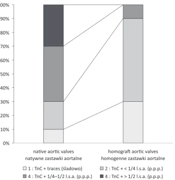
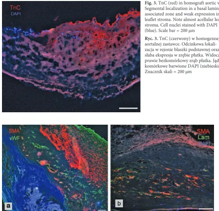
The authors found the presence of TnC in both groups of valves with significantly higher im-munoreactivity in native aortic valves than in ho-mograft valves (p = 0.007) (Fig. 1). In the cases of native valves (Fig. 2a) as well as homografts (Fig. 3), tissue distribution of TnC was similar: strongly TnC positive areas were seen within focal calci-fications (Fig. 2b), positive immunostaining was also visible in the walls of blood vessels located in tissue adjacent to the calcifying areas (Fig. 2c). In most cases (except for one homograft and two na-tive valves), segmental TnC immunoreactivity was also observed along both leaflet surfaces, in a basal lamina-associated zone (Fig. 2a, 3).
The native aortic valves revealed significantly higher density of interstitial cells than the ho-mografts (p = 0.003).
The proportion of myofibroblasts/activated interstitial cells (alpha-SMA positive) in the total interstitial cell population was more than twice as high in native valves as compared with homograft valves (FSMA = 7.5 and 3, respectively), however
Table 1. Primary and secondary antibodies used in immunofluorescentic examination. MC = monoclonal; PC = polyclonal
Tabela 1. Przeciwciała pierwotne i wtórne użyte w badaniach immunofluorescencyjnych. MC = monoklinalne; PC = poliklonalne
Antibody
(Przeciwciało) Type (Typ) Dilution(rozcieńczenie) Manufacturer(Producent) Code(Kod) Primary (Pierwotne)
mouse anti-human tenascin mouse anti-actin, smooth muscle mouse anti-human CD68 mouse anti CD34
rabbit anti von Willebrand Factor rabbit anti-laminin
Secondary (Wtórne)
Cy3-conjugated goat anti-mouse Cy2-conjugated goat anti-rabbit
MC MC MC MC PC PC 1:200 1:200 1:1 1:50 1:200 1:50 1:400 1:400
Chemicon, Temecula, CA Chemicon, Temecula, CA Chemicon, Temecula, CA Novocastra, Newcastle, UK Novocastra, Newcastle, UK Sigma, Saint Louis, Mo
Jackson Ir, West Grave, PA Jackson Ir, West Grave, PA
Fig. 1. TnC in the examined groups of valves. Graph shows the percentage of cases with TnC immunoreactivity at the particular score values (1–4). l.s.a. – leaflet section area
Ryc. 1. TnC w badanych grupach zastawek. Wykres przedstawia odsetek przypadków wykazujących dodatnią reakcję w danym przedziale (1–4). p.p.p. – powierzchnia przekroju płatka
0% 10% 20% 30% 40% 50% 60% 70% 80% 90% 100%
na�ve aor�c valves natywne zastawki aortalne
homogra� aor�c valves homogenne zastawki aortalne
1 : TnC + traces (śladowo) 2 : TnC + < 1/4 l.s.a. (p.p.p.) 4 : TnC + 1/4–1/2 l.s.a. (p.p.p.) 4 : TnC + > 1/2 l.s.a. (p.p.p.)
Fig. 2. TnC (red) in native stenotic aortic valve leaflet.
a: immunoreactivity in the leaflet stroma and segmen-tally along the surface in a basal lamina-associated zone;
b: high immunoreactivity in the vicinity of focal calci-fications (asterisk); c: immunoreactivity in the wall of blood vessel located in tissue adjacent to the calcifying areas (→). Green fluorescence: laminin in basal lamina.
a,b,c: cell nuclei stained with DAPI (blue). Scale bar: a, b = 200µm, c = 50 µm
Ryc. 2. TnC (czerwony) w płatku natywnej stenotycznej zastawki aortalnej; a: immunoreaktywność w zrębie płatka i odcinkowo wzdłuż powierzchni w rejonie blasz-ki podstawnej; b: wysoki poziom immunoreaktywno-ści w sąsiedztwie wapniejących złogów (gwiazdka); c:
Fig. 3. TnC (red) in homograft aortic valve. Segmental localization in a basal lamina-associated zone and weak expression in leaflet stroma. Note almost acellular leaflet stroma. Cell nuclei stained with DAPI (blue). Scale bar = 200 µm
Ryc. 3. TnC (czerwony) w homogennej aortalnej zastawce. odcinkowa lokali-zacja w rejonie blaszki podstawnej oraz słaba ekspresja w zrębie płatka. Widoczny prawie bezkomórkowy zrąb płatka. Jądra komórkowe barwione DAPI (niebieskie). Znacznik skali = 200 µm
Fig. 4. Myofibroblastic/activated valvular interstitial cells (red). a: in native stenotic aortic valve leaflet. Note blood vessels with SMA positive cells in their walls in deeper areas of valve stroma, close to focal calcifications (*). Endothelial cells (green). b: in homograft valve stroma. Cell nuclei stained with DAPI (blue). Scale bar = 200 µm
Ryc. 4. Komórki miofibroblastyczne/aktywowane komórki śródmiąższowe (czerwone). a: w płatku natywnej zastawki stenotycznej. W głębszych warstwach w sąsiedztwie wapniejących złogów (*) widoczne naczynia z komórkami SMA-pozytywnymi w ścianie. Komórki śródbłonkowe (zielony). b: w zrębie homograftu. Jądra komórkowe barwione DAPI (niebieskie). Znacznik skali = 200 µm
Fig. 5. Macrophages (red) arranged in epithelial-like fashion on the surface of homograft valve. Note vir-tually acellular leaflet stroma. Cell nuclei stained with DAPI (blue). Scale bar = 50 µm
this difference did not reach statistical significance (p = 0.07). In five native valves and two homografts, alpha-SMA positive cells were also found in the walls of small blood vessels (Fig. 4).
In all native and homograft valves, macrophag-es were located in the areas of focal calcifications. They were also present in other stromal areas in all homografts and in six native valves. In eight ho-mografts and in only one native valve, we found macrophages distributed along the leaflet surface in an epithelial-like manner (Fig. 5).
In almost all (nine) native valves, newly formed blood vessels were located in areas of focal calcifications (in three cases also beneath the leaf-let surface in the form of relatively wide vascular channels with variable diameter, partly lined with endothelial cells). In these areas, inflammatory infiltrations were also observed, predominantly composed of mononuclear cells. Blood vessels were found in only four homograft valves.
In the majority of homografts (six), the lay-ered structure of the valves was almost completely obscured, which was only partly observed in heav-ily stenotic native valves. The valves showed a sub-stantial loss of endothelial cells, more prominent in homografts. In these groups of valves, intersti-tial cells were unevenly distributed, and in most cases acellular areas were prominent (Fig. 3). Small mononuclear (in one case mixed) inflammatory infiltrations were seen in half of the homograft valves.
Discussion
TnC is associated with the morphogenesis of vascular, skeletal and nervous systems. In normal adult tissue, TnC expression remains at a very low level only in some areas of the connective tissue. In the course of inflammation, in malignant tumors or during intense tissue regeneration its expres-sion is significantly elevated [7].
Healthy valve stroma, as evidenced by immu-nohistochemistry and in-situ hybridization meth-ods in non-stenotic valves, shows only minimal TnC expression in basement-membrane-associat-ed zones [8]. Present study has demonstratbasement-membrane-associat-ed the presence of TnC in both native stenotic and in in-sufficient homograft aortic valves. In cases of na-tive stenotic valves, this finding is consistent with the observations of Satta et al. [8] who reported in-creased expression of TnC correlating with the de-gree of valve pathology. In authors’ material, par-ticularly strong TnC immunoreactivity appeared in leaflet areas involved in intensified remodeling processes leading to massive fibrosis, nodular cal-cification and blood vessel formation.
These results and previous observations of other authors show that TnC expressed in patho-logically altered aortic valves is produced by valvu-lar interstitial cells (including alpha-SMA positive myofibroblasts, which are regarded as a subpopu-lation of activated valvular interstitial cells involved in matrix remodeling and osteoblastic valvular interstitial cells responsible for the production of matrix components characteristic for bone tissue), as well as by endothelial cells and macrophages [8,9]. TnC inducing factors include cytokines (e.g. interleukin 1, TGF-beta 1) released by inflamma-tory cells, as well as mechanical/hemodynamic stress [10].
The impact of mechanical stress on the initia-tion of tissue mineralizainitia-tion is widely recognized [11, 12]. It seems to be a more important player in the pathology of the heart valves than of arteries, contributing to the earlier and more prominent calcification observed in the former ones, particu-larly in places of maximal leaflet flexion (near the base), which are exposed to the largest loads [13].
A notably accelerated degenerative process with prominent calcification has been demonstrat-ed in homograft aortic valves [14–16]. Although explanted homografts can show similar pathologi-cal changes to those observed in native stenotic valves (including interstitial cell transdifferentia-tion and productransdifferentia-tion of ground substance elements typical for bone tissue) [3], they also reveal some specific pathological features. Important factors modifying the process of homograft degeneration include a variable (but always substantial) level of endothelial and interstitial cell damage, depen-dent on the applied graft preimplantation preser-vation method (as “fresh”, antibiotic-preserved or cryopreserved), the source of the valve (multior-gan donor, cardiac transplantation or nonbeating heart donor), as well as host and donor immune responses [17–19].
Present study has demonstrated for the first time the participation of TnC in homograft valve pathology. In view of the fact that homograft du-rability is limited by the degenerative processes analogous to those observed in native stenotic valves, the TnC immunoreactivity found by us in homografts as well as its similar location in leaf-let tissue, provide further evidence for a common pathobiology of both homograft and native aortic valve degenerative processes. However, the sub-stantially lower level of TnC immunoreactivity observed in the homografts suggests some differ-ences between both processes.
cells), in particular those originating directly from the bloodstream, than the native valves [20, 21]. Localization of macrophages on the surface of the homograft leaflets, very often observed in authors’ material, indicate that they originate from blood monocytes adhering to the areas of the valve de-void of endothelial lining.
In the aortic homografts the authors examined in this and the previous study [22], they found completely acellular areas in valve stroma. The to-tal number of interstitial cells was also significant-ly lower. Studies on the viability of the homograft cells have shown that many of them are lost (partly due to apoptosis) in the months immediately fol-lowing the grafting, which significantly reduces valve regenerative abilities [19]. Thus, it seems probable that the chances of successful prevention of valve degeneration (e.g. by controlling the ac-tivity of the cells involved) are limited in cases of homografts. Also the lower number of homograft
valves revealing formation of new blood vessels and limited myofibroblastic transdifferentiation of interstitial cells in these valves, as compared with native stenotic valves, seem to support this opinion.
The relatively small number of valves exam-ined is a limitation of the present study, therefore our results are preliminary and require confirma-tion on a larger group of valves.
The authors conclude that the appearance of TnC in native stenotic aortic valves and in aortic valve homografts indicates the involvement of ac-tive remodeling mechanisms in the pathogenesis of the degeneration in both types of valves. In ho-mograft aortic valves, however, significantly lower TnC immunoreactivity and limited total as well as SMA positive interstitial cell number suggests that mechanisms leading to their dysfunction are also – to some extent – passive, which should be taken into account in determining preventive strategies.
References
[1] Stewart BF, Siscovick D, Lind BK, Gardin JM, Gottdiener JS, Smith VE, Kitzman DW, Otto CM: Clinical factors associated with calcific aortic valve disease. Cardiovascular Health Study. J Am Coll Cardiol 1997, 29, 630–634.
[2] O’Brien KD: Pathogenesis of calcific aortic valve disease: a disease process comes of age (and a good deal more). Arterioscler Thromb Vasc Biol 2006, 26, 1721–1728.
[3] Shetty R, Pepin A, Charest A, Perron J, Doyle D, Voisine P, Dagenais F, Pibarot P, Mathieu P: Expression of bone-regulatory proteins in human valve allografts. Heart 2006, 92, 1303–1308.
[4] Caira FC, Stock SR, Gleason TG, McGee EC, Huang J, Bonow RO, Spelsberg TC, McCarthy PM, Rahimtoola SH, Rajamannan NM: Human degenerative valve disease is associated with up-regulation of low-density lipopro-tein receptor-related prolipopro-tein 5 receptor-mediated bone formation. J Am Coll Cardiol 2006, 47, 1707–1712.
[5] Lis GJ, Litwin JA, Kapelak B, Furgal-Borzych A, Gajda M, Cichocki T, Sadowski J: Development of mature lamel-lar bone with a hematopoietic compartment in an aortic valve homograft. J Heart Valve Dis 2009, 18, 578–580.
[6] Goldbarg SH, Elmariah S, Miller MA, Fuster V: Insights into degenerative aortic valve disease. J Am Coll Cardiol 2007, 50, 1205–1213.
[7] Hsia HC, Schwarzbauer JE: Meet the tenascins: multifunctional and mysterious. J Biol Chem 2005, 280, 26641– 26644.
[8] Satta J, Melkko J, Pöllänen R, Tuukkanen J, Pääkkö P, Ohtonen P, Mennander A, Soini Y: Progression of human aortic valve stenosis is associated with tenascin-C expression. J Am Coll Cardiol 2002, 39, 96–101.
[9] Liu AC, Joag VR, Gotlieb AI: The emerging role of valve interstitial cell phenotypes in regulating heart valve pathobiology. Am J Pathol 2007, 171, 1407–1418.
[10] Jones FS, Jones PL: The tenascin family of ECM glycoproteins: structure, function, and regulation during embry-onic development and tissue remodeling. Dev Dyn 2000, 218, 235–259.
[11] Buch WS, Kosek JC, Angell WW: The role of rejection and mechanical trauma on valve graft viability. J Thorac Cardiovasc Surg 1971, 62, 696–706.
[12] Chanda J: Prevention of calcification of heart valve bioprostheses: An experimental study in rat. Ann Thorac Surg 1995, 60, S339–S342.
[13] Vesely I, Macris N, Dunmore PJ, Boughner D: The distribution and morphology of aortic valve cusp lipids. J Heart Valve Dis 1994, 3, 451–456.
[14] Angell WW, Oury JH, Duran CG, Infantes Alcon C: Twenty years comparision of the human homograft and porcine xenograft. Ann Thorac Surg 1989, 48, 89–90.
[15] Vogt PR, Stallmach T, Niederhäuser U, Schneider J, Zünd G, Lachat M, Künzli A, Turina MI: Explanted cryopreserved allografts: a morphological and immunohistochemical comparison between arterial allografts and allograft heart valves from infants and adults. Eur J Cardiothorac Surg 1999, 15, 639–645.
[16] Yap CH, Yii M: Factors influencing late allograft valve failure. Scand Cardiovasc J 2004, 38, 325–333.
[17] Crescenzo DG, Hilbert SL, Barrick MK, Corcoran PC, St Louis JD, Messier RH, Ferrans VJ, Wallace RB, Hopkins RA: Donor heart valves: Electron microscopic and morphometric assessment of cellular injury induced by warm ischemia. J Thorac Cardiovasc Surg 1992, 103, 253–258.
[19] Niwaya K, Sakaguchi H, Kawachi K, Kitamura S: Effect of warm ischemia and cryopreservation on cell viability of human allograft valves. Ann Thorac Surg 1995, 60, S114–S117.
[20] Rokita E: Physicochemical studies of aortic wall mineralization. Life Chem rep 1991, 8, 185–189.
[21] Mitchell RN, Jonas RA, Schoen FJ: Structure-Function correlations in cryopreserved allograft cardiac valves. Ann Thorac Surg 1995, 60, S108–S113.
[22] Lis GJ, Rokita E, Podolec P, Pfitzner R, Dziatkowiak A, Cichocki T: Mineralization and organic phase modifica-tions as contributory factors of accelerated degeneration in homograft aortic valves. J Heart Valve Dis 2003, 12, 741–751.
Address for correspondence:
Grzegorz J. Lis
Chair and Department of Histology Jagiellonian University Medical College Kopernika 7
31-034 Kraków Poland
Tel.: +48 12 411 13 69 E-mail: [email protected]
Conflict of interest: None declared