Hulya Ozturk
1, Elcin Hakan Terzi
2, Ufuk Ozgen
3, Arif Duran
4,
Hayrettin Ozturk
1Lithospermic Acid and Ischemia/Reperfusion Injury
of the Rat Small Intestine Prevention
Kwas litospermowy a zapobieganie niedokrwieniu/uszkodzeniu
reperfuzyjnemu jelita cienkiego u szczura
1 Department of Pediatric Surgery, Abant Izzet Baysal University, Medical School, Bolu, Turkey
2 Department of Histology and Embryology, Abant Izzet Baysal University, Medical School, Bolu, Turkey 3 Department of Pharmacognosy, Faculty of Pharmacy, Atatürk University, Erzurum, Turkey
4 Department of Emergency, Abant Izzet Baysal University, Medical School, Bolu, Turkey
Abstract
Background. Intestinal ischemia and reperfusion (I-R) injury of different causes, including cardiac insufficiency, sepsis, vasodepressant and cardiodepressant drugs, and complications of long-lasting surgery, represents a major clinical problem.
Objectives. The purpose of the present study was to investigate whether lithospermic acid (LA) can reduce oxida-tive stress and histological damage in the rat small bowel subjected to mesenteric I-R injury.
Material and Methods. The study was performed on three groups of animals, each composed of 7 rats: the SO (sham operation) group, the I-R/Untreated group and the I-R/LA (I-R plus LA pretreatment) group. Intestinal ischemia for 45 minutes and reperfusion for 60 minutes were applied. Ileum specimens were obtained to determine the tissue level of malondialdehyde (MDA), superoxide dismutase (SOD), glutathione peroxidase (GPx), catalase (CAT) and myeloperoxidase (MPO) activities and histological changes.
Results. Untreated intestinal I-R resulted in increased tissue MDA and MPO levels and diminished SOD and GPx activities. These changes were found to be almost reversed in the LA treatment group. Histopathologically, the intestinal injury in rats treated with LA was less than the untreated I-R group.
Conclusions. Lithospermic acid attenuates mesenteric ischemia reperfusion injury in rat intestines by increasing tissue SOD and GPx activities and decreasing MDA and MPO levels. Lithospermic acid also improves morphologi-cal alterations which occurred after periods of reperfusion (Adv Clin Exp Med 2012, 21, 4, 433–439).
Key words: small bowel, ischemia and reperfusion injury, lithospermic acid.
Streszczenie
Wprowadzenie. Uszkodzenie jelit spowodowane niedotlenieniem i reperfuzją (IR) wynikające z różnych przyczyn, w tym niewydolności serca, posocznicy, stosowania leków wazodepresyjnych i kardiodepresyjnych oraz powikłań dużych zabiegów chirurgicznych, jest ważnym problemem klinicznym.
Cel pracy. Zbadanie, czy kwas litospermowy (LA) może zmniejszyć stres oksydacyjny i uszkodzenie histologiczne jelita cienkiego u szczurów, u których wywołano niedotlenienie i reperfuzję krezki.
Materiał i metody. Badanie zostało przeprowadzone na trzech grupach zwierząt, z których każda składała się z 7 szczu-rów: grupa SO (operacja pozorowana), grupa IR nieleczona i grupa IR / LA (IR oraz podanie LA przed leczeniem). Niedokrwienie jelit utrzymywano przez 45 minut i reperfuzję przez 60 minut. Pobrano wycinki jelita krętego w celu określenia stężenia aldehydu malonowego (MDA), dysmutazy ponadtlenkowej (SOD), peroksydazy glutationowej (GPx), katalazy (CAT) i mieloperoksydazy (MPO) w tkankach i zmian histologicznych.
Wyniki. Nieleczone IR jelit spowodowało zwiększenie stężenia tkankowego MDA i MPO oraz mniejszą aktywność SOD i GPx. Zmiany te okazały się niemal odwrotne w grupie leczonej LA. Histopatologicznie uszkodzenie jelit u szczurów leczonych LA było mniejsze niż w grupie IR nieleczonej.
Wnioski. Podawanie kwasu litospermowego zmniejsza uszkodzenie jelit spowodowane niedotlenieniem i reperfuzją u szczurów przez zwiększenie tkankowej SOD i aktywności GPx, zmniejszenie stężenia MDA i MPO. Kwas litospermo-wy zmniejsza zmiany morfologiczne, które nastąpiły po okresie reperfuzji (Adv Clin Exp Med 2012, 21, 4, 433–439).
Słowa kluczowe: jelito cienkie, uszkodzenie spowodowane niedotlenieniem i reperfuzją, kwas litospermowy.
Adv Clin Exp Med 2012, 21, 4, 433–439 ISSN 1899–5276
ORIGINAL PAPERS
Intestinal ischemia and reperfusion (I-R) in-jury plays an important role in some clinical con-ditions, such as acute mesenteric ischemia, midgut volvulus, shock, cardiac surgical interventions and small bowel transplantation [1–4]. I-R injury of the intestine not only leads to loss of intestinal barrier functions but also may results in bacterial translo-cation and a systemic inflammatory response that causes multiple organ failure [5–7].
The development mechanisms of I-R injury are not fully known. However, it is known that re-active oxygen species (ROS) play a very important role in the pathophysiology of I-R injury. Hypoxia occurs during the period of ischemia, but reper-fusion injury occurs after reconstitution of blood flow [8]. Reperfusion of ischemic tissue increases the effects of early ischemic injury by release of ROS and accumulation of activated neutrophils [6]. Superoxide, hydrogen peroxide, and hydroxyl radicals are generated and may cause cellular dam-age by direct action and by secondary activation of polymorphonuclear neutrophils (PMN) [9, 10]. Various anti-oxidant agents have been studied to prevent reperfusion injury after ischemia. Lith-ospermic acid (LA) is a biologically active compo-nent isolated from the aqueous extract of danshen, the dried root and rhizome of Salvia miltiorrhiza
Bge (Labiatae) [11]. LA has a strong antioxidant effect in preventing the production of superoxide radical and lipid peroxide [12, 13].
In the present study, the authors aimed to de-termine if lithospermic acid has protective effects against mucosal injury induced by I-R in the rat small intestine.
Material and Methods
Animal Model and Experimental
Design
Twenty-one Sprague-Dawley rats weighing between 200 and 225 g were divided into 3 groups, each containing 7 rats: the SO (sham operation) group, the I-R/Untreated (untreated ischemia-rep-erfusion) group, and the I-R/LA (ischemia-reper-fusion plus LA pretreatment) group (10 mg/kg LA was administered intraperitoneally 30 min before the induction of ischemia).
The rats were kept under standardized condi-tions for food, water, light and temperature. After overnight fasting, each rat was anesthetized with ketamine (50 mg/kg) and xylazine (14 mg/kg) and underwent a midline laparotomy and bowel evis-ceration. After ligating collateral arcades from the right colic artery and the jejunal arteries, the su-perior mesenteric artery (SMA) was occluded with
an atraumatic microvascular clamp as described by Megison et al. [14]. During this period of warm intestinal ischemia, each rat was cannulated via the jugular vein with a 24-gauge venula needle and lac-tated Ringer’s solution (LR) was given (1 ml/kg/h) using an infusion pump. The bowel was placed in the abdominal cavity, and then the incision was closed. After 45 minutes of ischemia, the abdomi-nal cavity was reopened. The occluding clamp was removed and the intestine was returned to the peritoneal cavity, then the abdomen was reclosed. After 60 minutes reperfusion period, the abdomi-nal wall was opened once more and samples of ile-um were obtained for biochemical and histopatho-logical analysis. The animals were sacrificed when the procedure was completed. In the sham group, only laparotomy and preparation of SMA was per-formed, and SMA was not occluded with a clip.
Samples for biochemical analysis were fro-zen in liquid nitrogen and stored at –80°C until processing. Samples for histological analysis were fixed in (%10) formaldehyde.
Analytical Procedures of Oxidative
Stress-Associated Parameters
All tissues were homogenized in ice-cold buf-fer(0.25 M sucrose, 10 mM Tris-HCl, and 0.25 mM
for measuring neutrophil accumulation in tissue samples, because it is closely correlated with the number of neutrophils present in the tissue [22]. The homogenates werethen centrifuged at 17,000 × g at 4°C for 15 min, andMPO activity (U/g pro-tein) in the supernatant was measured as previ-ously described [23].
Histopathology
The tissue specimens were fixed in 10% form-aldehyde, then dehydrated and embedded in par-affin wax. The samples were sectioned and stained with hematoxylin and eosin (H&E) and assessed in a blinded fashion by pathologists. Mucosal le-sions were graded on a system described by Chiu et al. [24].
Statistical Analysis
Values are expressed as the means ±SEM. To evaluate differences among the groups studied, a two-way analysis of variance (ANOVA) with Fisher’s post hoc test was used. Differences were considered statistically significant when P < 0.05.
Results
The MDA, GPx, SOD and MPO values for the different groups are shown in Fig. 1a, b, c and d. The levels of MDA and MPO were significantly increased in the intestinal tissue of the I-R/Un-treated group (p < 0.001) compared to the control group. However, in the I-R/LA treatment group, intestinal tissue MDA and MPO levels were signif-icantly decreased compared to the I-R/Untreated group (p < 0.5). I/R caused a significant decrease in intestinal tissue GSH and SOD levels (p < 0.05) when compared to the control group. In the LA-treated I-R group, intestinal tissue GPx and SOD levels were increased (p < 0.05) compared to the untreated I-R/Untreated group.
Quantitative evaluation of hematoxylin and eosin stained histologic sections showed that the intestinal injury score increased significantly in the I-R/Untreated and I-R/LAgroup rats compared to the SO group (p < 0.05 and p < 0.05, respectively). On the other hand, the intestinal injury score was decreased in I-R/LA group rats compared to the I-R/Untreated (p < 0.05) (Fig. 2).
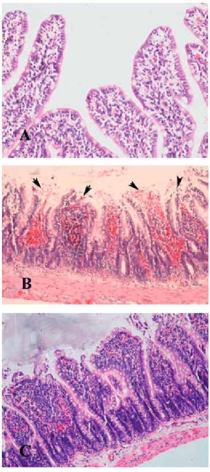
Using the Chiu scoring system, the ileal sec-tion of the sham-operated group revealed normal architecture (Fig. 3A), whereas in the rats sub-jected to I-R, significant mucosal injury with loss of villus, hemorrhage, and ulceration was seen (Fig. 3B). The injury rate of treatment groups was
found to be minimized. Ileal sections of the I-R/ LA group showed minimal alterations character-ized with moderate lifting of the epithelial layer from lamina propria (Fig. 3C).
Discussion
Seven phenolic compounds isolated from
Salvia miltiorrhiza as active components possess
potent antioxidative activity against oxidative in-jury. Lithospermic acid B in these compounds is the most important component [25]. Lithosper-mic acid B hasbeen shown to possess strong ef-fects on antioxidative and freeradical scavenging, on protecting renal dysfunction, hepatitis, lung fibrosis, and on improving blood circulation[13, 26–29]. Furthermore, lithospermic acid B showed endothelium-dependent vasodilator effects and an ameliorative effect on ischemia reperfusion induced acuterenal failure in rats [13, 30, 31]. In this experimental study, we investigated the ef-fects of lithospermic acid in intestinal I-R injury. As discussed below, the results of the present study demonstrate that treatment with lithospermic acid markedly attenuates the intestinal damage of rats subjected to I-R injury.
with the expression ofaquaporin 2 (AQP 2) and Na, K-ATPase in ischemia-reperfusion induced acute renal failure (ARF) in rats. Lithospermic acid B showed strong antioxidant activity against the production of reactive oxygen species (ROS), ROS-induced hemolysis,and production of lipid peroxide in a dose-dependent manner.They sug-gested that lithospermic acid B ameliorates renal
defects in rats with ischemia-reperfusion induced ARF, most likely via scavenging of ROS.
Under physiological situations, the damaging effects of ROS are prevented by antioxidant en-zymes such as SOD and GPx [36]. However, dur-ing reperfusion of the intestine, the oxidant/anti-oxidant balance may change. In the present study, SOD and GPx activities were determined to
evalu-MDA
0 1 2 3 4 5 6 7
Sham I-R I-R/LA
Groups
nmol/g
*
* †
GPx
0 5 10 15 20 25 30 35 40
Sham I-R I-R/LA
Groups
micromol/mg protei
n
*
* †
SOD
0 0,5 1 1,5 2 2,5
Sham I-R I-R/LA
Groups
U/g protein *
* †
MPO
0 0,5 1 1,5 2 2,5 3 3,5
Sham I-R I-R/LA
Groups
U/g protein
*
* †
Fig. 1. (a) Malondialdehyde (MDA) and (b) glutathione peroxidase (GPx) levels, (c) superoxide dismutase (SOD) and (d) myeloperoxidase (MPO) activity in the intestinal tissue of sham-operated control groups, I-R/Untreated groups and I-R/lithospermic acid-treated groups. Each group consists of seven animals. * p < 0.05, compared to con-trol group. † p < 0.05, compared to I-R/Untreated group. Group 1: sham-operated concon-trol; Group 2: I-R/Untreated; Group 3: I-R/lithospermic acid-treated
Ryc. 1. (a) Stężenie aldehydu malonowego (MDA) i (b) peroksydazy glutationowej (GPx), (c) dysmutazy ponadtlen-kowej (SOD) i (d) mieloperoksydazy (MPO) w tkance jelit w grupie pozornie operowanej kontrolnej, IR / nieleczonej i IR / LA. Każda grupa składa się z siedmiu zwierząt. * P < 0,05 w porównaniu z grupą kontrolną. † p < 0,05 w porów-naniu do grupy IR / nieleczonej. Grupa 1: kontrolna operowana pozornie; Grupa 2: IR / nieleczona, Grupa 3: IR / LA
A
B
C
ate the tissue antioxidant system; the treatment group had reversely enhanced activities of SOD and GPx, suggesting an attenuation of intestinal I-R injury by detoxifying the oxygen free radicals. As mentioned above, LA is a well known ROS scavenger and contributes to the effects of antioxi-dant enzymes. Hence, in I-R/LA group rats, SOD and GPx may be higher. Additionally, we found that I-R caused a considerable increase in the in-testinal tissue levels of oxidative stress markers, MDA and MPO. Moreover, the increase in these biomarkers after I-R was significantly ameliorated by treatment with lithospermic acid.
In conclusion, lithospermic acid attenuates mesenteric I-R injury in rat intestine by increas-ing tissue SOD and GPx activities and decreasincreas-ing MDA and MPO levels. Lithospermic acid also improves morphological alterations which occur after periods of reperfusion. This study first of-fers evidence that LA protects the ileum from I-R induced injury, likely acting by multiple mecha-nisms including antioxidative, free radical scav-enging and endothelium-dependent vasodilator effects. LA treatment may offer a new therapeutic alternative for I-R injury in some clinical entities, such as acute mesenteric ischemia, midgut volvu-lus, shock, cardiac surgical interventions and small bowel transplantation.
histologic damage
0 0,5 1 1,5 2 2,5 3 3,5 4 4,5
sham I-R I-R/LA
groups
score
* † *
Fig. 2. Comparative histologic score measurements at the groups. *p < 0.05 compared with group 1. †p < 0.05 compared with group 3. Values are mean ± SEM. Group 1: sham-operated control; Group 2: I-R/ Untreated; Group 3: I-R/lithospermic acid-treated
Ryc. 2. Porównawcze pomiary histologiczne w gru-pach. * p < 0,05 w porównaniu z grupą 1. † p < 0,05 w porównaniu z grupą 3. Wartości są średnią ± SEM. Grupa 1: kontrolna operowana pozornie; Grupa 2: IR / nieleczona, Grupa 3: IR / LA
Fig. 3. Photomicrographs of hematoxylin and eosin stained sections of rat small intestine. (A) Normal appearance of mucosa in SO group. (B) Note that the mucosa is almost completely destroyed in the speci-mens from rats with untreated I-R (arrows). Massive subepithelial lifting, a denuded tip with lamina propria and increased cellularity (C) Moderate epithelial lifting confined to the tips of the villi is observed in animals treated with LA. (H&E, original × 10)
References
[1] Schoenberg MH, Beger HG: Reperfusion injury after intestinal ischemia. Crit Care Med 1993, 21, 1376–1386.
[2] Higa OH, Parra ER, Ab’Saber AM, Farhat C, Higa R, Capelozzi VL: Protective effects of ascorbic acid pretreat-ment in a rat model of intestinal ischemia-reperfusion injury: a histomorphometric study. Clinics (Sao Paulo) 2007, 62, 315–320.
[3] Teke Z, Kabay B, Aytekin FO, Yenisey C, Demirkan NC, Sacar M, Erdem E, Ozden A: Pyrrolidine dithiocarbam-ate prevents 60 minutes of warm mesenteric ischemia/reperfusion injury in rats. Am J Surg 2007, 194, 255–262.
[4] Berland T, Oldenburg WA: Acute mesenteric ischemia. Curr Gastroenterol Rep 2008, 10, 341–346.
[5] Cicalese L, Sileri P, Green M, Abu-Elmagd K, Kocoshis S, Reyes J: Bacterial translocation in clinical intestinal transplantation. Transplantation 2001, 71, 1414–1417.
[6] Cerqueira NF, Hussni CA, Yoshida WB: Pathophysiology of mesenteric ischemia/reperfusion: a review. Acta Cir Bras 2005, 20, 336–343.
[7] Linfert D, Chowdhry T, Rabb H: Lymphocytes and ischemia/reperfusion injury. Transplant Rev (Orlando) 2009, 23, 1–10.
[8] Karatepe O, Gulcicek OB, Ugurlucan M, Adas G, Battal M, Kemik A, Kamali G, Altug T, Karahan S: Curcumin nutrition for the prevention of mesenteric ischemia-reperfusion injury: an experimental rodent model. Transplant Proc 2009, 41, 3611–3616.
[9] Parks DA, Williams TK, Beckman JS: Conversion of xanthine dehydrogenase to oxidase in ischemic rat intestine: a reevaluation. Am J Physiol 1998, 254, G768–G774.
[10] Grotz MR, Deitch EA, Ding J, Xu D, Huang Q, Regel G: Intestinal cytokine response after gut ischemia: role of gut barrier failure. Ann Surg 1999, 229, 478–486.
[11] Zhou C, Luo H, Niwa MJ: Studies on isolation and identification of water-soluble constituents of Salvia miltior-rhiza. Chin Pharm Univ 1999, 30, 411–416.
[12] Fung KP, Wu J, Zeng LH, Wong HN, Lee CM, Hon PM, Chang HM, Wu TW: Lithospermic acid B as an antioxidant-based protector of cultured ventricular myocytes and aortic endothelial cells of rabbits. Life Sci 1993, 53, PL189–93.
[13] Kang DG, Oh H, Sohn EJ, Hur TY, Lee KC, Kim KJ, Kim TY, Lee HS: Lithospermic acid B isolated from Salvia miltiorrhiza ameliorates ischemia/reperfusion-induced renal injury in rats. Life Sci 2004, 75, 1801–1816.
[14] Megison SM, Horton JW, Chao H, Walker PB: Prolonged survival and decreased mucosal injury after low-dose enteral allopurinol prophylaxis in mesenteric ischemia. J Pediatr Surg 1990, 25, 917–921.
[15] Dhalla NS, Elmoselhi AB, Hata T, Makino T: Status of myocardial antioxidants in ischemia-reperfusion injury. Cardiovasc Res 2000, 47, 446–456.
[16] Wall J: Antioxidants in prevention of reperfusion damage of vascular endothelium. TSMJ 2000, 1, 67.
[17] Cetin C, Köse AA, Aral E, Colak O, Erçel C, Karabağli Y, Alataş O, Eker A: Protective effect of fucoidin (a neu-trophil rolling inhibitor) on ischemia reperfusion injury: experimental study in rat epigastric island flaps. Ann Plast Surg 2001, 47, 540–546.
[18] Gurlek A, Celik M, Parlakpinar H, Aydogan H, Bay-Karabulut A: The protective effect of melatonin on isch-emia-reperfusion injury in the groin (inferior epigastric) flap model in rats. J Pineal Res 2006, 40, 312–317.
[19] Wall J: Antioxidants in prevention of reperfusion damage of vascular endothelium. TSMJ 2000, 1, 67.
[20] Del Rio D, Stewart AJ, Pellegrini N: A review of recent studies on malondialdehyde as toxic molecule and bio-logical marker of oxidative stress. Nutr Metab Cardiovasc Dis 2005, 15, 316–328.
[21] Beers RF Jr., Sizer IW: A spectrophotometric method for measuring the breakdown of hydrogen peroxide by catalase. J Biol Chem 1952, 195, 133–140.
[22] Mullane KM, Kraemer R, Smith B: Myeloperoxidase activity as a quantitative assessment of neutrophil infiltra-tion into ischemic myocardium. J Pharmacol Meth 1985, 14, 157–167.
[23] Schierwagen C, Bylund-Fellenius AC, Lundberg C: Improved method for quantification of tissue PMN accumu-lation measured by myeloperoxidase activity. J Pharmacol Meth 1990, 23, 179–186.
[24] Chiu CJ, Scott HJ, Gurd FN: Intestinal mucosal lesion in low-flow states. II. The protective effect of intraluminal glucose as energy substrate. Arch Surg 1970, 101, 484–488.
[25] Zupko I, Hohmann J, Redei D, Falkay G, Janicsak G, Mathe I: Antioxidant activity of leaves of Salvia species in enzyme-dependent and enzyme-independent systems of lipid peroxidation and their phenolic constituents. Planta Med 2001, 67, 366–368.
[26] Soung DY, Rhee SH, Kim JS, Lee JY, Yang HS, Choi JS, Yokozawa T, Han YN, Chung HY: Peroxynitrite scav-enging activity of lithospermate B from Salvia miltiorrhiza. J Pharm Pharmacol 2003, 55, 1427–1432.
[27] Wu XJ, Wang YP, Wang W, Sun WK, Xu YM, Xuan LJ: Free radical scavenging and inhibition of lipid peroxida-tion by magnesium lithospermate B. Acta Pharmacol Sin 2000, 21, 855–858.
[28] Au-Yeung KK, Zhu DY, O K, Siow YL: Inhibition of stress-activated protein kinase in the ischemic/reperfused heart: role of magnesium tanshinoate B in preventing apoptosis. Biochem Pharmacol 2001, 62, 483–493.
[29] Hase K, Kasimu R, Basnet P, Kadota S, Namba T: Preventive effect of lithospermate B from Salvia miltiorhiza on experimental hepatitis induced by carbon tetrachloride or D-galactosamine/lipopolysaccharide. Planta Med 1997, 63, 22–26.
[31] Kamata K, Iizuka T, Nagai M, Kasuya Y: Endothelium-dependent vasodilator effects of the extract from SalViae miltiorrhizae radix. A study on the identification of lithospermic acid in the extracts. Gen Pharmacol 1993, 24, 977–981.
[32] Brath E, Nemeth N, Kiss F, Sajtos E, Hever T, Matyas L, Toth L, Miko I, Furka I: Changes of local and systemic hemorheological properties in intestinal ischemia-reperfusion injury in the rat model. Microsurgery 2010, 30, 321–326.
[33] Yokozawa T, Chung HY, Oura H, Nonaka G, Nishioka I: Isolation of a renal function-facilitating constituent from the Oriental drug, Salviae Miltiorrhizae Radix. Jpn J Nephrology 1989, 31, 1091–1098.
[34] Yokozawa T, Zhou JJ, Oura H, Tanaka T, Nonaka G, Nishioka I: Effects on blood pressure of caffeic acid ana-logues isolated from Salviae Miltiorrhizae Radix in rats with adenine-induced renal hypertension. Phytother Res 1995, 9, 105–109.
[35] Liu X, Chen R, Shang Y, Jiao B, Huang C: Lithospermic acid as a novel xanthine oxidase inhibitor has anti-inflammatory and hypouricemic effects in rats. Chem Biol Interact 2008, 176, 137–142.
[36] Tunc T, Uysal B, Atabek C, Kesik V, Caliskan B, Oztas E, Ersoz N, Oter S, Guven A: Erdosteine and ebselen as useful agents in intestinal ischemia/reperfusion injury. J Surg Res 2009, 155, 210–216.
Address for correspondence:
Hayrettin OzturkAbant Izzet Baysal University, Medical School Department of Pediatric Surgery
14280 Bolu Turkey
E-mail: [email protected] Tel.: +90 374 2534656 ext. 3220
Conflict of interest: None declared