Marcin Frączek
1, C, D, F, Beata Rostkowska-Nadolska
1, A–C, E,
Małgorzata Kapral
2, A–C, Justyna Szota
3, B, E, Tomasz Kręcicki
1, E,
Urszula Mazurek
3, A, C, EMicroarray Analysis of NF-κB-dependent Genes
in Chronic Rhinosinusitis with Nasal Polyps*
Analiza profilu ekspresji genów zależnych od czynnika transkrypcyjnego
NF-κB w przewlekłym zapaleniu zatok przynosowych z polipami
1. Department of Otolaryngology, Wroclaw Medical University, Poland
2. Department of Biochemistry, Medical University of Silesia, Sosnowiec, Poland 3. Department of Molecular Biology, Medical University of Silesia, Sosnowiec, Poland
A – research concept and design; B – collection and/or assembly of data; C – data analysis and interpretation;
D – writing the article; E – critical revision of the article; F – final approval of article; G – other
Abstract
Background. The inflammatory process underlying nasal polyposis is induced and perpetuated by the enhan-ced activity of several agents including transcription factors. It has recently been demonstrated that one of them, named nuclear factor-kappa B (NF-κB), is implicated in the regulation of multiple pro-inflammatory genes.
Objectives. The aim of the study was to identify using microarray technology which NF-κB-dependent genes are activated in nasal polyp (NP) samples compared to the control mucosa.
Material and Methods. The transcriptional activity of genes was analyzed using an oligonucleotide microarray on 15 NPs and 8 cases of normal nasal mucosa.
Results. Gene expression patterns obtained in NPs were significantly different from those in normal mucosa. NPs and control cases clustered separately, each of them with large homogeneity in gene expression. Among 582 human NF-κB-dependent genes 25 showed a significantly higher expression in NPs compared to the control. The largest increase focused on gene encoding TFF3 (a 5-fold higher expression) followed by NOS2A (5x), SERPINA1 (4x),
UCP2 (4x), OXTR (4x) and IL8 (3x) (p < 0.05). In healthy mucosa 19 genes presented increased transcription acti-vity compared to NPs. The most significantly enhanced levels were shown in case of LTF gene (20 fold) followed by KRT6B (7x), LYZ (7x), SD11B2 (5x) and MMP3 (4x) (p < 0.05).
Conclusions. DNA microarray technology highlights the involvement of many unsuspected pathologic pathways which could be involved in NP growth. The identification of novel disease-related genes may help to understand the biology of NPs and elaborate new targeted therapy (Adv Clin Exp Med 2013, 22, 2, 209–217).
Key words: chronic rhinosinusitis, nasal polyps, DNA microarray, NF-κB.
Streszczenie
Wprowadzenie. W złożonej etiologii przewlekłego zapalenia zatok przynosowych z polipami (PZZPzP) istotną rolę pełni zwiększona aktywność niektórych czynników transkrypcyjnych. Jednym z nich jest czynnik transkrypcji jądrowej NF kappa B (NF-κB), który reguluje ekspresję wielu genów kodujących białka o właściwościach proza-palnych.
Cel pracy. Ocena profilu ekspresji genów zależnych od aktywności NF-κB w PZZPzP w porównaniu z niezmienio-ną błoniezmienio-ną śluzową jamy nosa osób z wykluczonym procesem zapalnym.
Adv Clin Exp Med 2013, 22, 2, 209–217 ISSN 1899–5276
ORIGINAl PAPERS
© Copyright by Wroclaw Medical University
During chronic inflammation within parana-sal sinuses the growth of naparana-sal polyps (NPs) is in-duced and perpetuated by the complex interaction of various cytokines produced by structural and infiltrating cells. It is known that chemokines ex-pression during that process is inspired by several transcription factors. It has recently been demon-strated that one of them, named nuclear factor-kappa B (NF-κB), is implicated in the regulation of multiple pro-inflammatory genes likely to par-ticipate in the etiology of chronic rhinosinusitis.
The inactive form of NF-κB resides in the cy-toplasm and is constituted by a heteromeric com-plex of p50, p65 and an inhibitor of the NF-κB (IκB) family of proteins. Activation of NF-κB, in response to a variety of extracellular signals, leads to degradation of IκB. Released p50-p65 heterodi-mer translocates to the nucleus where it binds to specific consensus sequences within the promoter of NF-κB target genes [1]. NF-κB is activated at sites of inflammation in different diseases where it is im-plicated in the regulation of more than 500 genes involved in important biological processes [2].
Takeno et al. [3] reported a correlation be-tween an increase in p50 subunit level and the ex-pression of Il-8, Il-16 and eotaxin which suggests that NF-κB may have an important role in the per-petuation of the inflammatory process in NPs. NF-κB acts as anti-apoptotic molecules in eosinophils and may be responsible for the recruitment and enhanced survival of inflammatory cells. More-over, it was observed that mice deficient in the p50 have shown a lack of eosinophilic accumulation in the airway inflammation compared with wild-type animals [4].
Since NF-κB coordinates and amplifies the im-mune and inflammatory response, selective inhibi-tion of its activity could be useful, either as single agents or associated with conventional therapy. Several reports have pointed to NF-κB as an inter-esting new therapy targeting the inactivation which leads to tumor cell death or growth inhibition [5].
The aim of the present study was to indi-cate using microarray technology which NF-κB-dependent genes are activated in NP samples com-pared to the control mucosa.
Material and Methods
Subjects
Fifteen patients (9 males and 6 females; mean age 51.23 ± 18.21) with NP treated surgically at the Department of Otolaryngology, Wroclaw Medical University were included into the studied group according to diagnostic criteria for chronic rhinos-inusitis established by the Task Force on Rhinosi-nusitis (AAO-HNS). Patients had been free of any medication for at least 4 weeks before surgery and had bilateral polyps in the nasal cavities. Patients with an established immunodeficiency, allergic fungal sinusitis, ciliary dyskinesias, sinonasal tu-mor, bronchial asthma, pollen allergy or cystic fi-brosis were excluded from the study.
The control group included 8 healthy individ-uals (5 males and 3 females; mean age 39.4 ± 11.2). All control tissue samples were taken from un-changed nasal mucosa of uncinate process during nasal trauma surgery or DCRs. The absence of NP was assessed through clinical history, endoscopic examination and imaging. Negative prick tests and an absence of allergies in the personal history were required. Other diseases were also excluded.
Tissue samples were flash frozen in liquid ni-trogen immediately after harvesting and stored at –70oC before RNA extraction. A portion of each
sample was routinely elaborated for subsequent immunohistochemical examination to visualize inflammatory cells and to exclude other patholo-gies. The presence of edema or inflammation was ruled out in healthy mucosa. The diagnosis of eo-sinophilic NP was determined if the percentage of eosinophils was greater than 80% of all leuko-cytes or the presence of clusters of eosinophils was Materiał i metody. Aktywność transkrypcyjna genów została oceniona metodą mikromacierzy oligonukleotydo-wych w grupie 15 pacjentów z PZZPzP i w 8-osobowej grupie kontrolnej.
Wyniki. Profil ekspresji genów w PZZPzP różnił się istotnie w porównaniu z profilem w niezmienionej błonie śluzowej jamy nosa. Analiza skupień wyróżniła dwie oddzielne grupy pacjentów (z PZZPzP oraz grupę kontrolną). Obie charakteryzowały się dużą jednorodnością ekspresji genów. Spośród 582 genów zależnych od czynnika trans-krypcyjnego NF-κB 25 miało istotnie zwiększoną ekspresję w PZZPzP w porównaniu z grupą kontrolną. Były to geny kodujące białka: TFF3 (5x większa ekspresja), NOS2A (5x), SERPINA1 (4x), UCP2 (4x), OXTR (4x) i Il8(3x) (p < 0.05). W grupie kontrolnej 19 genów charakteryzowała zwiększona ekspresja w stosunku do grup z PZZPzP. Największą różnicę ekspresji wykazały geny: LTF (20x większa ekspresja), KRT6B (7x), LYZ (7x), SD11B2 (5x) i MMP3 (4x) (p < 0.05).
Wnioski. Analizowanie profilu ekspresji genów z użyciem mikromacierzy oligonukleotydowych sugeruje istnienie niezbadanych dotąd szlaków patologicznych, które mogą mieć znaczenie w etiologii PZZPzP. Identyfikacja nowych genów związanych z PZZP może pomóc w zrozumieniu biologii tego schorzenia, a w przyszłości opracowaniu nowej celowanej terapii (Adv Clin Exp Med 2013, 22, 2, 209–217).
noted. When the dominant cells in the tissue were lymphocytes and plasmocytes, the diagnosis of non-eosinophilic NP was established. On the basis of that criterion 9 (60%) cases were classified as eosinophilic NP and 6 (40%) as non-eosinophilic ones.
Ethical Considerations
The study was approved by the local Ethics Committee of Wroclaw Medical University. All patients were informed about the research and signed an informed consent form.
RNA Extraction
Total RNA was extracted from frozen tissue specimens (about 40 mg) with the use of TRI-zol® reagent (Invitrogen, USA) according to the
producer’s protocol. Tissue samples were ho-mogenized in 1 ml TRIZOl® reagent using the Polytron (Kinematica AG, Switzerland) tissue homogenizer. All RNA extracts were treated with DNA-se I and cleaned by Mini-spin column us-ing an Rneasy Mini Total RNA Purification kit (Qiagen, USA).
The RNA extracts were qualitatively checked by electrophoresis in 1.0% agarose gel stained with ethidium bromide. RNA concentration was deter-mined spectrophotometrically using a GeneQuant pro (Biochrom, UK). RNA purity was judged by the ratio of absorbance at 260 and 280 nm (A260/A280)
(ratios between 1.9 and 2.1 were acceptable). An RNA integrity test was performed using agarose gel electrophoresis and staining the RNA with ethidium bromide.
Microarray Procedure
Gene expression profiles were determined using commercially available oligonucleotide mi-croarrays HG-U133A (Affymetrix, USA) as de-scribed previously [6]. Each gene chip contains 22 238 probe sets that correspond to more than 18 400 transcripts and 14 500 well-characterized human genes. Briefly, 8 μg of total RNA from ev-ery sample were used for the cDNA synthesis us-ing SuperScript Choice System (Gibco BRl life Technologies, UK). In the next step cDNA were templated to generate biotin-labeled cRNA using a BioArray HighYield RNA Transcript labeling Kit (Enzo life Sciences, USA). cRNA was purified on Rneasy Mini Kit columns (Qiagen). Next, 16 µg biotin-labeled cRNA was fragmented by using a Fragmentation Buffer (Qiagen) at 94°C before hybridization to gene chips for 16h at 45°C. The hybridized cRNA probe was stained with
strepta-vidin-phycoerythrin conjugate and scanned using a G2500A GeneArray Scanner.
Microarray Data Analysis
The resulting images were analyzed using Microarray Suite 5.0 software (Affymetrix). Re-sults were normalized with RMAExpress version 0.5 Release (http://rmaexpress.bmbolstad.com). Statistical analysis was performed with the use of Statistica 6.0 software (StatSoft, Poland). Clus-ter analysis (Ward’s method) was applied to the normalized data. To analyze the microarray data the t test for two unpaired groups was used. The criterion for estimating the differentially expressed genes was fold change (FC) of ≥ 2 or greater. Fold change is calculated from the signal log ratio (SlR) (values following log2 used as the ratio between 2 groups). Genes are up-regulated if SlR ≥ 0 and FC = 2SlR and down-regulated by SlR< 0 and FC
= 2-(SlR),respectively. Genes were considered as
po-tentially differencing if p < 0.05 and there was at least a 2-fold change in the mean expression level between NP and the controls.
Results
In this study oligonucleotide microarrays were used to investigate the expression of human NF-κB-dependent genes in NP tissues and normal healthy mucosa. On the basis of the Affymetrix database (http://www.affymetrix.com/analysis/in-dex.affx) 582 genes controlled by NF-κB transcrip-tion factor were chosen.
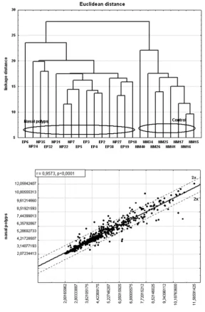
First, hierarchical clustering analysis was performed to find similarities in gene expression profiles. Grouping based on NF-κB-dependent gene expression fingerprints revealed two distinct groups. NP patients clustered together indicated large homogeneity in gene expression. Similarly, healthy mucosa cases presented a consistent gene expression profile but different to NPs (Fig. 1). Analysis of NF-κB-dependent gene using oligonu-cleotide microarrays failed to distinguish eosino-filic from non-eosinophilic nasal polyps.
In the next stage of analysis the authors identi-fied genes showing differential expression in NPs and normal nasal mucosa (Fig. 2). Among 582 human NF-κB-dependent genes, 25 showed a sig-nificantly higher expression in NPs compared to the control (Tabl. 1). The majority of the genes showed expression levels from 2 to 3-fold (19 genes) higher than in healthy mucosa. The larg-est increase was revealed in case of gene encod-ing TFF3 (5-fold higher expression) followed by
NOS2A (5x), SERPINA1 (4x), UCP2 (4x), OXTR
In healthy mucosa 19 genes presented increased transcription activity compared to NPs (Table 2). Most of the genes (11 genes) revealed only a 2 to 3-fold difference in expression. The most signifi-cantly enhanced level was shown in case of LTF
gene (20 fold) followed by KRT6B (7x), LYZ (7x),
SD11B2 (5x) and MMP3 (4x).
Discussion
In the present study DNA microarray tech-nology was applied to identify differential gene expression in NP tissues and in healthy nasal mucosa. DNA microarray consists of a matrix with attached sequences that allow analysis of the expression of whole panels of genes and in-dicate those which have been activated in the disease state.
Active fraction of NF-κB induces transcription of genes including IL-8, IL-16, iNOS, and eotaxin which upregulation has been previously implicated in the pathogenesis of NPs [4]. Higher transcrip-tional activity of these genes was also observed in our study. CCL11 (eotaxin 1) showed an almost 3 fold higher expression in NPs compared to the control. Eotaxins are the most potent chemoattrac-tant for eosinophils, and are thought to play a key role in tissue eosinophilia. In such a mechanism eotaxins increase local inflammatory status and are strongly involved in the development of NPs [7]. Il-8 was the most significantly up-regulated NF-κB dependent interleukin in our study. lo-cally produced Il-8 contributes to the progression of chronic inflammation in the sinus mucosa. It is postulated that Il-8 participates in the recruitment and activation of leukocytes, T lymphocytes and eosinophils [8]. However, when challenged with
Fig. 1. Dendrogram of 15 nasal polyps (eosinophilic polyps (EP), non-eosino-philic polyps (NP)) and 8 normal mucosa (NM) samples using cluster analysis based on the Euclidean distance with 582 human NF-κB-dependent genes repre-sented on the HG-U133A GeneChip
Ryc. 1. Dendrogram przedstawiający wynik analizy skupień 15 pacjentów z polipami nosa (polipy eozynofilowe (EP), polipy nieoeozynofilowe (NP)) oraz 8 wycinków niezmienionej błony śluzo-wej jamy nosa (NM) na podstawie odle-głości euklidesowej po ocenie ekspresji 582 genów zależnych od czynnika trans-krypcyjnego NF-κB. Analizę wykonano techniką mikromacierzy oligonukleoty-dowych z użyciem matrycy HG-U133A
Fig. 2. Scatter plots of gene expression in nasal polyps and normal tissues (x-axis represents the normalized signal value of normal nasal mucosa, and y-axis repre-sents the normalized signal value of NP)
Table 1. The list of NF-κB-dependent gene showing higher expression in nasal polyps compared to normal mucosa identi-fied by oligonuclotide microarray. *t test
Tabela 1. lista genów zależnych od czynnika transkrypcji jądrowej NF-κB charakteryzujących się wyższą ekspresją w tkan-kach polipów nosa niż w grupie kontrolnej. *t test
Probe set
(Zestaw próbek) Gene symbol(Symbol genu)
Gene name
(Nazwa genu) Fold change(Krotność zmiany)
p*
204623_at TFF3 trefoil factor 3 6.74 p < 0.0001
210037_s_at NOS2A nitric oxide synthase 2A 5.23 p = 0.0002 211429_s_at SERPINA1 serpin peptidase inhibitor, clade A (alpha-1
antipro-teinase antitrypsin), member 1 4.71 p < 0.0001 208998_at UCP2 uncoupling protein 2 4.16 p < 0.0001
202859_x_at Il8 interleukin 8 3.88 p = 0.0014
206825_at OXTR oxytocin receptor 3.55 p = 0.0008
204470_at CXCl1 chemokine (C-X-C motif) ligand 1 3.51 p = 0.0013 209875_s_at SPP1 Secreted phosphoprotein 1 2.86 p = 0.0025
201859_at PRG1 p53-responsive gene 1 2.78 p = 0.0002
210133_at CCl11 chemokine (C-C motif) ligand 11; eotaxin-1 2.74 p = 0.0056 201645_at TNC Tenascin C (hexabrachion) 2.66 p < 0.0001 208451_s_at C4A complement component 4A 2.57 p = 0.0018
201042_at TGM2 transglutaminase 2 2.56 p = 0.0002
202357_s_at CFB complement factor B 2.5 p = 0.0194
214211_at FTH1 ferritin, heavy polypeptide 1 2.4 p < 0.0001 204475_at MMP1 matrix metallopeptidase 1 2.39 p = 0.0223 204673_at MUC2 mucin 2, oligomeric mucus/gel-forming 2.39 p = 0.0404 201998_at ST6GAl1 ST6 beta-galactosamide alpha-2,6-sialyltranferase 1 2.29 p = 0.0002 204655_at CCl5 chemokine (C-C motif) ligand 5 2.17 p = 0.0009 201313_at ENO2 enolase 2 (gamma, neuronal 2.16 p < 0.0001
203828_s_at Il32 interleukin 32 2.11 p = 0.0052
202718_at IGFBP2 insulin-like growth factor binding protein 2 2.08 p < 0.0001 207076_s_at ASS1 argininosuccinate synthetase 1 2.06 p = 0.0005 201510_at ElF3 E74-like factor 3 (ets domain transcription factor,
epithelial-specific ) 2,06 p = 0.0186
205692_s_at CD38 CD38 molecule 2.05 p = 0.0006
214428_x_at C4B complement component 4B 2.03 p = 0.0024
217767_at C3 complement component 3 2.03 p = 0.0417
203936_s_at MMP9 matrix metallopeptidase 9 2.0 p = 0.0362
210136_at MBP myelin basic protein 2.0 p = 0.0475
Il-8 of primed nasal mucosa a significant influx of neutrophils but not eosinophils was induced [9]. In airway epithelium, Il-8 is one activator of MMP-9 and MMP-8 expression and secretion during airway remodeling [10]. Il-8 and MMP-8 may form a pivotal inductive cytokine-proteinase cascade in the pathogenesis of NP.
The next gene known for its involvement in NP growth showing enhanced transcriptional activity in the present study encodes nitric oxide synthase (NOS). NOS is the main source of nitric oxide (NO) in the respiratory tract. It was revealed that NO may cause microvascular leakage and stilation of glandular secretions contributing to mu-cosal edema formation characteristic for NPs [11]. Subsequent vascular dilatation in the nasal mucosa causes increased nasal airway resistance, which
could facilitate the local release and generation of mediators induced by inflammatory cells [12].
The expression of the majority of genes pre-senting higher transcriptional activity in the present study has not been widely investigated in NP to date. Among all the examined genes,
TFF3 (trefoil factor 3) was the most significantly expressed in NPs compared to the control. TFF3 is mainly synthesized by mucin-secreting epithe-lial cells and participate in mucosal surface pro-tection and repair after injury [13]. Since TFF3 inhibits apoptosis, it demonstrates prosurvival as well as proinvasive and proangiogenic activities [14]. It was found that Il-4 and Il-13 involved in NP growth induce TFF3 expression via a direct effect on epithelial cells [15]. In oral keratinocytes stimulated with TFF3 increased transcription of
Table 2. The list of NF-κB-dependent gene showing higher expression in normal mucosa than in nasal polyps identified by oligonuclotide microarray. * t test
Tabela 2. lista genów zależnych od czynnika NF-κB, których ekspresja była większa w grupie kontrolnej w porównaniu z polipami nosa. * t test
Probe set
(Zestaw próbek) Gene symbol(Symbol genu)
Gene name
(Nazwa genu) Fold change(Krotność zmiany)
p*
202018_s_at lTF lactoferrin 12.78 p = 0.0003
209126_x_at KRT6B keratin 6B 5.8 p = 0.0009
205828_at MMP3 matrix metallopeptidase 3 5.05 p < 0.0001 204130_at HSD11B2 hydroxysteroid (11-beta) dehydrogenase 2 4.3 p < 0.0001
219403_s_at HPSE heparanase 4.23 p < 0.0001
212657_s_at Il1RN interleukin 1 receptor antagonist 4.08 p = 0.0016
213975_s_at lYZ lysozyme 3.91 p = 0.0030
209189_at c-FOS FBJ murine osteosarcoma viral oncogene homolog 3.22 p = 0.0420 207206_s_at AlOX12 arachidonate 12-lipoxygenase 2.81 p < 0.0001 205392_s_at CCl14 chemokine (C-C motif) ligand 14 2.28 p = 0.0003 205440_s_at NPY1R neuropeptide Y receptor Y1 2.26 p = 0.0002
204734_at KRT15 keratin 15 2.25 p = 0.0225
202431_s_at MYC v-myc myelocytomatosis viral oncogene homolog 2.21 p = 0.0024 220987_s_at SNARK SNF1/AMP activated protein kinase 2.16 p < 0.0001 209396_s_at CHI3l1 chitinase 3-like 1 (cartilage glycoprotein-39) 2.15 p < 0.0001 201694_s_at EGR1 early growth response 1 2.05 p = 0.0354 202555_s_at MYlK myosin light chain kinase 2.0 p = 0.0072 202936_s_at SOX9 SRY (sex determining region Y)-box 9 2.0 p = 0.0004
207356_at DEFB4 defensin, beta 4 2.0 p = 0.0372
203234_at UPP1 uridine phosphorylase 1 2.0 p = 0.0397
genes related to cell survival, cellular growth and proliferation was showed [16].
SERPINA1 (alpha1-antitrypsin, AAT) activity has previously been found to be elevated in chron-ic sinusitis whchron-ich was presumed to reflect its pro-tective function, i.e. neutralization of neutrophil elastases which can be a driving force of mucosal inflammation [17]. SERPINA1, an acute phase protein, is one of the major serine protease inhibi-tors in the human body. AAT has been shown to increase fibroblast proliferation, to inhibit neutro-phil chemotaxis, superoxide production and cell apoptosis [18]. AAT specifically enhanced lPS induced RANTES expression in BAl and Il-10, Il-12 and Il-13 levels in lung homogenates [19]. All of this indicates that SERPINA1 plays an im-portant regulatory role in various inflammatory cascades.
UCP2 (uncoupling protein 2) is another gene which exhibits a distinctly higher expression in NP samples. UCP2 belongs to a newly discovered sub-group of mitochondrial carrier proteins. Its precise function is still unclear. UCPs regulate production of reactive oxygen species (ROS), inhibit inflam-mation as well as cell death [20]. UCP2 is expressed in many cells including infiltrating immune cells like macrophages, lymphocytes, dendritic cells and neutrophils. Since ROS modulates activation of T and B lymphocytes, UCP2 may influence the function of these cells [21]. As a regulator of mast cell UCP2 has potential implications for treatment of mast cell-mediated allergic and inflammatory diseases. It has been proved that UCP2 negatively regulates mast cell degranulation, histamine pro-duction, as well as Il-6 and PGD2 and release [22]. Thus, inhibition of UCP2 may rather worsen aller-gic and inflammatory diseases while its activation may act to reduce inflammatory responses.
Similar to the Il-8, CXCl1 is of particular in-terest as a chemoattractant and activator of neu-trophils. This growth-related oncogene is crucial for the recruitment of other inflammatory cells in-cluding monocytes, dendric and T cells. Both tran-scripts and protein levels for CXCl1 were noted to be significantly elevated in NP and also released by infiltrating eosinophils [23].
Besides Il-8, the authors revealed another up-regulated interleukin in NP tissues. Il-32 is a recent-ly described pro-inflammatory cytokine produced mainly by epithelial cells, T lymphocytes, natural killer cells and monocytes [24]. Il-32 induces the expression of other important pro-inflammatory cytokines (Il-1β, Il-6, TNF-α and Il-8) and may further amplify inflammatory reaction [24].
Secreted phosphoprotein 1 (SPP1; also known as early T lymphocyte activation protein 1) is a phosphorylated glycoprotein classified as a Th1
cytokine involved in the inflammatory and immu-nological processes. SSP1 has strong pro-inflam-matory properties and plays an important role in recruiting inflammatory cells and regulating the function of monocyte, macrophage, dendrit-ic cells, Th1 and B cells. Eosinophils are an im-portant source of SSP1 production in NP which implies a role for that cytokine in the etiology of NPs [25]. In sinonasal mucosa SSP1 could induce production of cytokines strictly involved in NP de-velopment like IFN-γ, Il-4, Il-5, Il-13, Il-1β, and TNF-a. This could be supported by the observa-tion that treating allergic mice with SSP1-specific antibodies deceased levels many of those chemok-ines [26].
Present investigation also found in NPs in-creased expression of C4A and CFB gene encod-ing elements of the complement pathway. C3a and C4a are potent chemotactic factors for inflamma-tory cells and are able to enhance degranulation of eosinophils and mast cells which indicates its potential role in the pathogenesis of rhinosinus-itis [27]. Previously Seppänen et al. [28] revealed up-regulation of complement in patients with CRS but simultaneously C4A deficient patients more commonly suffered from chronic or recurrent rhi-nosinusitis.
The role of other genes with higher transcrip-tional activity revealed by us in NP development is rather assumptive. The up-regulation of many of them in pathological conditions may either be the cause or a consequence of the disease. Tenascin-C is a pro-inflammatory extracellular matrix glyco-protein which appears in association with wound healing, cancer invasion, tissue remodeling and chronic inflammations [29]. Expression of tena-scin-C is induced upon activation of cell surface TlRs, but also subsequent activation of TlR4 by tenascin-C stimulates the synthesis of pro-inflam-matory cytokines and MMPs [30]. PRG1 (p53-re-sponsive gene 1) expression has been shown to be associated with accelerated cell growth, although its exact cellular function is still not clear [31]. p22/ PRG1 is a potential anti-apoptotic gene involved in NF-κB mediated resistance to apoptosis.
Attention should be paid to the function of transglutaminase 2 (TGM2). TGM2 causes a sus-tained increase in secretory phospholipase A(2) (sPlA(2)) activity indicating a novel mechanism by which increased expression of TGM2 may serve to amplify airway inflammation [32]. In Hallstrand et al.’s study both TGM2 gene expression and secret-ed protein were increassecret-ed in the airways of subjects with asthma. Although TGM2 and sPlA(2)s play a major role in inflammation, their importance in chronic rhinosinusitis is not well known.
healthy nasal mucosa, the one encoding lactofer-rin (lF) showed the highest (20-fold) level of tran-scription compared to NPs. lF represents one of the major components of mammalian secretions playing numerous roles in the innate and adap-tive immunity. Moreover, lF inhibits eotaxin-stimulated eosinophil migration and through the inhibition of pro-inflammatory cytokines acts as antiproliferative factor [33]. Reports on the ex-pression and presence of the protein in NP tissue and nasal secretion are scant and not consistent in indicating an increase or decrease in lF level in NP [34]. In view of the present outcomes and au-thors’ previous findings the authors suppose that decreased expression of lF in NP could be one of the etiological factors disturbing the local homeo-stasis of nasal mucosa.
In this study the microarray technique en-abled authors to identify the differential expres-sion of NF-κB-dependent genes in NP and control mucosa. The gene expression patterns the authors obtained were significantly different in normal mucosa in comparison with NP. Consequently, all examined tissues could be classified either as NPs or healthy mucosa. Simultaneously, an analysis
of NF-κB-dependent genes transcription by the described technique failed to differentiate eosino-philic from non-eosinoeosino-philic NPs, which suggests a similar expression pattern of analyzed genes in those types of NPs.
The attempt described above highlights the involvement of not only expected but, more inter-estingly, many unsuspected pathologic pathways. Up-regulated genes may be potentially associated with NP development. The identification of novel disease-related genes may help to understand the biology of chronic rhinosinusitis. Although NF-κB has been considered the “holy grail” in the targeting of new anti-inflammatory drugs, initial attempts did not provide satisfactory outcomes. It could be partially explained by the fact that transcription factor NF-κB influences the expression of a broad spectrum of genes, some of which may however have an opposite action during inflammation. The physiological roles of NF-κB are additionally both cell type- and stimulus-dependent. Further studies are warranted to learn more about transcriptional pathways downstream to NF-κB and the clinical usefulness of NF-κB inhibitory molecules.
References
[1] Baldwin AS: The NF-jB and IjB proteins: New discoveries and insights. Ann Rev Immunol 1996, 14, 649–681.
[2] Lawrence T: The NF-κB pathway in inflammation. Cold Spring Harb Perspect Biol 2009, 1(6), a001651.
[3] Takeno S, Hirakawa K, Ueda T, Furukido K, Ossada R, Yajin K: NF-κB activation in the nasal polyp epithelium: relationship to local cytokine gene expression. laryngoscope 2002, 112, 53–58.
[4] Yang L, Cohn L, Zhang DH, Homer R, Ray A, Ray P: Essential role of NF-κB in the induction of eosinophilia in allergic airway inflammation. J Exp Med 1998, 188, 1739–1750.
[5] Breccia M, Alimena G: NF-κB as a potential therapeutic target in myelodysplastic syndromes and acute myeloid leukemia. Expert Opin Ther Targets 2010, 14(11), 1157–1176.
[6] Rostkowska-Nadolska B, Kapral M, Fraczek M, Kowalczyk M, Gawron W, Mazurek U: A microarray study of gene expression profiles in nasal polyps. Auris Nasus larynx 2011, 38, 58–64.
[7] Yao T, Kojima Y, Koyanagi A, Yokoi H, Saito T, Kawano K, Furukawa M, Kusunoki T, Ikeda K: Eotaxin-1, -2, and -3 immunoreactivity and protein concentration in the nasal polyps of eosinophilic chronic rhinosinusitis patients. laryngoscope 2009, 119(6), 1053–1059.
[8] Warringa RA, Mengelers HJ, Raaijmakers JA, Bruijnzeel PL, Koenderman L: Upregulation of formyl-peptide and Il-8-induced eosinophil chemotaxis in patients with allergic asthma. J Allergy Clin Immunol 1993, 91(6), 1198–1205.
[9] Bocheńska-Marciniak M, Kupczyk M, Górski P, Kuna P: The effect of recombinant Il-8 on eosinophils’ and neutrophils’ migration in vivo and in vitro. Allergy 2003, 58(8), 795–801.
[10] Van den Steen P, Wuyts A, Husson S, Proost P, van Damme J, Opdenakker G: Gelatinase B/MMP-9 and neu-trophil collagenase/MMP-8 process the chemokines human GCP-2/CXCl6, ENA-78/CXCl5 and mouse GCP-2/ lIX and modulate their physiological activities. Eur J Biochem 2003, 270, 3739–3749.
[11] Furukawa K, Harrison DG, Saleh D, Shennib H, Chagnon FP, Giaid A: Expression of NOS in the human nasal mucosa. Am J Respir Crit Care Med 1996, 153, 847–850.
[12] Maniscalco M, Sofia M, Faraone S, Carratu L: The effect of PAF on nasal airway resistance in healthy subjects is not mediated by nitric oxide. Allergy 2000, 55, 757–761.
[13] Kjellev S: The trefoil factor family – small peptides with multiple functionalities. Cell Mol life Sci 2009, 66(8), 1350–1369.
[14] Babyatsky M, Lin J, Yio X, Chen A, Zhang JY, Zheng Y, Twyman C, Bao X, Schwartz M, Thung S, Lawrence Werther J, Itzkowitz S: Trefoil factor-3 expression in human colon cancer liver metastasis. Clin Exp Metastasis 2009, 26, 143–151.
[16] Storesund T, Schenck K, Osmundsen H, Røed A, Helgeland K, Kolltveit KM: Signal transduction and gene transcription induced by TFF3 in oral keratinocytes. Eur J Oral Sci 2009, 117(5), 511–517.
[17] Hamaguchi Y, Taya M, Suzumura H: lysosomal proteases and protease inhibitors in nasal allergy and non-atopic sinusitis. Am J Otolaryngol 1990, 11, 37–43.
[18] Dabbagh K, Laurent GJ, Shock A, Leoni P, Papakrivopoulou J, Chambers RC: Alpha-1-antitrypsin stimulates fibroblast proliferation and procollagen production and activates classical MAP kinase signalling pathways. J Cell Physiol 2001, 186, 73–81.
[19] Subramaniyam D, Steele C, Köhnlein T, Welte T, Grip O, Matalon S, Janciauskiene S: Effects of alpha 1-anti-trypsin on endotoxin-induced lung inflammation in vivo. Inflamm Res 2010, 59(7), 571–578.
[20] Mattiasson G, Sullivan PG: The emerging functions of UCP2 in health, disease, and therapeutics. Antioxid Redox Signal 2006, 8, 1–38.
[21] Negre-Salvayre A, Hirtz C, Carrera G, Cazenave R, Troly M, Salvayre R, Pénicaud L, Casteilla L: A role for uncoupling protein-2 as a regulator of mitochondrial hydrogen peroxide generation. FASEB J 1997, 11, 809–815.
[22] Tagen M, Elorza A, Kempuraj D, Boucher W, Kepley CL, Shirihai OS, Theoharides TC: Mitochondrial uncou-pling protein 2 inhibits mast cell activation and reduces histamine content. J Immunol 2009, 183(10), 6313– 6319.
[23] Payne SC, Han JK, Huyett P, Negri J, Kropf EZ, Borish L, Steinke JW: Microarray analysis of distinct gene transcription profiles in non-eosinophilic chronic sinusitis with nasal polyps. Am J Rhinol 2008, 22(6), 568–581.
[24] Kim SH, Han SY, Azam T, Yoon DY, Dinarello CA: Interleukin-32: a cytokine and inducer of TNFalpha. Immunity 2005, 22, 131–142.
[25] Lu X, Zhang XH, Wang H, Long XB, You XJ, Gao QX, Cui YH, Liu Z: Expression of osteopontin in chronic rhinosinusitis with and without nasal polyps. Allergy 2009, 64(1), 104–111.
[26] Xanthou G, Alissafi T, Semitekolou M, Simoes DC, Economidou E, Gaga M, Lambrecht BN, Lloyd CM, Panoutsakopoulou V: Osteopontin has a crucial role in allergic airway disease through regulation of dendritic cell subsets. Nat Med 2007, 13, 570–578.
[27] Welch TR, Frenzke M, Carroll MC, Witte DP: Evidence of a role for C4 in modulating interstitial inflammation in experimental glomerulonephritis. Clin Immunol 2001, 101, 366–370.
[28] Seppänen M, Suvilehto J, Lokki ML, Notkola IL, Järvinen A, Jarva H, Seppälä I, Tahkokallio O, Malmberg H, Meri S, Valtonen V: Immunoglobulins and complement factor C4 in adult rhinosinusitis. Clin Exp Immunol 2006, 145(2), 219–227.
[29] Mane DR, Kale AD, Naik VV: Immunohistochemical expression of Tenascin in embryogenesis, tumorigenesis and inflammatory oral mucosa. Arch Oral Biol 2011, 56(7), 655–663.
[30] Goh FG, Piccinini AM, Krausgruber T, Udalova IA, Midwood KS: Transcriptional regulation of the endogenous danger signal tenascin-C: a novel autocrine loop in inflammation. J Immunol 2010, 184(5), 2655–2662.
[31] Arlt A, Grobe O, Sieke A, Kruse ML, Fölsch UR, Schmidt WE, Schäfer H: Expression of the NF-kappa B target gene IEX-1 (p22/PRG1) does not prevent cell death but instead triggers apoptosis in Hela cells. Oncogene 2001, 20(1), 69–76.
[32] Hallstrand TS, Wurfel MM, Lai Y, Ni Z, Gelb MH, Altemeier WA, Beyer RP, Aitken ML, Henderson WR:
Transglutaminase 2, a novel regulator of eicosanoid production in asthma revealed by genome-wide expression profiling of distinct asthma phenotypes. PloS One 2010, 5(1), 8583–8586.
[33] Kruzel ML, Actor JK, Boldogh I, Zimecki M: lactoferrin in health and disease. Postepy Hig Med Dosw 2007, 61, 261–267.
[34] Nadolska B, Frączek M, Kręcicki T, Kocięba M, Zimecki M: lactoferrin inhibits the growth of nasal polyp fibro-blasts. Pharmacol Rep 2010, 62(6), 1139–1147.
Address for correspondence:
Marcin Frączek
Wroclaw Medical University Hospital Borowska 213
50-556 Wrocław Poland
E-mail: [email protected] Tel.: +48 668324061
Conflict of interest: None declared