6 | P a g e
International Journal of Phytopharmacology
Research Article
www.onlineijp.com
e- ISSN 0975 – 9328 Print ISSN 2229 – 7472
PHYTOCHEMICAL CONSTITUENTS, ANTIOXIDANT ACTIVITY
AND TOXICITY ASSESSMENT OF HYDROETHANOLIC LEAF
EXTRACT OF
GRIFFONIA SIMPLICIFOLIA
Ruby Ama Nyarko
1*, Christopher Larbie
2, Alexander Kofi Anning
1, Philip Kweku Baidoo
11
Department of Theoretical and Applied Biology, Kwame Nkrumah University of Science and Technology, Kumasi-Ghana.
2
Department of Biochemistry and Biotechnology, Kwame Nkrumah University of Science and Technology, Kumasi-Ghana.
ABSTRACT
Griffonia simplicifolia is an African legume of significant pharmacological activity, with its leaves commonly used for the treatment of bladder and kidney problems, relieving constipation, and as an aphrodisiac. However, the leaves have not been subjected to scientific scrutiny and safety assessment yet. In this study, the phytochemical constituents, in vitro
antioxidant activity, heavy metal concentration as well as the acute and sub-chronic toxicity of the hydroethanolic leaf extracts of G.simplicifolia were determined using standard methods. The results indicated the presence of glycosides, tannins, flavonoids, alkaloids, saponins and coumarins in the leaf extract of the plant.The highest total flavonoid and phenol contents recorded were 237.20 mg/100g QE in the hydro fraction and 8.71 mg/g GAE in the ethyl acetate, respectively. The highest 2, 2-diphenyl-1-picrylhydrazyl(DPPH) scavenging activity recorded was 24.82% in the hydro fraction. UV and FT-IR spectrometry also suggested the presence of phenolic compounds. Concentrations of all heavy metals analysed (Fe, Zn, Ni, Cu, Pb) were below WHO recommended limits. The median acute toxicity (LD50) of the extract was determined to be<5g/kg
body weight in mice. Sub-chronic use for 28 days resulted in significant weight gain, reduction in platelet large cell ratio and platelet count, and increase in low density lipoprotein (LDL) and blood glucose concentrations. The extract did not produce any toxic effects on vital organs except a slight decrease in liver weight of male rats. The hydroethanolic extract of G. simplicifolia could therefore be considered safe in moderate doses.
Key words: Griffonia simplicifolia, medicinal plants, phytochemicals, subchronic toxicity.
Corresponding Author : Ruby Ama Nyarko Email: rubynx06@yahoo.com
INTRODUCTION
Plants are a major source of medicine and form an integral part of primary health care for about 75-80% of the world‟s population (Abdel-daim et al., 2016). They are one of nature‟s gifts to humans and have dominated the world‟s pharmacopeia for thousands of years (Ernst, 2005). Based on the ethnobotanic knowledge of medicinal
Access this article online
DOI:
http://onlineijp.com/
DOI:
http://dx.doi.org/10.21276/ijp.2019.10.1.2
Quick Response code
Received:25.11.18 Revised:12.12.18 Accepted:15.12.18
7 | P a g e
plants to ensure their quality, efficacy and safety prior to their acceptance and use(Larbie et al., 2016).
Griffonia simplicifolia (M. Vahl ex DC.) Baill., commonly known as Griffonia, is an African legume of significant pharmacological activity. It is native to West and Central Africa, but it is primarily found in Ghana, Cote D‟Ivoire and Togo (Esposito et al., 2012). The seed of G. simplicifolia has occupied attention worldwide for several decades and has found modern therapeutic applications due to its high concentration of 5-hydroxyl-L-tryptophan (5-HTP), a direct precursor to serotonin (Weeks, 2009). It is used in the treatment of depression, obesity, insomnia, fibromyalgia, migraine, and improves cognitive functions (Esposito et al., 2012; Wang et al., 2017).
Apart from the seed, the leaf extract of G.
simplicifolia is also reportedly used for treating malaria, bladder and kidney problems, for relieving constipation, as an aphrodisiac and a remedy for cough (Esposito et al., 2012; Offoumou et al., 2018; Pathak et al., 2010). According to Pathak et al. (2010), the leaves of Griffonia contain volatile oils and coumarins, and about 0.1 % 5-HTP. Recent studies by Offoumou et al. (2018) also reported the presence of sterols, quinones, alkaloids and saponins in the aqueous and ethanolic extract of Griffonia leaf. Nonetheless, the toxicity and/or efficacy of the leaf extract have not been scientifically assessed. Primary studies are therefore required to help in clinical assessment and as a baseline for future research. The present study was, thus, aimed at determining the phytochemical constituents, antioxidant activity, heavy metal concentration, and UV-Vis and FT-IR spectra of the hydroethanolic leaf extract of G. simplicifolia, as well as its acute and sub-chronic toxicity in animals.
MATERIALS AND METHODS
Collection and Identification of Plant Materials
Leaves of G. simplicifolia were handpicked from the Kwame Nkrumah University of Science and Technology (KNUST) Botanic Garden, Kumasi-Ghana (latitude 6º35 N-6º40 N and longitude 1º30 W-1º35 W) before 9:00 am each sampling day, in November 2017. Plant identification was authenticated by a taxonomist at the Department of Theoretical and Applied Biology,
KNUST, and a voucher
specimen(KNUST/AB/2018/L9544) was deposited in the herbarium for reference purposes. The leaves were thoroughly washed with water and air-dried at room temperature under shade for three weeks. The dried samples were then milled, and packaged in zip-locks for storage.
Preparation of Plant Extract and Fractionation
A hydroethanolic extract of the milled leaves was prepared by suspending the leaves in 50% ethanol (50:50, ethanol: water, v/v) as previously described by Anim et al.
(2016). The extraction was done by cold maceration for 48 hours at room temperature on a shaker (Rocking Laboratory Shaker). The extract was filtered through cotton wool and the filtrate collected. The procedure was repeated twice. The extracts were concentrated using a rotary evaporator (Buchi R-205, Switzerland) under reduced pressure and freeze-dried (Labconco, England) to obtain the Griffonia simplicifolia ethanolic leaf extract (GSE). Forty grams (40 g) of the leaf extract was successively extracted with solvents of increasing polarity; petroleum ether, ethyl acetate and methanol. The residual portion was designated as hydro fraction. Each fractionation step was performed twice with 400 ml of solvent. The fractions were then concentrated by rotary evaporation, and air-dried at room temperature (25 ºC).
Phytochemical screening
Qualitative determination of glycosides, tannins, flavonoids, alkaloids, saponins, coumarins and sterols was performed on the raw powder and the hydroethanolic crude extract of G. simplicifolia using standard procedures described by Harborne (1998) and Trease & Evans (1989).
In vitro Antioxidant activity
The free radical scavenging activity of 2, 2-diphenyl-1-picrylhydrazyl (DPPH) method was used to determine the antioxidant activity as described by Brand-Williams et al. (1995) with some modifications. Equal volumes of different concentrations of each extract (concentration range 0-5 mg/mL) were prepared and 0.5 mM Methanolic solution of DPPH was added to each solution. The mixtures were incubated at room temperature for 20 minutes and the absorbance read at a wavelength of 517 nm (Tecan Infinite M200 Pro plate reader, Austria). The inhibition concentration at 50% (IC50) of each extract was calculated using the formula:
Where A0 is the mean absorbance of the wells containing
negative control; A1 is the mean absorbance of the wells
with the test sample. Butylatedhydroxytoluene (BHT) was used as standard control. Analyses were performed in triplicates.
Total phenolic content determination
Total phenolic content (TPC) was measured using the Folin–Ciocalteau assay as described by Marinova et al. (2005). Ten microliters (10 μL) of 5.0 mg/ml of extract was diluted with 790 μL distilled water. The diluted sample was mixed with 50 μL of Folin-Ciocalteau reagent and incubated in a dark place for 8 minutes. Afterwards, 150 μL of 7% Na2CO3 was added
8 | P a g e
was used as the standard phenolic compound. The results were expressed in milligrams of GA equivalents per gram dry mass (mg GAE/g DM).
Total flavonoid content determination
The total flavonoid content was measured with an aluminium chloride colorimetric assay as described by Marinova et al. (2005). An aliquot (1 ml) of extracts or a standard solution of (+)-catechin (20, 40, 60, 80 and 100 mg/L) was added to a 10 ml volumetric flask, containing 4 ml of distilled deionized water (dd H2O), and 0.3 ml 5 %
NaNO2 was added. After 5 min, 0.3 ml of 10% AlCl3 was
added. At the sixth minute, 2 ml of 1 M NaOH was added and the total volume was made up to 10 ml with dd H2O.
The solution was mixed well and the absorbance was measured against a prepared reagent blank at 510 nm with a UV-VIS Spectrophotometer Lambda 5. The data were expressed as milligrams of (+)-quercetin equivalents (QE) (mg QE/100 g DW). All samples were analysed in triplicates.
UV-Vis and Fourier transform infrared (FTIR) spectrophotometric analyses
The fractions of the leaf (10 mg/ml) were diluted in the ratio 1:10 using their respective solvents and analysed at a wavelength ranging from 200-800 nm. A double beam ultraviolet-visible spectrophotometer (Perkin Elmer, USA) was used to detect and record the characteristic peaks present in that range. Ten milligram (10mg) of the dried extract powder was encapsulated in 100 mg of KBr pellet to prepare translucent sample discs. The powdered sample of each extract was loaded in FTIR spectroscope (UATR Spectrum 2, Perkin Elmer) with a scan range from 400 to 4000 cm-1 and a resolution of 4 cm-1.
Heavy metal analysis
One gram (1.0 g) each of the extract and the raw sample was weighed into a 50 ml digestion tube. The sample was mixed with 1.0 ml of H2O, 2.0 ml of conc.
HCl, 5 ml of 1:1conc. HNO3: 60% HClO4 and 2.0 ml of
Conc. H2SO4. The mixture was allowed to stand for 20
minutes. At a temperature of 150 °C, the samples were heated in a digestion block. The digested samples were allowed to cool after which they were diluted with 50 ml of distilled water. All the samples were wet digested. The digests were analysed for the levels of lead, copper, nickel, zinc and iron using an Atomic Absorbtion Spectrometer (AnalytikjenanovAA 400P).
Animal selection and groupings
Swiss albino mice and Sprague-Dawley rats of both sexes were obtained from the animal house of the School of Medical Science, University of Ghana, Legon Accra. The animals were housed in aluminium rodent cages with bedding of wood shavings. They were
segregated according to sex to avoid any chance of mating, under standard conditions (25± 2°C, 40-60% humidity and ~12h light and dark cycle). They were fed with standard animal feed (AGRICARE, Kumasi, Ghana) and distilled water ad libitum throughout the period of the study except an overnight fast prior to sacrificing.
In grouping the animals, their body weights were taken into consideration to achieve approximately equal conditions among the groups. They were allowed to acclimatize to laboratory conditions for a week before the experiment begun. The animals were identified by tail marks made with permanent markers. Animal studies were conducted in the animal holding facility of the Department of Biochemistry and Biotechnology (KNUST, Ghana) and in accordance with the guidelines of the Committee for the Purpose of Control and Supervision of Experiment on Animals (CPCSEA, New Delhi, India).
Acute oral toxicity
The acute toxicity study was carried out using three female Swiss albino mice (20-25 g). After an overnight fast, the animals were treated once with 5000 mg/kg bwt of the GSE administered orally by gavage. The dose was selected based on the OECD (2001) fixed dose method. The animals were observed for signs of toxicity (including paw-licking, stretching, respiratory distress, diarrhoea) and mortality for the first 4 hours and subsequently, daily for 7 days.
Sub-chronic oral toxicity
Sub-chronic toxicity study was performed using 16 male and 16 female rats. For each sex, the animals were divided into four groups of four animals each. Group I served as the control and received 1.0 ml of distilled water once daily. Groups II, III and IV were treated with 100, 250 and 500 mg/kg bwt of the GSE once daily in 1.0 ml of distilled water for 28 days. They were observed daily for general signs of toxicity and mortality.
Effect of treatment on body weight
Rats in all groups were weighed on the first day (D0) and subsequently at the end of every fourth day (i.e., D4, D8, D12, D16, D20, D24 and D28). The percent change in body weight was calculated using the formula:
Where Wn = weight on day 4 (D4), D8, D12, D16, D20,
D24 and D28, and Wo = weight on D0.
Collection of blood, serum and isolation of organs
9 | P a g e
haematological analyses. Five millilitres of blood was then dispensed into gel-activated tubes and centrifuged at 3000 rpm for 5 minutes. The sera were aspirated with a Pasteur pipette into sample bottles for the various biochemical assays. The sacrificed animals were dissected and their liver, kidney, heart, stomach, spleen, testes or uterus were excised, freed of fat, washed with normal buffered saline, and blotted with clean tissue paper. They were observed macroscopically and weighed to obtain absolute organ weight (AOW). The relative organ weights (ROW) of the organs were calculated for each rat using the formula:
Biochemical and haematological analyses
Evaluation of biochemical parameters was done using the Cobas Integra 400 Clinical Chemistry Analyser (Roche, USA) and reagents from Fortress Diagnostics
(UK). Parameters analysed included alanine
aminotransferase (ALT), aspartate amino transferase (AST), total, direct and indirect bilirubin, creatinine, urea, sodium, potassium, chloride, total cholesterol (TChol), high density lipoproteins (HDL), total triglycerides (Trigs), glucose, and lactase dehydrogenase. Low density lipoprotein (LDL) concentration was calculated using the Friedewald‟s equation (Crook, 2006).
Haematological analyses were performed using Sysmex Haematology System (USA). Parameters included red blood cell count, haemoglobin concentration, and white blood cell count, mean corpuscular volume, mean
corpuscular haemoglobin and mean corpuscular
haemoglobin concentration (MCHC).
Data analyses
Data were analysed with GraphPad Prism 6.0 (GraphPad Software Inc., La Jolla, CA, USA). The percent change in body weight, haematological parameters and serum biochemistry were expressed as mean ± standard error of mean (SEM). Data were assessed by one-way ANOVA followed by Tukey‟s multiple comparison test. All analyses were conducted assuming a significance value of 5%.
RESULTS
Phytochemical constituents
The raw leaf powder and hydroethanolic leaf extract of GS both contained glycosides, tannins, saponins and coumarins. In addition, the extract showed the presence of alkaloids and flavonoids (Table 1). However, none of the samples were observed to possess sterols.
Total flavonoid and phenol content
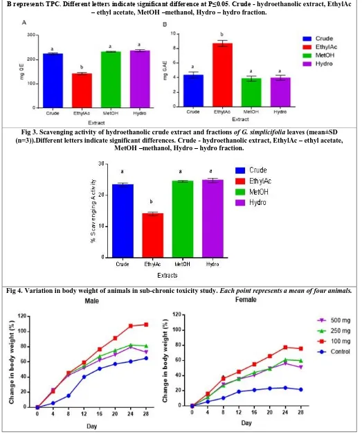
The total flavonoid content (TFC) did not differ among the various fractions analysed except the ethyl acetate fraction which was significantly lower compared to the other three extracts (Figure 2A; p<0.01). Mean TFC of the hydro fraction was 237.2 mg QE, whilst the methanol fraction, crude extract and ethyl acetate fractions recorded values of 232 mg QE, 224 mg QE and 142 mg QE, respectively. The ethyl acetate fraction recorded the highest TPC (8.71 mg GAE; P<0.05). The crude extract, hydro fraction and methanol fraction recorded mean TPC of 4.37 mg GAE, 3.98 mg GAE and 3.87 mg GAE, respectively. However, the TPC of the crude, methanol and hydro fractions were not statistically different from each other (Figure 2B).
In vitro antioxidant activity
There was no statistical difference in the scavenging activities of the hydro fraction, methanol fraction and crude extract. The hydro fraction recorded a scavenging activity of 24.82%. This was followed by the methanol fraction and crude extract with 24.49% and 23.30%, respectively. The ethyl acetate fraction had a lower scavenging activity compared to the other three extracts (14.07%; P<0.0001) as shown in Figure 3.
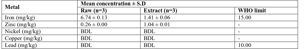
Heavy metal content
The raw leaf powder of G. simplicifolia contained 6.74 mg/L of iron and 0.26 mg/L of zinc (Table 2). The extract, on the other hand, contained 1.41 mg/L of iron and 1.04 mg/L of zinc. The raw powder had higher iron (6.74 mg/kg) but lower zinc (0.26 mg/kg) contents than those of the extract. The levels of nickel, copper and lead in both the raw powder and extract were below detectable limits (<0.00001mg/L). Similarly, iron and lead concentrations in the samples were below the WHO limit for medicinal plants (WHO, 1998, 2007).
UV-VIS spectroscopic analysis
The crude, ethyl acetate and hydro fractions of G. simplicifolia each recorded five (5) major absorption peaks whereas the methanol fraction recorded three (3) peaks (Table 3). The wavelength generally ranged from 201.00 to 666.90.
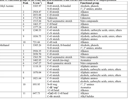
FT-IR Spectra of fractions of G. simplicifolia
The FTIR spectra of G. simplicifolia leaf fractions predicted the functional groups corresponding to each peak as well as the nature of their bonds (Table 4). The frequencies and nature of the peaks were used to identify the functional groups by comparing the values with standards. Ethyl acetate, methanol and the hydro fraction recorded 10, 13 and 5 peaks, respectively.
Safety evaluation of the hydroethanolic leaf extract of
G. simplicifolia
10 | P a g e
In the study, no untoward changes were observed in the rats when they were dosed orally with 5000 mg/kg GSE. No changes in the nature of stool, urine and eye colour were observed. Also, there was no stretching, paw liking or diarrhoea. No mortality was recorded. Orally, 5000 mg/kg of GS was well tolerated in mice even after 7days. Hence, the LD50 was estimated to be≤5000 mg/kg.
Sub-chronic toxicity
Effect of treatments on body weight
In the sub-chronic study, the animals generally showed no exterior signs of toxicity to the G. simplicifolia
treatment. As expected, the body weights of all test animals generally increased over time (Figure 4). There were significant increases in percent change in body weight in male rats treated with 100 mg/kg GS from days 8 to 28, in those treated with 250 mg/kg GS on day 8, 20 and 24, and in those treated with 500mg/kg GS on day 8 (p<0.001) and day 24 (p<0.5) compared to the control group. But, compared to the 100 mg group, significant decreases were observed in males treated with 250 mg and 500 mg from day 20 to 28 (p<0.5). In female rats, however, there were highly significant increases in change in body weight in the 100 mg/kg treated group from day 8 through to 28 compared to the normal (p<0.0001). Similarly, significant increases were observed in those treated with 250 mg and 500 mg from day 8 to 28. However, compared to the 100 mg group, they decreased in percent change in body weight from day 20 to 28 as observed in the male rats (p<0.5).
Effect of treatments on relative organ weight (ROW)
Compared to the control group, a significant decrease in relative liver weight was observed at all doses in male rats, whereas that of females increased at 250mg/kg (p<0.5). Uterus weight in females administered with 100 mg/kg GSE also decreased considerably (p<0.01). The treatment had no significant effect on any of the other organs (Table 5).
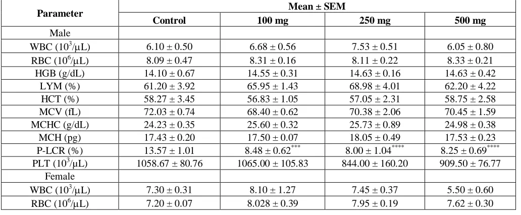
Effect of treatments on haematological indices
With the exception of P-LCR and platelet count, the GSE showed no significant effects on haematological indices (Table 6). P-LCRs for all doses were lower relative to the control (p<0.001). A significant decrease in platelet count was also observed in female rats (p<0.05). All other parameters did not differ among groups.
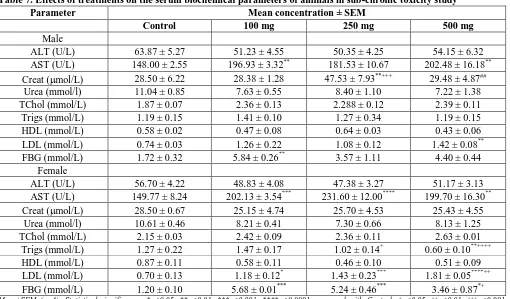
Effect of treatments on serum biochemistry
The effects of GSE treatment on the serum biochemical parameters of the animals are shown in Table 7. In male rats, compared to the control, AST concentration increased at 100 and 500 mg (P<0.01). Also, creatinine concentration increased significantly at 250 mg. Finally, significant increases were recorded in the concentrations of LDL at 500 mg and FBG at 100 mg. In the female rats, the levels of AST at all doses (P<0.01), triglycerides at 500 mg, and lastly, LDL and FBG at all doses increased significantly relative to the control. All other parameters in both males and females showed no significant changes compared to the control group.
Table 1. Phytochemical constituents of raw powder and hydroethanolic leaf extract of G. simplicifolia
Phytochemical Raw Extract
Glycosides + +
Tannins + +
Flavonoids + +
Alkaloids + +
Saponins + +
Coumarins + +
Sterols - -
‘+’ indicates presence and ‘-’ indicates absence.
Table 2. Level of heavy metals in the raw leaf and hydroethanolic leaf extract of G. simplicifolia
Metal Mean concentration ± S.D
Raw (n=3) Extract (n=3) WHO limit
Iron (mg/kg) 6.74 ± 0.13 1.41 ± 0.06 15.00
Zinc (mg/kg) 0.26 ± 0.00 1.04 ± 0.01 -
Nickel (mg/kg) BDL BDL -
Copper (mg/kg) BDL BDL -
Lead (mg/kg) BDL BDL 10.00
11 | P a g e
Table 3. UV-Vis peak characteristics of hydroethanolic crude extract and fractions of G. simplicifolialeaves.
Peak Wavelength (nm) Absorbance
Crude 1 666.90 0.01
2 341.90 0.37
3 332.00 0.41
4 329.00 0.42
5 201.00 2.83
Ethyl acetate 1 203.90 0.31
2 210.90 0.29
3 206.90 0.27
4 226.00 0.25
5 209.00 0.17
Methanol 1 664.00 0.02
2 204.20 2.07
3 202.10 2.16
Hydro 1 329.90 0.08
2 332.80 0.08
3 337.00 0.07
4 335.10 0.07
5 341.80 0.07
Table 4. Functional groups in the crude and hydroethanolic crude extract and fractions of G. simplicifolialeaf
Peak X (cm-1) Bond Functional group
Ethyl Acetate 1 3385.97 O-H stretch, H-bonded
N-H stretch
Alcohols, phenols 1º, 2º amines, amides
2 2924.47 C-H stretch Alkanes
3 2853.08 C-H stretch Alkanes
4 1712.90 Unknown Unknown
5 1515.84 N–O asymmetric stretch Nitro compounds
6 1463.37 C-H bend Alkanes
7 1377.24 C–H rock Alkanes
8 1240.33 C–O stretch
C–N stretch
Alcohols, carboxylic acids, esters, ethers Aliphatic amines
9 1036.75 C–O stretch
C–N stretch
Alcohols, carboxylic acids, esters, ethers Aliphatic amines
10 721.66 C–H rock Alkanes
Methanol 1 3305.26 O-H stretch, H-bonded
N-H stretch
Alcohols, phenols 1º, 2º amines, amides
2 2926.35 C-H stretch Alkanes
3 2850.70 C-H stretch Alkanes
4 1563.59 N–O asymmetric stretch Nitro compounds
5 1402.85 C–C stretch (in–ring) Aromatics
6 1347.27 N–O symmetric stretch Nitro compounds
7 1118.18 C-N stretch
C–O stretch
Aliphatic amines
alcohols, carboxylic acids, esters, ethers
8 1070.04 C-N stretch
C–O stretch
Aliphatic amines
alcohols, carboxylic acids, esters, ethers
9 1021.64 C-N stretch
C–O stretch
Aliphatic amines
alcohols, carboxylic acids, esters, ethers
10 893.95 N–H wag
C–H “oop” =C–H bend
1°, 2° amines Aromatics Alkenes
11 647.73 –C≡C–H: C–H bend
C–Br stretch
12 | P a g e
C–Cl stretch Alkyl halides
Methanol 12 617.94 –C≡C–H: C–H bend
C–Br stretch C–Cl stretch
Alkynes Alkyl halides Alkyl halides
13 508.32 Unknown Unknown
Hydro 1 3339.13 O–H stretch, H–bonded
N–H stretch
Alcohols, phenols 1º, 2º amines, amides
2 2108.12 –C≡C– stretch Alkynes
3 1634.4 N–H bend 1° amines
4 418.64 Unknown Unknown
5 403.53 Unknown Unknown
Table 5. Effect of GS on ROW of animals in sub-chronic toxicity study
Organ ROW (%)
Control 100 mg 250 mg 500 mg
Male
Liver 3.18 ± 0.22 2.86 ± 0.02* 2.67 ± 0.07*** 2.67 ± 0.06***
Testes 1.16 ± 0.01 1.32 ± 0.03 1.25 ± 0.02 1.26 ± 0.03
Lung 0.63 ± 0.07 0.68 ± 0.07 0.71 ± 0.06 0.63 ± 0.05
Kidneys 0.55 ± 0.02 0.60 ± 0.02 0.58 ± 0.02 0.59 ± 0.01
Stomach 0.60 ± 0.03 0.63 ± 0.01 0.62 ± 0.04 0.56 ± 0.01
Heart 0.32 ± 0.01 0.35 ± 0.01 0.35 ± 0.01 0.33 ± 0.01
Spleen 0.19 ± 0.01 0.25 ± 0.01 0.20 ± 0.02 0.20 ± 0.01
Female
Liver 2.95 ± 0.10 2.99 ± 0.04 3.11 ± 0.03* 2.76 ± 0.02
Lungs 0.73 ± 0.07 0.83 ± 0.03 0.78 ± 0.06 0.68 ± 0.01
Kidneys 0.58 ± 0.00 0.58 ± 0.01 0.61 ± 0.03 0.55 ± 0.03
Stomach 0.65 ± 0.04 0.70 ± 0.01 0.62 ± 0.03 0.58 ± 0.01
Heart 0.35 ± 0.02 0.37 ± 0.01 0.38 ± 0.01 0.33 ± 0.02
Uterus 0.26 ± 0.03 0.18 ± 0.01** 0.26 ± 0.01+++ 0.22 ± 0.00
Spleen 0.23 ± 0.01 0.25 ± 0.01 0.22 ± 0.01 0.21 ± 0.00
Mean±SEM (n=4); Statistical significance; *p<0.05, **p<0.01, ***p<0.001 compared with Control, +++p<0.001 compared with 100 mg group
Table 6. Effect of treatments on the haematological parameters of animals in sub-chronic toxicity study
Parameter Mean ± SEM
Control 100 mg 250 mg 500 mg
Male
WBC (103/L) 6.10 ± 0.50 6.68 ± 0.56 7.53 ± 0.51 6.05 ± 0.80
RBC (106/L) 8.09 ± 0.47 8.31 ± 0.16 8.11 ± 0.22 8.33 ± 0.21
HGB (g/dL) 14.10 ± 0.67 14.55 ± 0.31 14.63 ± 0.16 14.63 ± 0.42
LYM (%) 61.20 ± 3.92 65.95 ± 1.43 68.98 ± 4.01 62.20 ± 4.22
HCT (%) 58.27 ± 3.45 56.83 ± 1.05 57.05 ± 2.31 58.75 ± 2.58
MCV (fL) 72.03 ± 0.74 68.40 ± 0.62 70.38 ± 2.06 70.45 ± 1.59
MCHC (g/dL) 24.23 ± 0.35 25.60 ± 0.32 25.73 ± 0.89 24.98 ± 0.38
MCH (pg) 17.43 ± 0.20 17.50 ± 0.07 18.05 ± 0.49 17.53 ± 0.23
P-LCR (%) 13.57 ± 1.01 8.48 ± 0.62*** 8.00 ± 1.04**** 8.25 ± 0.69****
PLT (103/µL) 1058.67 ± 80.76 1065.00 ± 105.83 844.00 ± 160.20 909.50 ± 76.77
Female
WBC (103/L) 7.30 ± 0.31 8.10 ± 1.27 7.45 ± 0.37 5.50 ± 0.60
13 | P a g e
HGB (g/dL) 13.43 ± 0.09 13.90 ± 0.24 14.28 ± 0.35 14.35 ± 0.55
LYM (%) 63.47 ± 0.48 69.98 ± 1.89 69.88 ± 1.69 66.10 ± 5.10
HCT (%) 52.07 ± 0.88 55.30 ± 1.85 54.00 ± 1.27 54.30 ± 1.80
MCV (fL) 72.30 ± 0.60 69.13 ± 2.10 68.00 ± 1.62 71.25 ± 0.45
MCHC (g/dL) 25.80 ± 0.32 25.23 ± 0.96 26.45 ± 0.27 26.45 ± 0.15
MCH (pg) 18.63 ± 0.12 17.43 ± 0.87 18.00 ± 0.34 18.85 ± 0.05
P-LCR (%) 11.90 ± 0.56 7.65 ± 0.72**** 6.50 ± 0.32**** 8.70 ± 1.40*
PLT (103/µL) 1323.00 ± 156.52 1052.50 ± 84.19 891.75 ± 46.22* 821.50 ± 66.50*
Mean±SEM (n=4); Statistical significance; *p<0.05, ***p<0.001, ****p<0.0001 compared with Control. WBC – white blood cell, RBC-red blood cell, HGB-haemoglobin, LYM-lymphocyte, HCT-haematocrit, MCV-mean corpuscular volume, MCH-mean corpuscular haemoglobin, MCHC-mean corpuscular haemoglobin concentration, P-LCR-platelet large cell volume, PCT-plateletcrit, PLT-platelet
Table 7. Effects of treatments on the serum biochemical parameters of animals in sub-chronic toxicity study
Parameter Mean concentration ± SEM
Control 100 mg 250 mg 500 mg
Male
ALT (U/L) 63.87 ± 5.27 51.23 ± 4.55 50.35 ± 4.25 54.15 ± 6.32
AST (U/L) 148.00 ± 2.55 196.93 ± 3.32** 181.53 ± 10.67 202.48 ± 16.18**
Creat (mol/L) 28.50 ± 6.22 28.38 ± 1.28 47.53 ± 7.93**+++ 29.48 ± 4.87##
Urea (mmol/l) 11.04 ± 0.85 7.63 ± 0.55 8.40 ± 1.10 7.22 ± 1.38
TChol (mmol/L) 1.87 ± 0.07 2.36 ± 0.13 2.288 ± 0.12 2.39 ± 0.11
Trigs (mmol/L) 1.19 ± 0.15 1.41 ± 0.10 1.27 ± 0.34 1.19 ± 0.15
HDL (mmol/L) 0.58 ± 0.02 0.47 ± 0.08 0.64 ± 0.03 0.43 ± 0.06
LDL (mmol/L) 0.74 ± 0.03 1.26 ± 0.22 1.08 ± 0.12 1.42 ± 0.08**
FBG (mmol/L) 1.72 ± 0.32 5.84 ± 0.26** 3.57 ± 1.11 4.40 ± 0.44
Female
ALT (U/L) 56.70 ± 4.22 48.83 ± 4.08 47.38 ± 3.27 51.17 ± 3.13
AST (U/L) 149.77 ± 8.24 202.13 ± 3.54*** 231.60 ± 12.00**** 199.70 ± 16.30**
Creat (mol/L) 28.50 ± 0.67 25.15 ± 4.74 25.70 ± 4.53 25.43 ± 4.55
Urea (mmol/l) 10.61 ± 0.46 8.21 ± 0.41 7.30 ± 0.66 8.13 ± 1.25
TChol (mmol/L) 2.15 ± 0.03 2.42 ± 0.09 2.36 ± 0.11 2.63 ± 0.01
Trigs (mmol/L) 1.27 ± 0.22 1.47 ± 0.17 1.02 ± 0.14+ 0.60 ± 0.10**++++
HDL (mmol/L) 0.87 ± 0.11 0.58 ± 0.11 0.46 ± 0.10 0.51 ± 0.09
LDL (mmol/L) 0.70 ± 0.13 1.18 ± 0.12* 1.43 ± 0.23*** 1.81 ± 0.05****++
FBG (mmol/L) 1.20 ± 0.10 5.68 ± 0.01*** 5.24 ± 0.46*** 3.46 ± 0.87*+
Mean±SEM (n=4); Statistical significance; *p<0.05, **p<0.01, ***p<0.001, ****p<0.0001 compared with Control; +p<0.05, ++p<0.01, +++p<0.001, ++++
p<0.0001 compared to the 100 mg group; ##p<0.01 with respect to 250 mg group. ALT-alanine aminotransferase, AST-aspartate aminotransferase, Creat-creatinine, TChol-total cholesterol, Trigs-triglycerides, HDL-high density lipoproteins, LDL-low density lipoproteins, FBG-fasting blood glucose.
14 | P a g e
Fig 2. Total polyphenol content (mean±SD; n=3)) of the extract and fractions of Griffonia leaves. A represents TFC, B represents TPC. Different letters indicate significant difference at P≤0.05. Crude - hydroethanolic extract, EthylAc
– ethyl acetate, MetOH –methanol, Hydro – hydro fraction.
Fig 3. Scavenging activity of hydroethanolic crude extract and fractions of G. simplicifolia leaves (mean±SD (n=3)).Different letters indicate significant differences. Crude - hydroethanolic extract, EthylAc – ethyl acetate,
MetOH –methanol, Hydro – hydro fraction.
15 | P a g e
DISCUSSION
The presence of phytochemicals in a plant material is an indication of the medicinal value of the plant. Phytochemicals such as phenols and polyphenolic compounds like flavonoids, tannins, saponins and alkaloids have been shown to possess good antioxidant and anti-inflammatory properties (Anim et al., 2016; Iwalewa et al., 2007). In this study, the crude leaf extract of G. simplicifolia was shown to possess phytochemicals such as glycosides, tannins, flavonoids, alkaloids and saponins, and coumarins. These phytochemicals could be partly responsible for the ethnomedicinal activity of the leaves of this plant species (Miller et al., 2010). A recent study by Offoumou et al. (2018) in Cote d‟Ivoire reported the presence of alkaloids and saponins in the ethanolic extract of Griffonia leaves. Contrary to the results of this present study, these authors reported the presence of sterols but the absence of flavonoids in the extracts. These variations could reflect differences in the extracts used, geographical sources, genetic makeup and/or level of maturity of the leaves used (Giurleo, 2017). Similarly, the varying concentrations of polyphenols in the different fractions in the present study could be accounted for the different solvents used and their polarity (Medini et al., 2014). This observation is also consistent with the findings of Chebil et al. (2007) that the solubility of polyphenols and bioactive compounds differ in different solvents. TFC ranged from 142.3 mg QE in ethyl acetate fraction to 237.20 mg QE in the hydro fraction and TPC ranged from 3.87 mg GAE in methanol fraction to 8.71 mg GAE in ethyl acetate fraction. A study conducted in Ghana by Giurleo (2017) reported a total polyphenol content range of 9.78 to 29.83 GAE/gram in 60% methanol/water extract of G. simplicifolia leaves, which differed considerably among the four different regions of the country studied. Thus, the relatively lower TPC observed in our study might reflect differences in sampling location. However, few reports exist on the polyphenol content of leaves related to Griffonia, making effective comparisons difficult at this time.
Free radicals play vital physiological roles in the human body, including signal transduction, smooth muscle relaxation, cell growth and immune response. However, at high concentrations, they result in oxidative stress, which leads to the deterioration of cellular biomolecules (Verma
et al., 2017). The antioxidant defence system helps eliminate the excess free radicals to protect the body from oxidative insults. The antioxidant activities of medicinal plants are thus measured to determine their ability to scavenge free radicals. In the current study, the antioxidant activity of Griffonia leaves ranged from 14.07 % in the ethyl acetate fraction to 24.82 % in the hydro fraction which is much lower than the scavenging activity of ethanolic extract of the milk thistle (Silybummarianum) seeds, the source of silymarin (a nephroprotective drug), which has a scavenging activity of 92.0 %. This suggests
that Griffonia might not be a very potent antioxidant as compared to Silybum. Giurleo (2017) also measured the antioxidant activity of Griffonia leaves in his study. However, he used the ABTS assay instead of the DPPH method; thus his results are not comparable to those obtained from this present study.
Heavy metals such as iron, copper, zinc and nickel function as micronutrients in human body, although excessive concentrations in food or medicine may cause health risks (Darko, 2010). The generally low levels of heavy metals observed both in the raw powder and the hydroethanolic extracts of Griffonia leaves relative to the WHO guideline values in this study suggest that there is no potential negative side effects. Nonetheless, given the likely spatio-temporal variation in heavy metal concentrations in a plant species (Annan et al., 2013; Haider et al., 2004), continuous monitorings of these contaminants in plant materials is absolutely essential to ensure thequality of herbal medicine.
Most phenolic compounds such as flavones and flavonols, exhibit two major absorption bands in the ultraviolet-visible region; one in the 300-500 nm range and the other below 280 nm (Markham, 1989).The UV-Vis spectra of all the Griffonia extracts showed similar bands, suggesting the presence of flavones and flavonols.
The Organisation of Economic Co-operation and Development of France recommends four classes of acute systemic toxicity based on oral LD50 values (OECD,
2001), namely: very toxic (≤ 5 mg/kg), toxic (> 5 ≤ 50 mg/kg), harmful (> 50 ≤ 500 mg/kg), and no label (> 500 ≤ 2000 mg/kg). Based on this classification, the estimated LD50 of the extract of ≤ 5 g/kg bwt suggests no
behavioural changes and no adverse gastrointestinal effects. This indicates that the leaf extract of Griffonia is safe for oral use. The gram equivalence of an LD50 ≤ 5g/kg
bwt in an average adult man with an approximate body weight of 60 kg would translate to 750 g dose of the extract.
One of the key indicators of the general health status of an animal is the changes in its body weight (Salawu et al., 2009). In this study, there was a general weight gain in both males and females at all doses, indicating that the extract did not interfere with normal metabolism and nutritional benefits such as weight gain and stability of appetite expected of animals supplied with food and water ad libitum. However, at higher doses, the extract may interfere with gastric functions and decrease food conversion efficiency (Arthur et al., 2011). Thus, GSE can be used for weight gain, but it will be more effective at a lower dose.
16 | P a g e
although the male rates showed considerable reduction in liver weight during the same period. This does not conclusively indicate liver or uterus toxicity in females, but it would be safer to avoid using the extract during pregnancy.
In both males and females, the extract caused a marked reduction in platelet-large cell ratio (LCR). P-LCR is defined as the percentage of platelets that exceed the normal platelet volume of 12 fL in the total platelet count (Gawlita et al., 2016). P-LCR is a prognostic factor for coronary artery disease and myocardial infarctions; the lower the percentage, the lower the chance of developing cardiovascular problems. Furthermore, the extract caused a reduction in platelet count. This indicates that GSE is a potent blood thinner and can reduce the incidence of thrombotic complications such as myocardial infarction and sudden cardiac arrest (Gawlita et al., 2016). GS should therefore be further investigated for its potential as a treatment for cardiovascular diseases.
The GSE treatment increased plasma
concentration of AST. Unlike ALT, AST is not specific to the liver because it is also found in the heart, skeletal muscles, kidneys, brain and red blood cells and thus, an increased concentration is not necessarily an indication of liver damage. When the plasma concentration of AST is greater than ALT, the injury may be due to muscle necrosis. However, this can only be confirmed if there is an increase in creatine kinase concentration (Aragon & Younossi, 2010). Treatment with GS extract also resulted in hyperbilirubinemia especially at the lowest dose. The extract also caused an increase in LDL (low-density
lipoproteins, commonly known as „bad cholesterol‟) at the highest dose in males and at all doses in females. High concentration of LDL in the blood is often associated with an increased risk of atherosclerosis and heart diseases (Ai
et al., 2010). This means that the extract could potentially be dangerous for patients who already have high blood cholesterol levels, especially females. It may also cause hypercholesterolemia in normal individuals. Finally, the increase in blood glucose in both males and females suggests the presence of hyperglycaemic components in the GS extract.
CONCLUSION
In general, the study demonstrates that G. Simplicifolia leaf extract is safe for oral use due to the high LD50. It can also be used for weight gain and for
reducing platelet large cell ratio. Sub-chronic use may, however, cause side effects such as blood thinning and an increase in LDL and blood glucose concentration.
AUTHOR CONTRIBUTIONS
This work was carried out by collaboration among all authors. Authors AKA and CL conceived of the study. Authors RAN and CL did the experimental design, crude extract preparation, laboratory work, data and statistical analyses. Author RAN drafted the manuscript. Author AKA also participated in the design and coordination of the study, as well as the data analyses. He also helped to draft the manuscript. Author PKB participated in the design and coordination of the study. All authors read and approved the final manuscript.
REFERENCES
Abdel-daim MM, Aly SM, Abo-el-sooud K, Giorgi M, Ursoniu S. Role of natural products in ameliorating drugs and
chemicals toxicity. Evidence-Based Complementary and Alternative Medicine, 2016, 2–4.
https://doi.org/http://dx.doi.org/10.1155/2016/7879406
Ai M, Otokozawa S, Asztalos BF, Ito Y, Nakajima K, White CC, Schaefer EJ. Small dense LDL cholesterol and coronary heart disease: results from the Framingham offspring study. Clinical Chemistry, 56(6), 2010, 967–976. https://doi.org/10.1373/clinchem.2009.137489
Anim MT, Larbie C, Appiah-Opong R, Tuffour I, Owusu BAK, Aning A. Phytochemical, antioxidant and cytotoxicity of hydroethanolic extracts of Crotalaria restusa L. World Journal of Pharmaceutical Research, 5(2), 2016, 162–179. Annan K, Dickson R, Nooni I, Amponsah I. The heavy metal contents of some selected medicinal plants sampled from
different geographical locations. Pharmacognosy Research, 5(2), 2013, 103. https://doi.org/10.4103/0974-8490.110539
Aragon G, Younossi ZM. When and how to evaluate mildly elevated liver enzymes in apparently healthy patients. Cleveland Clinical Journal of Medicine, 77(3), 2010, 195–204. https://doi.org/10.3949/ccjm.77a.09064
Arthur FKN, Woode E, Terlabi EO, Larbie C. Evaluation of acute and subchronic toxicity of Annona muricata ( Linn .) aqueous extract in animals. European Journal of Experimental Biology, 1(4), 2011, 115–124.
Bailey SA, Zidell RH, Perry RW. Relationships between organ weight and body/brain weight in the rat: what is the best analytical endpoint? Toxicologic Pathology, 32(4), 2004, 448–466. https://doi.org/10.1080/01926230490465874 Brand-Williams W, Cuvelier ME, Berset CLWT. Use of a free radical method to evaluate antioxidant activity. LWT-Food
Science and Technology, 28(1), 1995, 25–30.
Chebil L, Humeau C, Anthony J, Dehez F, Engasser JM, Ghoul M. Solubility of flavonoids in organic solvents. Journal of Chemical and Engineering Data, 52(5), 2007, 1552–1556. https://doi.org/10.1021/je7001094
Crook M. Clinical Chemistry & Metabolic Medicine(7th ed.), Hodder Arnold, London, 2006.
17 | P a g e
University of Science and Technology, 2010. Retrieved from
http://ir.knust.edu.gh/handle/123456789/492%5Cnfiles/1095/CHEM - 2010 - HEAVY METALS CONTENT AND PHYTOCHEMICALS IN SOME SE.pdf%5Cnfiles/1119/492.html
DhananiT, Shah S, Gajbhiye NA,Kumar S. Effect of extraction methods on yield, phytochemical constituents and antioxidant
activity of Withania somnifera. Arabian Journal of Chemistry, 10, 2017, S1193–S1199.
https://doi.org/10.1016/j.arabjc.2013.02.015
Ernst E. The efficacy of herbal medicine - An overview. Fundamental and Clinical Pharmacology, 19(4), 2005, 405–409. https://doi.org/10.1111/j.1472-8206.2005.00335.x
Esposito M, Ruberto M, Pascotto A, Carotenuto M. Nutraceutical preparations in childhood migraine prophylaxis : effects on headache outcomes including disability and behaviour. Neurological Science, 33, 2012, 1365–1368. https://doi.org/10.1007/s10072-012-1019-8
Gawlita M, Wasilewski J, Osadnik T, Reguła R, Bujak K, Gonera M. Mean platelet volume and platelet-large cell ratio as prognostic factors for coronary artery disease and myocardial infarction. Folia Cardiologica, 10(6), 2016, 418–422. https://doi.org/10.5603/FC.2015.0079
Giurleo DA. Phytochemical Exploration of Griffonia simplicifolia Seeds and Leaves. The State University of New Jersey, 2017.
Haider S, Naithani V, Barthwal J, Kakkar P. Heavy metal content in some therapeutically important medicinal plants. Bulletin of Environmental Contamination and Toxicology, 2004. https://doi.org/10.1007/s00128-003-0249-0
Harborne JB. Phytochemical methods; a guide to modern techniques of plant analysis. Journal of Chemical Information and Modeling (Vol. 3), 1998. https://doi.org/10.1017/CBO9781107415324.004
Iwalewa E, McGaw L, Naidoo V, Eloff J. Inflammation: the foundation of diseases and disorders. A review of phytomedicines of South African origin used to treat pain and inflammatory conditions. African Journal of Biotechnology, 6(25), 2007, 2868–2885. https://doi.org/10.5897/AJB2007.000-2457
Kigen G, Rono HK, Kipkore WK, Rotich JK. Current trends of traditional herbal medicine practice in Kenya : A review.
African Journal of Pharmacology and Therapeutics, 2(1), 2013, 32–37.
Larbie C, Owusu KP, Torkornoo D, Asibey O. Acute and sub-chronic toxicity of aqueous ethanolic extract of Ficus pumila
leaves in rats. European Journal of Biomedical and Pharmaceutical Sciences, 3(8), 2016, 22–27.
Marinova D, Ribarova F, Atanassova M. Total phenolics and flavonoids in Bulgarian fruits and vegetables. Journal of the University of Chemical Technology and Metallurgy, 40(3), 2005, 255–260.
Markham KR. In: Dey PM, Harborne JB (Eds.), Plant Phenolics Vol. 1, 197. Academic Press, New York, 1989.
Medini F, Fellah H, Ksouri R, Abdelly C. Total phenolic, flavonoid and tannin contents and antioxidant and antimicrobial activities of organic extracts of shoots of the plant Limonium delicatulum. Journal of Taibah University for Science,
8(3), 2014, 216–224. https://doi.org/10.1016/j.jtusci.2014.01.003
Miller RP, Tadagavadi RK, Ramesh G, Reeves WB. Mechanisms of cisplatin nephrotoxicity. Toxins, 2, 2010, 2490–2518. https://doi.org/10.3390/toxins2112490
Neergheen-Bhujun VS. Underestimating the toxicological challenges associated with the use of herbal medicinal products in developing countries. BioMed Research International, 2013, 1–10. https://doi.org/10.1155/2013/804086
OECD. Acute Oral Toxicity – Acute Toxic Class Method. Oecd Guideline for Testing of Chemicals, (December), 2001, 1–14. https://doi.org/10.1787/9789264071001-en
Offoumou MR, Kipre GR, Kigbafori DS, Camara D, Djaman AJ,Zirihi GN. In vitro / ex vivo antiplasmodial activity and phytochemical screening of crude extracts Entandrophragma angolense (Welw.) C. DC., Griffonia simplicifolia (Vahl ex DC.) Baill. et Uapaca guineensis Müll. Arg. three plants of Ivorian pharmacopeia in treatment of malaria.
International Journal of Current Microbiology and Applied Sciences, 7(03), 2018, 2088–2095. https://doi.org/https://doi.org/10.20546/ijcmas.2018.703.245
Pathak SK, Tahilani P, Prakash JN, Jitendra B. A review on Griffonia simplicifollia - an ideal herbal anti-depressant.
International Journal of Pharmacy & Life Sciences, 1(3), 2010, 174–181.
Piao Y, Liu Y, Xie X. Change trends of organ weight background data in sprague dawley rats at different ages.Journal of Toxicologic Pathology, 26(1), 2013, 29–34. https://doi.org/10.1293/tox.26.29
Salawu OA, Chindo BA, Tijani AY, Obidike IC, Salawu TA, AkingbasoteAJ. Acute and sub-acute toxicological evaluation of the methanolic stem bark extract of Crossopteryx febrifuga in rats. African Journal of Pharmacy and Pharmacology,
3(12), 2009, 621–626. Retrieved from http://www.academicjournals.org/ajpp
Serçe A, Toptancı BÇ, Tanrıkut SE, Altaş S, Kızıl G, Kızıl S, Kızıl M. Assessment of the antioxidant activity of Silybum marianum extract and its protective effect against DNA oxidation, protein damage and lipid peroxidation. Food Technology and Biotechnology, 54(4), 2016, 455–461. https://doi.org/10.17113/ftb.54.04.16.4323
18 | P a g e
https://doi.org/10.1016/j.jep.2012.01.028
Verma PK, Raina R, Singh M, Wazir VS, Kumar P. Attenuating potential of Calendula officinalis on biochemical and antioxidant parameters in hepatotoxic rats. Indian Journal of Physiology and Pharmacology, 61(4), 2017, 398–410. Wang D, Wang H, Gu L. The antidepressant and cognitive improvement activities of the traditional chinese herb Cistanche.
Evidence-Based Complementary and Alternative Medicine, 2017, 1–9. https://doi.org/https://doi.org/10.1155/2017/3925903
Weeks BS. Formulations of dietary supplements and herbal extracts for relaxation and anxiolytic action: Relarian TM. Medical Science Monitor, 15(11), 2009, 256–262.
WHO. Quality Control Methods for Medicinal Plant Materials.World Health Organization Geneva. Geneva, Switzerland, 1998.
WHO. WHO guidelines for assessing quality of herbal medicines with reference to contaminants and residues. Geneva, Switzerland, 2007.