PRIMARY RESEARCH
Inactivation of the Wnt/β-catenin
signaling pathway underlies inhibitory role
of microRNA-129-5p in epithelial–mesenchymal
transition and angiogenesis of prostate cancer
by targeting ZIC2
Zhenming Jiang
1, Yuxi Zhang
1,2*, Xi Chen
3, Pingeng Wu
1and Dong Chen
4Abstract
Background: Prostate cancer (PCa) is a common disease that often occurs among older men and a frequent cause of malignancy associated death in this group. microRNA (miR)-129-5p has been identified as an essential regulator with a significant role in the prognosis of PC. Therefore, this study aimed to investigate roles of miR-129-5p in PCa. Methods: Microarray analysis was conducted to identify PCa-related genes. The expression of miR-129-5p and ZIC2 in PCa tissues was investigated. To understand the role of miR-129-5p and ZIC2 in PCa, DU145 cells were trans-fected with mimic or inhibitor of miR-129-5p, or si-ZIC2 and the expression of Wnt, β-catenin, E-cadherin, vimentin, N-cadherin, vascular endothelial growth factor (VEGF), and CD31, as well as the extent of β-catenin phosphorylation was determined. In addition, cell proliferation, migration, invasion, angiogenesis, apoptosis and tumorigenesis were detected.
Results: miR-129-5p was poorly expressed and ZIC2 was highly expressed in PCa tissues. Down-regulation of ZIC2 or overexpression of miR-129-5p reduced the expression of ZIC2, Wnt, β-catenin, N-cadherin, vimentin, and β-catenin phosphorylation but increased the expression of E-cadherin. Importantly, miR-129-5p overexpression significantly reduced cell migration, invasion, angiogenesis and tumorigenesis while increasing cell apoptosis.
Conclusions: The findings of the present study indicated that overexpression of miR-129-5p or silencing of ZIC2 could inhibit epithelial–mesenchymal transition (EMT) and angiogenesis in PCa through blockage of the Wnt/β-catenin signaling pathway.
Keywords: Prostate cancer, microRNA-129-5p, Zinc-finger protein of the cerebellum 2, Wnt/β-catenin signaling pathway, Epithelial–mesenchymal transition, Angiogenesis
© The Author(s) 2019. This article is distributed under the terms of the Creative Commons Attribution 4.0 International License (http://creat iveco mmons .org/licen ses/by/4.0/), which permits unrestricted use, distribution, and reproduction in any medium, provided you give appropriate credit to the original author(s) and the source, provide a link to the Creative Commons license, and indicate if changes were made. The Creative Commons Public Domain Dedication waiver (http://creativecommons.org/ publicdomain/zero/1.0/) applies to the data made available in this article, unless otherwise stated.
Open Access
*Correspondence: zyxfan@sina.com
1 Department of Urology, The First Hospital of China Medical University,
No. 155, Nanjing North Street, Heping District, Shenyang 110001, Liaoning, People’s Republic of China
Background
Prostate cancer (PCa) is a most frequently occurring malignancy among older men [1, 2]. Prostate tumors are usually indolent, but a considerable number of tumors are highly aggressive and often metastasize to bones and other organs, leading to high morbidity and mortality [3,
4]. In addition, PCa is typically marked by a high recur-rence rates, whereby about 40% of local PCa cases recur after initial treatment, and the tumor progresses to hor-mone refractory/castration resistance stage is basically untreatable [5, 6]. Epithelial–mesenchymal transition (EMT) and its reverse process are essential physiological processes during organogenesis and tissue differentia-tion of normal embryonic development [7, 8]. The EMT process is also a part of cancer pathogenesis including PCa [9]. Angiogenesis is also an important feature of malignancy, and is particularly relevant in the progres-sion to end-stage PCa [10]. There is a need to unravel the molecular events and players that are involved in these mechanisms.
Past studies have highlighted the regulatory role of microRNAs (miRNAs) in PCa pathogenesis. miRNAs regulate post-transcriptional gene expression and their dysregulation is implicated in the development of cancer [11, 12]. It has been previously reported that up-regu-lated miR-129-5p could reduce EMT and thus functions as a tumor suppressor [13]. Down-regulation of miR-129 has been demonstrated as a valuable prognostic biomarker of PCa proliferation [14]. Zinc-finger protein of the cerebellum (ZIC) 2, identified as a target gene of miR-129-5p in the present study, is the vertebrate homo-logues of the Drosophila odd-paired (OPA) gene, includ-ing ZIC1, ZIC2, ZIC3, ZIC4 and ZIC5, and has been implicated in multiple diseases including cancer [15, 16]. Another study has proved that the RNA levels of ZIC1, ZIC2, ZIC4 and ZIC5 are all induced in Gleason grade 3 embedded in Gleason score (GS) 4 + 3 = 7 PCa [17].
miRNAs have recently become important regulators of EMT in diversity cancers [18]. miRNAs appear to regu-late EMT by modulating posttranscriptional components such as EMT-transcription factors, epithelial and mes-enchymal genes, or through regulation of key signaling pathways, which in turn modulate cancer progression and metastasis [7, 18]. For instance, overexpression of miR-129-5p attenuated EMT and proliferation in gas-tric cancer by downregulating the expression of HMGB1 [19]. The canonical Wnt signaling pathway, extensively conserved in the animal kingdom, is essential for embry-onic development and adult tissue homeostasis [20]. Moreover, miR-129-5p has been reported to hamper pro-liferation and invasion of chondrosarcoma cells by block-ing the Wnt/β-catenin signalblock-ing pathway [21]. Based on the aforementioned evidences, we hypothesize that
miR-129-5p played a significant role in PCa pathogen-esis via its regulation of ZIC2-mediated Wnt/β-catenin signaling pathway. Therefore, the current study aimed to examine if miR-129-5p could impact EMT and angiogen-esis in PCa by regulating ZIC2-mediated Wnt/β-catenin signaling pathway.
Materials and methods Ethical statement
The study was approved by the Institutional Review Board of the First Hospital of China Medical Univer-sity. Written informed consents were obtained from all patients or their guardians. All study procedures were conducted in accordance with the Declaration of Hel-sinki. All animal experiments were conducted under the approval of guidelines for the protection and use of experimental animals issued by the National Institutes of Health (NIH), and strictly complied with the principles of completing the experiments with the minimum number of animals and minimizing pain.
Microarray analysis
The Gene Expression Omnibus (GEO) database (https ://
www.ncbi.nlm.nih.gov/geo/) was used to identify
PCa-related microarray datasets. The “limma” package in the R language was used to analyze differential expression with |log foldchange| > 2 and p < 0.05 as the screening threshold of differentially expressed genes (DEGs). The “pheatmap” package was used to construct a heat map of the DEGs. Next, PCa-related genes were selected using the MalaCards database (http://www.malac ards.org/). The STRING database (https ://strin g-db.org/) was used to analyze the correlation between known PCa genes and the DEGs obtained. A gene interaction network was constructed using Cytoscape. The TargetScan database (http://www.targe tscan .org/vert_71/), miRDB database (http://mirdb .org/miRDB /index .html), mirDIP database (http://ophid .utoro nto.ca/mirDI P/index .jsp#r), miRNA-path database (http://lgmb.fmrp.usp.br/mirna path/tools .php) and starBase database (http://starb ase.sysu.edu. cn/) were used to predict the miRNAs that regulated the ZIC2 gene, and then the intersection of the results was obtained. The intersection of the results was searched in the microRNA.org database (http://34.236.212.39/micro rna/home.do).
Study subjects
pathological examination). All subjects had no missing clinical data. The patients included were aged between 54 and 76 years, old, with 26 patients ≥ 70 years old and 34 patients < 70 years old; 46 patients of prostate verse diameter > 35 mm and 14 patients of prostate trans-verse diameter < 35 mm; 43 patients of Gleason score ≤ 7 points and 17 patients of Gleason score > 7 points. And 38 patients in I + II stage, 22 patients in IIIA stage of tumor, node, metastases staging [22]. All the 60 cases of PCa patients were diagnosed as primary tumors. Moreover, all the patients had no previous history of PCa-related chemotherapy or radiotherapy. The adjacent normal tis-sues were pathologically confirmed to be with no tumor cell infiltration and no obvious inflammatory reaction. The collected samples were fixed with 10% formaldehyde, routinely dehydrated, paraffin-embedded, and cut into 4 μm sections for subsequent experiments.
Immunohistochemistry
The SP-9001 kit (Beijing noble Ryder Technology Co., Ltd., Beijing, China) was used for Immunohistochem-istry. The normal and PCa paraffin tissues were allowed to stand at room temperature for 30 min, fixed by 4 °C acetone for 10 min followed by dewaxing and hydration. Next, samples were soaked with 3% H2O2 for 5–10 min to inhibit endogenous peroxidase activity and sealed with 5% normal goat serum working solution (C1771, Beijing Applygen Technology Co., Ltd., Beijing, China). After incubation for 10–15 min at 37 °C, the sections were probed with rabbit anti-human antibodies to Wnt3a (ab19925, 1:200, Abcam, Cambridge, UK) and β-catenin (ab16051, 1:100, Abcam, Cambridge, UK) overnight at 4 °C. Next, the sections were allowed to stand at room temperature for 30 min and incubated with the second-ary antibody, biotinylated goat anti-rabbit antibody to immune globulin (IgG; 1:1000, ab6721, Abcam, Cam-bridge, UK) for 1 h at 37 °C. Following that, the sections were reacted with horseradish peroxidase (HRP)-labeled streptavidin (0343-10000U, Imunbio Biotechnology Co., Ltd., Beijing, China) for 1 h at 37 °C and with diamin-obenzidine (DAB; ST033, Guangzhou Whiga Science and Technology Co., Ltd., Guangzhou, Guangdong, China) for 3–10 min. Subsequently, the sections were counter-stained by hematoxylin (Shanghai Fusheng Industrial Co., Ltd., Shanghai, China) for 1 min, immersed in 1% hydro-chloric acid alcohol for 10 s, soaked with tap water, and stained for 10 s with 1% ammonia to obtain a blue color. The sections were then dehydrated with conventional gradient alcohol, cleared by xylene, and sealed with neu-tral balsam. Phosphate buffer solution (PBS) instead of the primary antibody was used as the blank control. Five high-power fields (200×) were randomly selected from each section and 100 cells were counted in each field.
Scores were determined as the proportion of positive cells [23]. The positive cells/total cells > 10% was consid-ered as positive (+) and positive cells ≤ 10% was consid-ered as negative (−). Normally, the β-catenin+ cells in PCa tissues were mainly in the cytoplasm and nucleus, and poorly expressed in the membrane. The Wnt+ cells were mainly in the cytoplasm. The positively stained cells were expressed as brown or tan. Each experiment was carried out three times.
Dual‑luciferase reporter gene assay
Biological prediction website (https ://cm.jeffe rson.edu/ rna22 /Inter activ e/) was applied to conduct the target gene analysis for ZIC2 and miR-129-5p. A dual-lucif-erase reporter gene assay was used to verify whether ZIC2 was a target gene of miR-129-5p. Based on the predicted binding sequence between the 3′untranslated region (3′UTR) of ZIC2 mRNA and miR-129-5p, the target sequence and the mutant sequence were each designed. The target sequence was chemically synthe-sized and digested by XhoI and NotI restriction sites. The synthesized fragment was cloned into the PUC57 vector (HZ0087, Shanghai Huzheng Industrial Co., Ltd., Shang-hai, China), and recombinant plasmids were identified by DNA sequence assay once positive clones were identified. Next, the recombinant plasmids were subcloned into the psiCHECK-2 vector (HZ0197, Shanghai Huzhen Indus-trial Co., Ltd., Shanghai, China), transferred into Escheri-chia coli DH5α cells and amplified. All the plasmids were extracted in accordance with the instructions of plasmid mini-extracting kit Omega (D1100-50T, Beijing Solar-bio Science & technology Co., Ltd., Beijing, China). The cells were seeded in a 6-well plate (2 × 10⁵ cell/well) and transfected once they were adherent to the wall. After 48 h, the effect of miR-129-5p on the luciferase activity of FGF3 3′-UTR in cells was detected using the dual-lucif-erase reporter assay kit (D0010, Beijing Solarbio Science & technology Co., Ltd., Beijing, China). The fluores-cence intensity was measured using a Promega Glomax 20/20 luminometer fluorescence detector (E5311, Shanxi Zhongmei Bio-technology Co., Ltd., Xian, Shaanxi, China). Each experiment was repeated three times.
Cell culture, grouping and transfection
70–80% confluence, the following experiments were car-ried out.
The cells at passage three were treated with trypsin, seeded in 24-well plates, and cultured until they grew into monolayers. These cells were then grouped as fol-lows: the blank group (DU145 cells transfected without any sequence), the negative control (NC) group (DU145 cells transfected with scramble siRNA), the miR-129-5p mimic group (DU145 cells transfected with miR-129-5p mimic plasmid), the miR-129-5p inhibitor group (DU145 cells transfected with miR-129-5p inhibitor plasmid), the si-ZIC2 group (DU145 cells transfected with si-ZIC2 plasmid), and the miR-129-5p inhibitor + si-ZIC2 group (DU145 cells transfected with miR-129-5p inhibitor and si-ZIC2 plasmid). The transfection sequences were con-structed by Shanghai Sangon Biotech Company (Shang-hai, China). Before transfection, cells were cultured in 6-well plates for 24 h. When cell density reached 30–50%, the cells were transfected following the manufacturer’s instructions of lipofectamin 2000 (11668-019, Invitro-gen, New York, CA, USA). A total of 250 μL serum-free Opti-minimal essential medium (MEM; 51985042, Gibco, Gaithersburg, MD, USA) was used for dilution of 100 pmol plasmid (the final concentration was 50 nM), mixed gently and incubated for 5 min. Another 250 μL serum-free medium Opti-MEM was used to dilute 5 μL lipofectamin 2000, mixed gently and incubated for 5 min. These two mixtures were incubated for 20 min, added to the cell culture wells, and cultured within 5% CO2 at 37 °C for 6–8 h. Next, a complete medium was used for incubation for 24–48 h for following experiments.
Reverse transcription quantitative polymerase chain reaction (RT‑qPCR)
The total RNA was extracted from the transfected cells in strict accordance with the instructions of the TRIZOL kit (15596-018, Beijing Solarbio Science & technology Co., Ltd., Beijing, China), and RNA concentration was deter-mined. The primers used in this study were synthesized by Dalian TaKaRa Corporation (Dalian, Liaoning, China) (Table 1). The reverse transcription was conducted fol-lowing the instructions of cDNA Reverse Transcription Kit (K1622, Beijing Reanta Biotechnology Co., Ltd., Bei-jing, China). RT-qPCR was performed using a fluores-cence quantitative PCR instrument (ViiA 7, Sun Yat-sen University DAAN GENE Co., Ltd., Guangzhou, Guang-dong, China). U6 was used as the internal reference gene and the relative expression of miR-129-5p was calcu-lated. With glyceraldehyde-3-phosphate dehydrogenase (GAPDH) used as the primer of internal reference, the relative expression of target genes (ZIC2, Wnt, β-catenin, E-cadherin and vimentin) was calculated by relative quantitative method (2−ΔΔCt) [25].
Western blot analysis
After 48 h of culture, cells of each group were lysed with a protein lysis buffer for 30 min at 4 °C and shaken once every 10 min. After centrifugation at 25,764×g for
20 min at 4 °C, the supernatant was collected and used as the protein extraction solution. The protein concentra-tion was determined using a bicinchoninic acid (BCA) kit (20201ES76, YEASEN Biotech Co., Ltd., Shanghai, China). The protein was separated by polyacrylamide gel electrophoresis (PAGE), transferred onto a polyvi-nylidene fluoride (PVDF) membrane by wet transfer method, and blocked with 5% bovine serum albumin (BSA) for 1 h. The membrane was probed with the pri-mary antibodies; rabbit anti-human antibodies to ZIC2 (1:2000, ab150404), Wnt3a (1:1000, ab28472), β-catenin (1:4000, ab6302), p-β-catenin (1:500, ab75777), E-cad-herin (1:20,000, ab40772), N-cadE-cad-herin (1:1000, ab76057), vimentin (1:2000, ab92547), VEGF (1:2000, ab32152), CD31 (1:5000, ab76533), and GAPDH (1:500, ab9485) overnight at 4 °C. After being washed three times with tris-buffered saline tween (TBST) (each time for 5 min), the membrane was probed with HRP-labeled goat anti-rabbit IgG (1:10,000, ab6721) for 1 h at room tem-perature. All antibodies were bought from Abcam (Cam-bridge, UK). Subsequently, the membrane was washed three times with TBST (each time for 5 min) and devel-oped. The ImageJ 1.48u software (National Institutes of Health, Bethesda, MD, USA) was used for protein quan-titative analysis by computing the ratio of gray value of each protein to that of the internal reference. Each exper-iment was repeated three times independently.
Table 1 Primer sequences for RT-qPCR
RT-qPCR reverse transcription quantitative polymerase chain reaction, miR-129-5p micro RNA-129-5p, ZIC2 zinc-finger protein of the cerebellum 2, GAPDH glyceraldehyde-3-phosphate dehydrogenase, F forward, R reverse
Gene Sequence (5′–3′)
miR-129-5p F: CAA AAA GCG GAC AGG R: CAG TGC GTG TCG TGG AGT ZIC2 F: GAG GGC ACC TTG TGA TCA TGT
R: ACA GGG TGG GAA AGA ACG TG Wnt F: CAG AAG GAC CTT GTT TGC CAGG
R: CCT CAG GGT ATT GCT GGA CAAC β-Catenin F: CAA GAC CTC GTG CTC CAG TTAG R: GAC CAA AAG GTG ATG CTG GACAG E-Cadherin F: TGC CCA GAA AAT GAA AAA GG
R: GTG TAT GTG GCA ATG CGT TC Vimentin F: GAG AAC TTT GCC GTT GAA GC
R: GCT TCC TGT AGG TGG CAA TC U6 F: CTC GCT TCG GCA GCACA
R: AAC GCT TCA CGA ATT TGC GT GAPDH F: ACC CAG AAG ACT GTG GAT GG
3‑(4,5‑Dimethylthiazol‑2‑yl)‑2,5‑diphenyltetrazolium bromide (MTT) assay
After transfection for 48 h, the cells were counted and seeded in 96-well plates at a ratio of 3 × 10³–6 × 10³ cells/ well (100 μL/well). Six replicate wells were prepared. At the 24th h, 48th h, and 72nd h, cells were incubated with 20 μL prepared 5 mg/mL MTT solution at 37 °C for 2 h. Next, 15 μL Dimethyl Sulphoxide (DMSO; WBBB011a, Beijing Qiangxin Biorepublic Co., Ltd., Beijing, China) was then added to each well. The optical density (OD) value was obtained at 570 nm using a microplate reader (NYW-96M, Beijing Nuoyawei Instrument Co., Ltd., Bei-jing, China). Each experiment was conducted for three times. A cell viability curve was plotted using the time points at 24th h, 48th h, and 72nd h as abscissa and OD value as ordinate. The cell viability was calculated as fol-lows = OD value of treated cells/OD value of control cells × 100% [26].
Transwell assay
Cells were starved in serum-free medium for 24 h and detached. Next the cells were resuspended in serum-free Opti-MEMI (31985008, Nanjing SenBeiJia Biological Technology Co., Ltd., Nanjing, Jiangsu, China) contain-ing 10 g/L BSA, and the cell density was adjusted into 3 × 10⁴ cells/mL. A transwell chamber was placed in a 24-well plate, and the bottom membrane on the apical chamber was coated with diluted Matrigel (40111ES08, YEASEN Biotech Co., Ltd., Shanghai, China) at a ratio of 1:8, and then air-dried. Totally, 200 μL of cell suspension was added into the apical chamber coated with Matrigel, and 600 μL of Roswell Park Memorial Institute (RPMI) 1640 culture medium with 20% FBS was added to the basolateral chamber. After 24 h of routine culture, the cells on the apical chamber were removed using cotton swabs, fixed using 4% paraformaldehyde for 15 min, and stained with methanol-prepared 0.5% crystal violet solu-tion for 15 min. Five visual fields were randomly selected and photographed (200×) under an inverted microscope (XDS-800D, Shanghai CIKONG Optical Instrument Co., Ltd., Shanghai, China). The number of cells that had pen-etrated the membrane was counted and the number of cells in each field was calculated to determine cell migra-tion and invasion. Three replicates were set for all groups. This experiment was repeated for three times to obtain an average value.
Matrigel angiogenesis assay
Matrigel (356234, Shanghai ShanRan Biotech Co., Ltd., Shanghai, China) was placed in a freezer at 4 °C overnight to melt into a yellow gel. A total of 70 μL (0.5 mmol/L thickness) of the yellow gel was rapidly added to a pre-cooled 96-well plate with a pre-chilled micropipette.
Next, the plate was incubated for about 30 min at 37 °C until the Matrigel was frozen. After 48 h of infection, the cells were dissociated into a cell suspension. The cell suspension at 1 × 10⁵ cells/mL was seeded into the wells coated with Matrigel and cell culture medium for the corresponding group was added to each well. The plates were incubated for 18 h, and then photographed under a low power microscopy system. Three fields were ran-domly selected from each well and the number of blood vessels formed in each field was calculated. Each experi-ment was carried out three times.
Flow cytometry
After transfection, the DU145 cells were treated with ethylene diamine tetraacetic acid (EDTA)-free trypsin, collected in a flow tube and centrifuged. The cells were centrifuged again, and then the supernatant was removed. According to the instructions of Annexin-V-fluorescein isothiocyanate (FITC) Cell Apoptosis Detection Kit (40302ES60, YEASEN Biotech Co., Ltd., Shanghai, China), the Annexin-V-FITC/propodium iodide (PI) staining solution was prepared by dilution of the Annexin-V-FITC, PI, and 4-(2-hydroxyethyl)-1-pip-erazineethanesulfonic acid (HEPES) buffer solution at a ratio of 1:2:50. Every 100 μL staining solution was used to re-suspend 1 × 10⁶ cells, and the cells were shaken fully, incubated for 15 min, added with 1 mL HEPES buffer and mixed well. Cell apoptosis was determined by using 525 nm and 620 nm band pass filters to detect the fluo-rescence of FITC and PI at an excitation wavelength of 488 nm.
In vivo xenograft assay
Male BALB/c nude mice (aged 4–6 weeks and weighing 16–22 g) were used. All mice were housed in a humidity-controlled (50–60%) room on a 12/12 h light/dark cycle with ad libitum access to chow and drinking water. After 24 h of transfection, stably transfected DU-145 cells were detached with trypsin, resuspended in serum-free 1640 medium and counted. A total of 1.5 × 10⁶ DU-145 cells were implanted subcutaneously on the back of the nude mice (200 μL suspension). The growth of the resultant tumor was observed every 7 days starting from the 7th day. The volume (V) of the tumor was calculated using the formula V (mm3) = (D2× L)/2, where L is the length, and D is the width of the tumor. The development of solid tumors was monitored for up to 35 days post xenotrans-plantation. All mice were euthanized, and the tumor was excised and weighed.
Statistical analysis
enumeration data were expressed as the number of cases or percentages. The comparisons were carried out using a Chi square test or Fisher’s exact test. The measurement data were summarized as mean ± standard deviation and. Data between 2 groups were analyzed using a t test, while those among multiple groups were compared using one-way analysis of variance (ANOVA). Pairwise compari-sons of mean values were made using Tukey test. α = 0.05 was taken as the test level, and p < 0.05 was considered as statistically significant.
Results
miR‑129‑5p affects PCa by regulating ZIC2 and the Wnt/ β‑catenin signaling pathway
The GEO database was used to identify 3 PCa-related microarray datasets (GSE45016, GSE55945 and GSE46602). Differential analysis of gene expression in PCa samples and normal control samples on these three
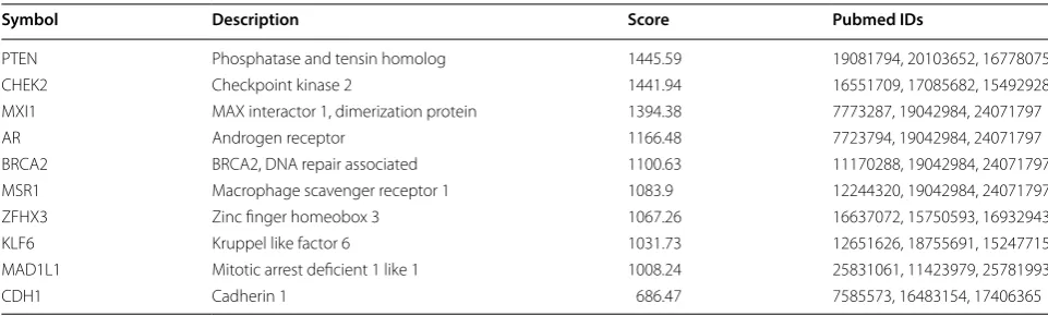
microarray datasets identified 667 DEGs in GSE45016, 33 DEGs in GSE55945, and 759 DEGs in GSE46602. Fig-ure 1a, b present the heat maps of 50 most significant DEGs in GSE46602 and GSE45016, respectively, and Fig. 1c is the heat map of all DEGs in GSE55945. A Venn diagram analysis was performed for the DEGs obtained from the three datasets to identify the overlapping DEGs (Fig. 1d). It was found that 8 genes were overlapping. Based on MalaCards database, 10 PCa-related known genes with highest scores were obtained (Table 2). In order to further identify the PCa-related genes among the DEGs, 8 obtained DEGs and 10 known PCa-related genes were used for correlation interaction analysis, and a gene interaction network map was plotted (Fig. 1e). The results revealed that the 10 known PCa-related genes were closely clustered and there was a replicating inter-action between them. Among the 8 DEGs, ZIC2 and HOXC6 genes not only interacted directly with AR in the
MSR1 CHEK2 PTEN BRCA2 MXI1 MAD1L1 KLF6 HOXC4 AR CYP3A5 DLX1 ABP1 ZIC2 HOXC6 CDH1 ZFHX3 GSM113315 0 GSM113315 1 GSM113316 3 GSM113316 6 GSM113317 6 GSM113317 7 GSM113317 8 GSM113317 9 GSM113318 0 GSM113318 1 GSM113318 2 GSM113318 3 GSM113318 4 GSM113318 5 GSM113313 6 GSM113313 7 GSM113313 8 GSM113313 9 GSM113314 0 GSM113314 1 GSM113314 2 GSM113314 3 GSM113314 4 GSM113314 5 GSM113314 6 GSM113314 7 GSM113314 8 GSM113314 9 GSM113315 2 GSM113315 3 GSM113315 4 GSM113315 5 GSM113315 6 GSM113315 7 GSM113315 8 GSM113315 9 GSM113316 0 GSM113316 1 GSM113316 2 GSM113316 4 GSM113316 5 GSM113316 7 GSM113316 8 GSM113316 9 GSM113317 0 GSM113317 1 GSM113317 2 GSM113317 3 GSM113317 4 GSM113317 5 VEGFA HPN GJB1 VWA5A FAM107A SNAI2 GGA2 PIK3R1 NTNG2 CPLX3 ZBTB20 NIPAL3 CD177 KRT15 KRT5 SLC14A1 CSTA ZIC2 DMKN SV2B LURAP1 TCF7L1 KRT14 AJUBA TP63 FAM83B VSNL1 MIR205HG TRIM29 AVPI1 COL4A6 FGFR2 MEG3 PPARGC1A AOX1 LINC00844 ZNF711 GPRC5B PGAP1 CYP3A5 FLRT3 TRAF3IP2 MYOF SLC18A2 SUPT3H MIR205 KRT23 LAMB3 LSAMP LOC100130232 type type Normal Tumor 4 6 8 10 12 GSM109587 6 GSM109587 7 GSM109587 8 GSM109587 9 GSM109588 0 GSM109588 1 GSM109588 2 GSM109588 3 GSM109588 4 GSM109588 5 GSM109588 6 FCN1 SULT1B1 FAM166B HIST1H4I DACT3 RAB39A COX6A2 CLDN9 LOC101929454 NKAIN2 MND1 LINC00326 ZNF280A PSTPIP1 PRNT LOC283435 CYP11A1 LOC100506119 CYTL1 BPIFB1 CYP3A7−CYP3AP1 DPEP3 SOSTDC1 EPHA5−AS1 COL9A1 GPR87 FLRT3 CYP3A5 ACSS3 ARHGEF28 WFDC2 SOCS3 KRT15 SLC14A1 FBXO17 KIF7 TP63 HOXC6 ZIC2 VWF TACC3 CCDC106 DUSP1 LGALS3 BCAS1 ATF3 NR4A1 ZFP36 JUNB EVA1C type type Normal Tumor 2 4 6 8 10 12 14 GSM134894 6 GSM134894 7 GSM134894 8 GSM134894 9 GSM134895 0 GSM134895 1 GSM134895 2 GSM134895 3 GSM134893 3 GSM134893 4 GSM134893 5 GSM134893 6 GSM134893 7 GSM134893 8 GSM134893 9 GSM134894 0 GSM134894 1 GSM134894 2 GSM134894 3 GSM134894 4 GSM134894 5 CRISP3 INSM1 KCNG3 ZIC2 FOXD1 PCA3 MS4A8 AMACR TMEM45B F5 MATR3 ACSM1 MMP26 HOXC6 HOXC4 DLX2 DLX1 KLK12 CTA−246H3.12 SLIT1 SLC45A2 LOC100996425 HPN SIM2 DNAH5 AOC1 PENK SLC2A5 LIX1 TMEM35 LINC00844 CYP3A5 CD177 type type Normal Tumor 4 6 8 10 12
a b c
590 7 516 143 18 0 8 GSE46602 GSE45016 GSE55945 108 16 0 2 1 3 5 2 0 0 0 5 0 5 2 1 35 2 23 12 3 1 0 0 0 0 0 3
1 1 4
Targ etSca
n miRDB
mi
rD
IP
miRNApath
starBase
d e f
Fig. 1 miR-129-5p influences PCa by regulating ZIC2 via the Wnt/β-catenin signaling pathway. a–c The heat maps depicting DEGs in PCa-related microarray datasets, with the X label indicating the sample number, the Y label indicating the gene name; the left-side dendrogram indicating gene expression clustering, with each small square in the figure representing the expression of one gene in one sample, and the histogram in the upper right representing the color gradation; d venn diagram analysis for DEGs in PCa-related microarray datasets, the 3 circles with different colors in the figure respectively represent the number of DEGs in the 3 PCa microarray datasets, the overlapping area indicates the intersection among the three microarray datasets; e gene interaction network map, each small ball in the figure denotes one gene, and the line between two small balls indicates an interaction; f predicted miRNAs that regulate ZIC2, and results from five databases are included. Five colors represent the prediction outcomes of five databases, the overlapping area indicates the intersecting results. PCa prostate cancer, ZIC2 zinc-finger protein of the cerebellum 2, DEGs
known genes, but also interacted with other DEGs. After further retrieving the literatures concerning ZIC2 and HOXC6 genes, it was found that the effect of HOXC6 on PCa had been reported in numerous studies [27–29]. However, the role of ZIC2 in PCa remained unclear. By retrieval of PCa-related signaling pathways, the Wnt/β-catenin signaling pathway was found to have a role in development of PCa [30, 31]. In order to understand the upstream regulatory mechanism of ZIC2 and predict the miRNAs that regulated ZIC2, five databases including TargetScan and miRDB were applied to obtain overlap-ping results (Fig. 1f). Finally, there were 5 miRNAs in the intersection of the five database predictions. After retrieval of the binding of these 5 miRNAs with ZIC2 in the microRNA.org database, miR-181a, miR-181b, and miR-181c were all found to bind to ZIC2 at the same location, and they were found to have multiple binding sites on ZIC2. The mirSVR score of miR-96-5p and ZIC2 was − 0.3957, whereas miR-129 only bound to ZIC2 at
180 locations with mirSVR score of − 0.7863. Hence,
miR-129 was selected for following study. Based on the above analysis and related reports, it was evident that miR-129-5p was likely to affect the development of PCa by targeting ZIC2 and regulating the Wnt/β-catenin sign-aling pathway.
PCa tissues show increased ZIC2 expression and activated Wnt/β‑catenin signaling pathway while decreasing miR‑129‑5p expression
RT-qPCR was used quantify the expression of miR-129-5p, ZIC2, Wnt, and β-catenin in adjacent normal tissues and PCa tissues, and the results (Fig. 2a) showed that compared with the control group, the expression of miR-129-5p in the PCa group was markedly decreased, but the mRNA expression of ZIC2, Wnt, and β-catenin was significantly increased (all p < 0.05). These results
suggested that PCa tissues showed poorly expressed miR-129-5p while highly expressed ZIC2, Wnt, and β-catenin. Immunohistochemistry was used to analyze the expres-sion of Wnt3a and β-catenin in PCa tissues (Fig. 2b, c), which revealed that the positive expression rates of Wnt3a and β-catenin in adjacent normal tissues were 13.33% and 36.67%, respectively, whereas these rates in the PCa tissues were 73.33% and 93.33%, respectively. The number of positive cells was obviously increased. All these results indicated that the positive expression rates of Wnt3a and β-catenin in the PCa tissues were signifi-cantly increased (all p < 0.05).
miR‑129‑5p targets ZIC2
Based on bioinformatic analysis, there was a specific binding between the ZIC2 gene sequence and the 129-5p sequence, and ZIC2 was a target gene of miR-129-5p (Fig. 3a). The dual-luciferase reporter gene assay was used to verify that ZIC2 was a target of miR-129-5p (Fig. 3b). The experimental results showed that the lucif-erase signal of ZIC2-Wt in the miR-129-5p mimic group was decreased (p < 0.05) as compared with the NC group, with no significant difference in the ZIC2-mut (p > 0.05). Therefore, miR-129-5p was demonstrated to specifically bind to ZIC2.
Overexpression of miR‑129‑5p dampens the Wnt/β‑catenin signaling pathway and EMT process by targeting ZIC2
miR-129-5p expression, mRNA and protein expression of ZIC2, Wnt, β-catenin, E-cadherin and vimentin as well as the extent of β-catenin phosphorylation in cells were evaluated by RT-qPCR (Fig. 4a) and Western blot analysis (Fig. 4b, c). And the results showed that the expression of miR-129-5p in the miR-129-5p mimic group increased (p < 0.05), with no significant difference of expression of miR-129-5p in the si-ZIC2 group (p > 0.05) when
Table 2 PCa-related genes
Symbol gene abbreviations, Description the gene description or gene full name, Score this score originates from Solr based GeneCards search engine score, obtained by querying the disease in GeneCards, Pubmed IDs PMID number of the related references, PCa prostate cancer
Symbol Description Score Pubmed IDs
PTEN Phosphatase and tensin homolog 1445.59 19081794, 20103652, 16778075 CHEK2 Checkpoint kinase 2 1441.94 16551709, 17085682, 15492928 MXI1 MAX interactor 1, dimerization protein 1394.38 7773287, 19042984, 24071797
AR Androgen receptor 1166.48 7723794, 19042984, 24071797
BRCA2 BRCA2, DNA repair associated 1100.63 11170288, 19042984, 24071797 MSR1 Macrophage scavenger receptor 1 1083.9 12244320, 19042984, 24071797 ZFHX3 Zinc finger homeobox 3 1067.26 16637072, 15750593, 16932943 KLF6 Kruppel like factor 6 1031.73 12651626, 18755691, 15247715 MAD1L1 Mitotic arrest deficient 1 like 1 1008.24 25831061, 11423979, 25781993
compared with the blank and NC groups. The expres-sion of ZIC2, Wnt, β-catenin and vimentin, as well as the extent of β-catenin phosphorylation were significantly decreased, while that of E-cadherin was significantly increased in the miR-129-5p mimic and si-ZIC2 groups (p < 0.05). Compared with the blank and NC groups, the miR-129-5p expression and expression of E-cadherin in the miR-129-5p inhibitor group were significantly lower, while the expression of ZIC2, Wnt, β-catenin and vimen-tin as well as the extent of β-catenin phosphorylation were significantly enhanced (p < 0.05). Relative to the blank and NC groups, the expression of miR-129-5p in the miR-129-5p inhibitor + si-ZIC2 group was markedly
decreased (p < 0.05), with no significant difference in the expression of ZIC2, Wnt, β-catenin, E-cadherin, and vimentin as well as the extent of β-catenin phosphoryla-tion (p > 0.05). Taken together, these results indicated that overexpressed miR-129-5p might hinder the activation of the Wnt/β-catenin signaling pathway by targeting ZIC2.
Inhibited proliferation of PCa cells is observed
after overexpression of miR‑129‑5p or down‑regulation of ZIC2 treatment
MTT assay was applied to investigate effects of miR-129-5 on the proliferation of PCa cells (Fig. 5a). There was no difference in OD value obtained at 24 h between
Wn
t
b
a c
β-cateni
n
Adjacent normal tissues PCa tissues
Positive rate (%
)
Wnt β-catenin
0 20 40 60 80
100 *
*
Adjacent normal tissues PCa tissues Adjacent normal tissues
PCa tissues
Relative expression
miR-129-5p ZIC2 Wnt β-catenin
0 1 2 3 4
* *
*
*
25μm 25μm
25μm 25μm
Fig. 2 miR-129-5p is poorly-expressed while ZIC2, Wnt3a and β-catenin are highly-expressed in PCa tissues. a The miR-129-5p expression and mRNA expression of ZIC2, Wnt3a and β-catenin in PCa tissues and adjacent normal tissues determined by RT-qPCR. Data are summarized as mean ± standard deviation and compared with paired t test (n = 60). b Immunohistochemical staining of Wnt3a and β-catenin in PCa tissues and adjacent normal tissues, in which brown or yellowish staining represents positive cells. c The statistical analysis of b. *p < 0.05 vs. the adjacent normal tissues. Positive rates are count data and were compared with the Chi square test
has-miR-129-5p/ZIC2 Alignment
3’ cgUUCGGGUCUGGCGUUUUUc 5’ hsa-miR-129-5p mirSVR score: -0.7863 180:5’ cuAGGCAUGAAGAGCAAAAAu 3’ ZIC2: :: PhastCons score: 0.5391 C
a
b
Luciferase activity
ZIC2-WT ZIC2-MUT
0.0 0.5 1.0 1.5
NC
miR-129-5p mimic
*
groups (p > 0.05). However, the OD values of the miR-129-5p mimic and si-ZIC2 groups at 48 h and 72 h were significantly decreased (p < 0.05), while those of the miR-129-5p inhibitor group were significantly increased (p < 0.05) as compared with the blank and NC groups. No difference in the OD values of the miR-129-5 inhibitor + si-ZIC2 group at 48 h and 72 h after transfection was observed (p > 0.05). Cell survival rate in all groups showed consistent trends (Fig. 5b). These results suggested the proliferation of PCa cells might be
inhibited by over-expressed miR-129-5p or down-regu-lated ZIC2.
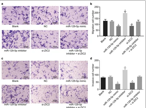
Cell migration and invasion PCa cells are repressed after overexpression of miR‑129‑5p or down‑regulation of ZIC2 treatment
Transwell assay was used to evaluate the effect of miR-129-5p on migration (Fig. 6a, b) and invasion (Fig. 6c, d) of PCa cells. Compared with the blank and NC groups, cell migration and invasion in the miR-129-5p mimic and
Relative expression
miR-129-5p
ZIC2 Wn
t
β-cateni n
E-Cadheri n
Vimentin
a Blank PC-3
NC
miR-129-5p mimic miR-129-5p inhibitor si-ZIC2
miR-129-5p inhibitor + si-ZIC2
Blank NC
miR-129-5p mimic miR-129-5p inhibitor si-ZIC2
miR-129-5p inhibitor + si-ZIC2
0.0 0.5 1.0 1.5 2.0 2.5
*
* * * * * * *
*
* * * * *
* * *
*
Blan
k NC
miR-129-5p mimicmiR-129-5p inhibito
r si-ZIC
2
miR-129-5p inhibitor + si-ZIC
2 ZIC2
Wnt
β-catenin
p-β-catenin
E-Cadherin
N-Cadherin
Vimentin
GAPDH
c b
Relative protein expressio
n
ZIC2 Wn
t
β-catenin p-β-cateni n
E-Cadheri n
N-Cadheri n
Vimentin 0.0
0.5 1.0 1.5 2.0
PC-3
*
* *
*
* *
* *
* *
* * * *
* * * *
* *
*
Fig. 4 Overexpression of miR-129-5p suppresses the activation of ZIC2-dependent Wnt/β-catenin signaling pathway and EMT in PCa. DU-145 cells were treated with miR-129-5p mimic, miR-129-5p inhibitor or/and si-ZIC2. a The miR-129-5p expression and the mRNA expression of ZIC2, Wnt3a, β-catenin, E-cadherin and vimentin in DU-145 cells determined by RT-qPCR. b, c The protein expression of ZIC2, Wnt3a, β-catenin, E-cadherin and vimentin, and extent of β-catenin phosphorylation in DU-145 cells determined by western blot analysis; *p < 0.05 vs. the blank and NC groups; the experimental data are summarized as mean ± standard deviation; the one-way analysis of variance (ANOVA) was employed to analyze data among groups. This experiment was repeated three times
OD value (570 nm
)
24 h 48 h 72 h 0.0
0.5 1.0 1.5 2.0
k n a l B k
n a l B NC
miR-129-5p mimic miR-129-5p inhibitor si-ZIC2
miR-129-5p inhibitor + si-ZIC2
NC
miR-129-5p mimic miR-129-5p inhibitor si-ZIC2
miR-129-5p inhibitor + si-ZIC2
* * *
*
* *
Cell survival rate %
0 50 100 150
*
* *
a b
si-ZIC2 groups were significantly decreased (all p < 0.05) while those in the miR-129-5p inhibitor group were sig-nificantly elevated (all p < 0.05). No significant difference in cell migration and invasion in the miR-129-5p inhibi-tor + si-ZIC2 group was observed (p > 0.05). The results indicated that cell migration and invasion of PCa was attenuated by over-expression of miR-129-5p or down-regulation of ZIC2.
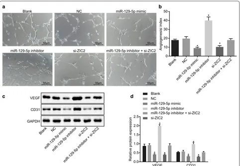
Repressed angiogenesis of PCa cells is attributed to overexpressed miR‑129‑5p or down‑regulated ZIC2
Matrigel assay was used to analyze the impact of miR-129-5p expression on angiogenesis of PCa cells, and the results (Fig. 7a, b) showed that, the miR-129-5p mimic and si-ZIC2 groups showed significantly decreased angiogenesis (p < 0.05), while miR-129-5p inhibitor group showed significantly enhanced angiogenesis
(p < 0.05) when compared to the blank and NC groups. No significant difference in angiogenesis was found in the miR-129-5p inhibitor + si-ZIC2 group (p > 0.05).
Western blot analysis was used to analyze the expres-sion of VEGF and CD31 in PCa cells (Fig. 7c, d). The protein expression of VEGF and CD31 in the miR-129-5p mimic and si-ZIC2 groups was found to be significantly decreased (p < 0.05), while that in the miR-129-5p inhibitor group was significantly higher (p < 0.05) as compared with the blank and NC groups. No significant difference, however, was found in the protein expression of VEGF and CD31 in the miR-129-5p inhibitor + si-ZIC2 group (p > 0.05). In sum-mary, these findings indicated that overexpressed miR-129-5p or down-regulated ZIC2 suppressed angio-genesis in PCa cells.
c
C N k
n a l B
miR-129-5p inhibitor si-ZIC2
miR-129-5p mimic
miR-129-5p inhibitor + si-ZIC2
C N k
n a l B
miR-129-5p inhibitor si-ZIC2
miR-129-5p mimic
miR-129-5p inhibitor + si-ZIC2
d
Migration cell numbe
r
0 50 100 150 250
200
Blank NC
miR-129-5p mimi c
miR-129-5p inhibito r
si-ZIC 2
miR-129-5p inhibitor + si-ZIC 2
Invasive cell number
0 50 100 150 200
*
* *
Blank NC
miR-129-5p mimi c
miR-129-5p inhibitor si-ZIC
2
miR-129-5p inhibitor + si-ZIC 2
a b
50μm 50μm 50μm
50μm 50μm 50μm
50μm 50μm 50μm
50μm 50μm 50μm
* *
*
Overexpressed miR‑129‑5p or down‑regulated ZIC2 leads to PCa cell apoptosis
Flow cytometry was used to examine the effect of miR-129-5p on apoptosis of PCa cells (Fig. 8a, b). The apop-tosis rate of the miR-129-5p mimic and si-ZIC2 groups was significantly potentiated (p < 0.05), while that of the miR-129-5p inhibitor group was significantly diminished (p < 0.05), as compared with the blank and NC groups. No statistically significant difference was evident in the apoptosis rate of the miR-129-5p inhibitor + si-ZIC2 group (p > 0.05). These results showed overexpressed miR-129-5p or down-regulated ZIC2 might promote the apoptosis of PCa cells.
miR‑129‑5p overexpression or ZIC2 silencing promotes cell tumorigenesis in PCa
Finally, we examined the function of miR-129-5p over-expression or ZIC2 silencing on tumorigenesis. As shown in Fig. 9, both the tumor volume and weight were
significantly lower in the miR-129-5p mimic and si-ZIC2 groups than that in the blank and NC groups (p < 0.05). Conversely, the tumor volume and weight were signifi-cantly higher in the miR-129-5p inhibitor group than that in the blank and NC groups (p < 0.05). These data illus-trated that ZIC2 served as an oncogene in PCa and miR-129-5p played a tumor-suppressive role.
Discussion
PCa is one of the most prevalent carcinomas among men, resulting in a high number of cancer-related deaths [32]. miRNAs have been implicated in biological processes such as cell proliferation, differentiation, development, apoptosis, and metabolism, and their alterations have been found in various cancers and participate in patho-genesis of cancers [33, 34]. Importantly, miR-129-5p has been demonstrated to express aberrantly in primary tumors of human PCa and prostate control specimens [35]. Therefore, this study investigated the function of C
N k
n a l B
miR-129-5p inhibitor si-ZIC2
miR-129-5p mimic
miR-129-5p inhibitor + si-ZIC2
a b
d c
Angiogenesisi index
0
10 20 30 40 50
Blan
k NC
miR-129-5p mimi c
miR-129-5p inhibitor si-ZIC
2
miR-129-5p inhibitor + si-ZIC2 *
* *
50μm 50μm 50μm
50μm 50μm 50μm
Relative protein expression
VEGF CD31
VEGF
0.0 0.5 1.0 1.5 2.0 2.5
*
* * * *
*
Blan
k NC
miR-129-5p mimi c
miR-129-5p inhibito r
si-ZIC 2
miR-129-5p inhibitor + si-ZIC 2
CD31
GAPDH
Blank NC
miR-129-5p mimic miR-129-5p inhibitor
si-ZIC2
miR-129-5p inhibitor + si-ZIC2
miR-129-5p in PCa, and it was found that up-regulated miR-129-5p could inhibit EMT and angiogenesis in PCa.
Initially, the results obtained from present study revealed that up-regulation of miR-129-5p could attenu-ate EMT and angiogenesis of PCa. A previous study reported that miR-129-5p modulated EMT in tubu-lar epithelial cells by targeting the gene PDPK1 [36]. β-Catenin, p-β-catenin, N-cadherin and vimentin are well established as indicators of EMT, and accordingly, the poor expression of β-catenin in chondrosarcoma
cells of the miR-129-5p group has been observed, which further suppressed the cell proliferation, migration and promoted apoptosis [21]. Similarly, over-expression of miR-129-5p in the breast cancer cell line MCF-7 was found to significantly induce E-cadherin and suppress N-cadherin and vimentin expression [37]. Corroborat-ing these findCorroborat-ings, we noted that poor expression of β-catenin, N-cadherin, and vimentin and high expression of E-cadherin were markers of PCa inhibition. Addition-ally, up-regulation miR-195 has been found to reduce
a Blank b
miR-129-5p inhibitor si-ZIC2
104
miR-129-5p mimic
miR-129-5p inhibitor + si-ZIC2
Apoptosis rate (%
)
Blank NC
miR-129-5p mimi c
miR-129-5p inhibito r
si-ZIC 2
miR-129-5p inhibitor + si-ZIC 2 0
10 20 30 40
*
* *
NC
PI
100 101 102 103 104
10
0
10
1
10
2
10
3
10
4
FITC
10
0
10
1
10
2
10
3
PI
10
4
100 101 102 103 104 FITC
PI
104 100 101 102 103
10
1
10
0
10
2
10
3
10
4
FITC
PI
10
1
10
0
10
2
10
3
104 100 101 102 103
10
4
FITC
PI
100 101 102 103
10
1
10
0
10
2
10
3
10
4
FITC
PI
100 101 102 103 104
10
1
10
0
10
2
10
3
10
4
FITC
Fig. 8 Overexpressed miR-129-5p or down-regulated ZIC2 plays a positive role in apoptosis of PCa cells. DU-145 cells were treated with miR-129-5p mimic, miR-129-5p inhibitor or/and si-ZIC2. a The apoptosis in each group; b the apoptosis rate in each group. *p < 0.05 vs. the blank and NC groups. The data are summarized as mean ± standard deviation and the one-way analysis of variance was used for comparison. This experiment was performed three times
Tumor volume (mm
3)
7 d 14 d 21 d 28 d 35 d 0
500 1000 1500 2000
* * *
*
* *
*
* *
Tumor weight (g
)
0.0 0.5 1.0 1.5 2.0
*
* *
b a Blank
NC
miR-129-5p mimic miR-129-5p inhibitor si-ZIC2
miR-129-5p inhibitor + si-ZIC2
Blank NC
miR-129-5p mimic miR-129-5p inhibitor si-ZIC2
miR-129-5p inhibitor + si-ZIC2 Blank NC miR-129-5p mimic
miR-129-5p inhibitor si-ZIC2 miR-129-5p inhibitor + si-ZIC2
VEGF level by blocking VEGF receptor 2 signaling in endothelial cells and consequently inhibiting angiogen-esis, consistent with our findings [38].
Additionally, miR-129 might play a role in inhibition of viability, proliferation, migration and invasion of PCa cells via directly suppressing E26 transformation spe-cific-1 (ETS1), which was also provided new train of thought for us to popularize the carcinogenesis of PCa [39]. In laryngeal cancer, miR-129-5p can mediate cell proliferation, invasiveness, and migration by suppressing the expression of STAT3 [40]. Another study suggested that miR-129-5p mediates FNDC3B to suppress pro-liferation, migration and invasion of glioblastoma cells U87 cells [41]. Overexpression of miR-129-5p alone has been found sufficient to promote apoptosis [42]. Consist-ent with these findings, the currConsist-ent study demonstrated that up-regulated miR-129-5p could inhibit proliferation, migration, and invasion while promoting apoptosis of PCa cells.
Here, we showed that an operant mechanism of miR-129-5p in PCa involves impairment of the Wnt/β-catenin signaling pathway via down-regulation of ZIC2. The Wnt/β-catenin signaling pathway, central to tis-sue development in embryos and tistis-sue maintenance in adults, is a major up-regulated signaling pathway in cas-tration-resistant PCa [43]. Vimentin and E-cadherin are established as Wnt/β-catenin signaling pathway-related factors [44]. The Wnt/β-catenin signaling pathway is
blocked upon down-regulation of the EMT marker vimentin [45]. The up-regulated E-cadherin expression in microwells was found following with a downregulation of the Wnt signaling pathway and the deficiency of nuclear β-catenin as well [46]. Consistent with former results, our data indicated over-expressed miR-129-5p and down-regulated ZIC2 reduced the expression of Wnt, β-catenin and vimentin, but restored the expression of E-cadherin, which further indicated the inhibitory role of over-expressed miR-129-5p or down-regulated ZIC2 on the Wnt/β-catenin signaling pathway.
Conclusion
In a conclusion, elevated miR-129-5p was found to block the activation of the Wnt/β-catenin signaling pathway in PCa, consequently inhibiting EMT and angiogenesis via targeting ZIC2 (Fig. 10). miR-129-5p can be considered as a new therapeutic target for PCa therapy. The down-regulation of miR-129-5p can promote ZIC2 expres-sion via activating the Wnt/β-catenin signaling pathway, and further enhanced the expression of Wnt, β-catenin, N-cadherin and vimentin and inhibited the expression of E-cadherin, thus resulting in cancer cell proliferation, invasion, migration along with EMT and angiogenesis and reduced apoptosis. Overexpression of miR-129-5p may reverse these events, thus limiting the growth of PCa. However, further studies with different disease models and larger cohorts are essential to validate these
miR-129-5p
Wnt β-catenin N-cadherin Vimentin
E-cadherin
Apoptosis Proliferation Migration Invasion EMT Prostate cancer
ZIC2
findings and expand the translational potential of this direction.
Abbreviations
PCa: prostate cancer; VEGF: vascular endothelial growth factor; EMT: epithe-lial–mesenchymal transition; ZIC: zinc-finger protein of the cerebellum; OPA: odd-paired; GS: Gleason score.
Acknowledgements
The authors would like to extend their sincere gratitude to the reviewers.
Authors’ contributions
ZJ, YZ and XC designed the study. DC collated the data, YZ and ZJ carried out data analyses and produced the initial draft of the manuscript. YZ, PW and DC contributed to drafting the manuscript. All authors read and approved the final manuscript.
Funding
This study was supported by Prostate Cancer Foundation China (PCF China) Young Investigator Award, National Natural Science Foundation Youth Fund (No. 81001143), Shenyang Science and Technology Program Key Science and Technology Research and Development Program (No. 17-230-9-18), the National Key Research and Development Program of China (No. 2017YFC0908003), the First Batch of Talents of Introduction of Top Health Talented Team of Qinghai Province and the First Batch of Talents of Xining “Absorbing 555 Talents Project”.
Availability of data and materials
The datasets generated/analyzed during the current study are available.
Ethics approval and consent to participate
The study was approved by the Institutional Review Board of the First Hospital of China Medical University. Written informed consents were obtained from all patients or their guardians. All study procedures were conducted in accord-ance with the Declaration of Helsinki. All animal experiments were conducted under the approval of guidelines for the protection and use of experimental animals issued by the National Institutes of Health (NIH), and strictly complied with the principles of completing the experiments with the minimum number of animals and minimizing pain.
Consent for publication
Consent for publication was obtained from the participants.
Competing interests
The authors declare that they have no competing interests.
Author details
1 Department of Urology, The First Hospital of China Medical University, No.
155, Nanjing North Street, Heping District, Shenyang 110001, Liaoning, Peo-ple’s Republic of China. 2 Department of Urology, People’s Hospital of Datong
Hui and Tu Autonomous County, No. 1, Wenhua Road, Qiaotou Town, Datong Hui and Tu Autonomous County, Xining 810100, Qinghai, People’s Republic of China. 3 Department of Pharmacy, The First Hospital of China Medical
University, Shenyang 110001, People’s Republic of China. 4 Central Lab, The
First Hospital of China Medical University, Shenyang 110001, People’s Republic of China.
Received: 7 March 2019 Accepted: 23 September 2019
References
1. Perner S, Cronauer MV, Schrader AJ, Klocker H, Culig Z, Baniahmad A. Adaptive responses of androgen receptor signaling in castration-resistant prostate cancer. Oncotarget. 2015;6(34):35542–55.
2. El-Haibi CP, Singh R, Gupta P, Sharma PK, Greenleaf KN, Singh S, Lillard JW Jr. Antibody microarray analysis of signaling networks regulated
by Cxcl13 and Cxcr5 in prostate cancer. J Proteomics Bioinform. 2012;5(8):177–84.
3. Jaiswal S, Sarmad R, Arora S, Dasaraju R, Sarmad K. Prostate cancer for the internist. N Am J Med Sci. 2015;7(10):429–35.
4. Loberg RD, Logothetis CJ, Keller ET, Pienta KJ. Pathogenesis and treat-ment of prostate cancer bone metastases: targeting the lethal pheno-type. J Clin Oncol. 2005;23(32):8232–41.
5. Grubb RL 3rd, Kibel AS. Prostate cancer: screening, diagnosis and man-agement in 2007. Mo Med. 2007;104(5):408–13 (quiz 13–4). 6. Barlow LJ, Shen MM. SnapShot: prostate cancer. Cancer Cell.
2013;24(3):400.
7. Zhang J, Ma L. MicroRNA control of epithelial–mesenchymal transition and metastasis. Cancer Metastasis Rev. 2012;31(3–4):653–62. 8. Lamouille S, Subramanyam D, Blelloch R, Derynck R. Regulation of
epithelial–mesenchymal and mesenchymal–epithelial transitions by microRNAs. Curr Opin Cell Biol. 2013;25(2):200–7.
9. Zhang J, Kuang Y, Wang Y, Xu Q, Ren Q. Notch-4 silencing inhibits prostate cancer growth and EMT via the NF-kappaB pathway. Apoptosis. 2017;22(6):877–84.
10. Mukherji D, Temraz S, Wehbe D, Shamseddine A. Angiogenesis and anti-angiogenic therapy in prostate cancer. Crit Rev Oncol Hematol. 2013;87(2):122–31.
11. Lin S, Gregory RI. MicroRNA biogenesis pathways in cancer. Nat Rev Cancer. 2015;15(6):321–33.
12. Pereira DM, Rodrigues PM, Borralho PM, Rodrigues CM. Deliver-ing the promise of miRNA cancer therapeutics. Drug Discov Today. 2013;18(5–6):282–9.
13. Yu Y, Zhao Y, Sun XH, Ge J, Zhang B, Wang X, Cao XC. Down-regulation of miR-129-5p via the Twist1-Snail feedback loop stimulates the epithelial– mesenchymal transition and is associated with poor prognosis in breast cancer. Oncotarget. 2015;6(33):34423–36.
14. Xu S, Yi XM, Zhang ZY, Ge JP, Zhou WQ. miR-129 predicts prognosis and inhibits cell growth in human prostate carcinoma. Mol Med Rep. 2016;14(6):5025–32.
15. Luo Z, Gao X, Lin C, Smith ER, Marshall SA, Swanson SK, Florens L, Wash-burn MP, Shilatifard A. Zic2 is an enhancer-binding factor required for embryonic stem cell specification. Mol Cell. 2015;57(4):685–94. 16. Ma G, Dai W, Sang A, Yang X, Li Q. Roles of ZIC family genes in human
gastric cancer. Int J Mol Med. 2016;38(1):259–66.
17. Hoogland AM, Bottcher R, Verhoef E, Jenster G, van Leenders GJ. Gene-expression analysis of gleason grade 3 tumor glands embedded in low- and high-risk prostate cancer. Oncotarget. 2016;7(25):37846–56. 18. Zaravinos A. The regulatory role of microRNAs in EMT and cancer. J Oncol.
2015;2015:865816.
19. Wang S, Chen Y, Yu X, Lu Y, Wang H, Wu F, Teng L. miR-129-5p attenuates cell proliferation and epithelial mesenchymal transition via HMGB1 in gastric cancer. Pathol Res Pract. 2019;215(4):676–82.
20. Kafka A, Basic-Kinda S, Pecina-Slaus N. The cellular story of dishevelleds. Croat Med J. 2014;55(5):459–67.
21. Zhang P, Li J, Song Y, Wang X. MiR-129-5p inhibits proliferation and invasion of chondrosarcoma cells by regulating SOX4/Wnt/beta-catenin signaling pathway. Cell Physiol Biochem. 2017;42(1):242–53.
22. Pasoglou V, Larbi A, Collette L, Annet L, Jamar F, Machiels JP, Michoux N, Vande Berg BC, Tombal B, Lecouvet FE. One-step TNM staging of high-risk prostate cancer using magnetic resonance imaging (MRI): toward an upfront simplified “all-in-one” imaging approach? Prostate. 2014;74(5):469–77.
23. Zhong H, De Marzo AM, Laughner E, Lim M, Hilton DA, Zagzag D, Buechler P, Isaacs WB, Semenza GL, Simons JW. Overexpression of hypoxia-inducible factor 1alpha in common human cancers and their metastases. Cancer Res. 1999;59(22):5830–5.
24. Yu H, Li X, Sun S, Gao X, Zhou D. c-Met inhibitor SU11274 enhances the response of the prostate cancer cell line DU145 to ionizing radiation. Biochem Biophys Res Commun. 2012;427(3):659–65.
25. Ayuk SM, Abrahamse H, Houreld NN. The role of photobiomodulation on gene expression of cell adhesion molecules in diabetic wounded fibroblasts in vitro. J Photochem Photobiol B. 2016;161:368–74. 26. Arunkumar R, Sharmila G, Elumalai P, Senthilkumar K, Banudevi S,
•fast, convenient online submission
•
thorough peer review by experienced researchers in your field
• rapid publication on acceptance
• support for research data, including large and complex data types
•
gold Open Access which fosters wider collaboration and increased citations maximum visibility for your research: over 100M website views per year
•
At BMC, research is always in progress.
Learn more biomedcentral.com/submissions
Ready to submit your research? Choose BMC and benefit from:
and proliferation of human prostate cancer cells in vitro and in silico approach through docking analysis. Phytomedicine. 2012;19(10):912–23. 27. McCabe CD, Spyropoulos DD, Martin D, Moreno CS. Genome-wide
analysis of the homeobox C6 transcriptional network in prostate cancer. Cancer Res. 2008;68(6):1988–96.
28. Waltregny D, Alami Y, Clausse N, de Leval J, Castronovo V. Overexpression of the homeobox gene HOXC8 in human prostate cancer correlates with loss of tumor differentiation. Prostate. 2002;50(3):162–9.
29. Vinarskaja A, Yamanaka M, Ingenwerth M, Schulz WA. DNA meth-ylation and the HOXC6 paradox in prostate cancer. Cancers (Basel). 2011;3(4):3714–25.
30. Weirick T, Militello G, Ponomareva Y, John D, Doring C, Dimmeler S, Uchida S. Logic programming to infer complex RNA expression patterns from RNA-seq data. Brief Bioinform. 2018;19(2):199–209.
31. Wang D, Lu G, Shao Y, Xu D. MiR-182 promotes prostate cancer progres-sion through activating Wnt/beta-catenin signal pathway. Biomed Pharmacother. 2018;99:334–9.
32. Quinn DI, Shore ND, Egawa S, Gerritsen WR, Fizazi K. Immunotherapy for castration-resistant prostate cancer: progress and new paradigms. Urol Oncol. 2015;33(5):245–60.
33. Deng K, Wang H, Guo X, Xia J. The cross talk between long, non-coding RNAs and microRNAs in gastric cancer. Acta Biochim Biophys Sin (Shang-hai). 2016;48(2):111–6.
34. Li Y, Deng X, Zeng X, Peng X. The Role of Mir-148a in Cancer. J Cancer. 2016;7(10):1233–41.
35. Valentino A, Calarco A, Di Salle A, Finicelli M, Crispi S, Calogero RA, Riccardo F, Sciarra A, Gentilucci A, Galderisi U, Margarucci S, Peluso G. Deregulation of MicroRNAs mediated control of carnitine cycle in prostate cancer: molecular basis and pathophysiological consequences. Oncogene. 2017;36(43):6030–40.
36. Li Y, An H, Pang J, Huang L, Li J, Liu L. MicroRNA profiling identifies miR-129-5p as a regulator of EMT in tubular epithelial cells. Int J Clin Exp Med. 2015;8(11):20610–6.
37. Luan QX, Zhang BG, Li XJ, Guo MY. MiR-129-5p is downregulated in breast cancer cells partly due to promoter H3K27m3 modification and regulates epithelial–mesenchymal transition and multi-drug resistance. Eur Rev Med Pharmacol Sci. 2016;20(20):4257–65.
38. Wang R, Zhao N, Li S, Fang JH, Chen MX, Yang J, Jia WH, Yuan Y, Zhuang SM. MicroRNA-195 suppresses angiogenesis and metastasis of
hepatocellular carcinoma by inhibiting the expression of VEGF, VAV2, and CDC42. Hepatology. 2013;58(2):642–53.
39. Xu S, Ge J, Zhang Z, Zhou W. MiR-129 inhibits cell proliferation and metastasis by targeting ETS1 via PI3K/AKT/mTOR pathway in prostate cancer. Biomed Pharmacother. 2017;96:634–41.
40. Shen N, Huang X, Li J. Upregulation of miR-129-5p affects laryngeal cancer cell proliferation, invasiveness, and migration by affecting STAT3 expression. Tumour Biol. 2016;37(2):1789–96.
41. Xu H, Hu Y, Qiu W. Potential mechanisms of microRNA-129-5p in inhibit-ing cell processes includinhibit-ing viability, proliferation, migration and inva-siveness of glioblastoma cells U87 through targeting FNDC3B. Biomed Pharmacother. 2017;87:405–11.
42. Brest P, Lassalle S, Hofman V, Bordone O, Gavric Tanga V, Bonnetaud C, Moreilhon C, Rios G, Santini J, Barbry P, Svanborg C, Mograbi B, Mari B, Hofman P. MiR-129-5p is required for histone deacetylase inhibitor-induced cell death in thyroid cancer cells. Endocr Relat Cancer. 2011;18(6):711–9.
43. Liu KX, Edwards B, Lee S, Finelli MJ, Davies B, Davies KE, Oliver PL. Neuron-specific antioxidant OXR1 extends survival of a mouse model of amyotrophic lateral sclerosis. Brain. 2015;138(Pt 5):1167–81.
44. Qiao B, He BX, Cai JH, Tao Q, King-Yin Lam A. MicroRNA-27a-3p modulates the Wnt/beta-catenin signaling pathway to promote epithelial-mesen-chymal transition in oral squamous carcinoma stem cells by targeting SFRP1. Sci Rep. 2017;7:44688.
45. Wang YP, Guo PT, Zhu Z, Zhang H, Xu Y, Chen YZ, Liu F, Ma SP. Pleomor-phic adenoma gene like-2 induces epithelial–mesenchymal transition via Wnt/beta-catenin signaling pathway in human colorectal adenocarci-noma. Oncol Rep. 2017;37(4):1961–70.
46. Azarin SM, Lian X, Larson EA, Popelka HM, de Pablo JJ, Palecek SP. Modula-tion of Wnt/beta-catenin signaling in human embryonic stem cells using a 3-D microwell array. Biomaterials. 2012;33(7):2041–9.
Publisher’s Note