R E S E A R C H
Open Access
flam
piRNA precursors channel from the
nucleus to the cytoplasm in a temporally
regulated manner along
Drosophila
oogenesis
Cynthia Dennis, Emilie Brasset and Chantal Vaury
*Abstract
Background:PIWI-interacting RNAs (piRNAs) are the effectors of transposable element silencing in the reproductive apparatus. InDrosophilaovarian somatic cells, piRNAs arise from long RNA precursors presumably processed within cytoplasmic Yb-bodies.
Results:Here we show that the nucleo-cytoplasmic traffic of piRNA precursors encoded by theflamencolocus is subjected to a spatio-temporal regulation. Precursor RNAs first gather in a single nuclear focus, Dot COM, close to the nuclear periphery, and transit through the membrane before being delivered to the cytoplasmic Yb-bodies. Early in oogenesis, flamenco transcripts are rapidly transferred to the cytoplasm making their initial nuclear gathering in Dot COM too transient to be visualized. As oogenesis proceeds, the cytoplasmic delivery steadily decreases concomitantly with the decrease in the protein levels of Armi and Yb, two components of the Yb-bodies. Both events lead to a reduction of Yb-body assembly in late stages of oogenesis, which likely results in a drop in piRNA production.
Conclusion:Our findings show a spatio-temporal regulation of the piRNA biogenesis in the follicle cells of
Drosophila ovaries, that involves coordinated control of both piRNA precursors and components of the piRNA
processing machinery. This newly unveiled regulation establishes another level of complexity in the production of piRNAs and suggests a stage-dependent involvement of the piRNA biogenesis in the mechanism of transposable elements silencing along oogenesis.
Keywords:Transposable elements, Silencing, piRNAs,Flamenco, Dot COM, Yb-body, Drosophila, Oogenesis
Background
Eukaryotic genomes are composed of a variable propor-tion of transposable elements (TEs) accumulated through-out evolution. These sequences are silenced by the host to protect itself and its progeny against potentially deleteri-ous mutations and genome invasion. In the gonads, where it is essential to ensure the maintenance of genome integ-rity for the next generation, the piRNA pathway is respon-sible for TE silencing in both somatic and germinal tissues [1–4]. This process involves small guide piRNAs of 23–29
nucleotides (nts) that originate from discrete genomic re-gions called piRNA clusters.
142 piRNA clusters have been identified inDrosophila
melanogaster [2], mostly located in pericentromeric and telomeric regions. These clusters vary considerably in size from a few kilobases (kb) to several hundred kb. They are enriched in full length or truncated TEs that are often nested within one another [2, 5]. In ovarian somatic sup-port cells surrounding the germline, piRNAs are mainly produced from two piRNA clusters:traffic jam[6] and
fla-menco(flam) [2,7]. Theflamcluster is located at the peri-centromeric region of the X-chromosome, spans over more than 200 kb and is strongly enriched in both ancient and recent retrotransposons mostly inserted in an
© The Author(s). 2019Open AccessThis article is distributed under the terms of the Creative Commons Attribution 4.0 International License (http://creativecommons.org/licenses/by/4.0/), which permits unrestricted use, distribution, and reproduction in any medium, provided you give appropriate credit to the original author(s) and the source, provide a link to the Creative Commons license, and indicate if changes were made. The Creative Commons Public Domain Dedication waiver (http://creativecommons.org/publicdomain/zero/1.0/) applies to the data made available in this article, unless otherwise stated. * Correspondence:chantal.vaury@uca.fr
orientation antisense to the TE transcription [5]. flam is transcribed from a polymerase II promoter as a long single-stranded RNA that is a substrate for piRNA biogen-esis.Flam transcripts undergo alternative splicing to gen-erate diverse piRNA precursors that all share the first exon at their 5′end [8]. These transcripts are then proc-essed into 23–29 nt piRNAs, presumably in cytoplasmic Yb-bodies [9,10]. Mature piRNAs associated with Piwi pro-tein form a piRNA-induced silencing complex (piRISC) that is delivered to the nucleus to target nascent TE mRNAs and initiate transcriptional gene silencing [11,12].
Two studies have reported that flam precursor tran-scripts, together with transcripts from other piRNA clus-ters, concentrate in 1 to 2 foci in ovarian follicle cells [13,14]. Analysis of the subcellular localization of these sites of accumulation at different stages of development have yielded varying findings. The first study focused on follicle cells in late stages of oogenesis and showed that
flam RNA precursors accumulate in a single nuclear substructure, named Dot COM, that faces the cytoplas-mic Yb-body to which theflamprecursors are channeled by nucleo-cytoplasmic transfer [13]. A subsequent re-port [14] visualized flam transcripts concentrated within the cytoplasm in 1 to 2 substructures named
flam bodies, located close to Yb-bodies. The authors worked mainly with a cultured Drosophila ovarian somatic stem cell line (OSS cells) derived from a somatic stem cell population of the germarium [15] that expresses a functional piRNA pathway [6].
We show here that flam transcripts are channeled to the cytoplasm in a temporally regulated manner. In early stages of oogenesis, they are mainly detected within the cytoplasm. As oogenesis proceeds,flamtranscripts accu-mulate in a focus detected within the nucleus or within the nuclear membrane as though their delivery to the cytoplasm was impeded. Combined with a drop in Armi and Yb protein levels after stage 8, this affects the as-sembly of Yb-bodies which then are very small and ab-sent from most cells. These findings emphasize the temporal regulation of Yb-body assembly, which re-quires both cytoplasmicflamdelivery and Armi and Yb proteins. In their absence, Yb-bodies fail to assemble correctly, which potentially can cause a decrease in the production of piRNAs.
Results and discussion
The cytoplasmic transfer offlamprecursor transcripts decreases during ovariole development
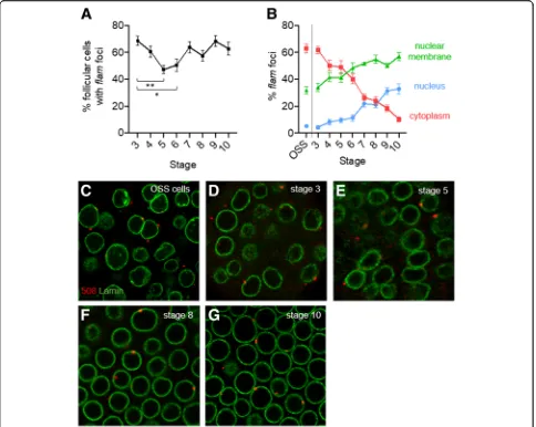
In a previous study [16], we performed a global quantita-tive analysis of the subcellular localization of flam tran-scripts within ovarian somatic follicle cells, independently of the developmental stages of oogenesis. Depending on the follicle cell observed, the focus whereflamprecursors gathered was visualized either completely within the
nucleus close to the nuclear periphery, or stretching across the nuclear membrane, or within the cytoplasm close to the nuclear membrane. To investigate whether this differential localization is somehow related to the stage of the developing egg chambers, we examinedflam RNA precursors in follicle cells within egg chambers in stages 3 to 10 of oogenesis. To do so, we performed an immuno-RNA FISH experiment on WTDrosophila ovar-ies using an anti-lamin antibody to label the nuclear mem-brane, and a specific flam RNA probe described in [13]. The same experiment was performed in parallel with OSS cells. The localization of flam foci was quantified as fol-lows. When positioned close to the inner side or outer side of the lamin signal, foci were considered as nuclear within Dot COM or cytoplasmic within the Yb-bodies re-spectively. When co-localized with the lamin signal (to-tally or partially), the flam focus was considered as stretching across the nuclear membrane.
Firstly we investigated egg chambers from stage 3 to 10 of oogenesis and quantified the proportion of cells in which a flam signal could be visualized (Fig. 1a). We found no significant difference between stages 3, 4 and 7–10 where flamfoci are detected in more than 60% of cells. This proportion appears slightly lower around 50% at stages 5 and 6 (Fig.1a).
Secondly, focusing on the follicle cells that harbored a
flam signal, we investigated its subcellular localization. We found thatflamtranscripts that accumulated mostly in a single focus, vary drastically in localization from early to late stages of oogenesis. In early stages, in a ma-jority of follicle cells, 1 to 2 flam foci are observed within the cytoplasm (Fig.1b, d, e). OSS cells, which de-rive from a somatic stem cell population of the germar-ium, and follicle cells from early stage 3, display a similar percentage of flam foci within the cytoplasm (around 62%) (Fig. 1b & c). This similarity may be ex-plained by the intrinsic nature of these two cell types; both are highly dividing cells required to rapidly encap-sulate the 16-cell germinal cyst. The percentage of cells with cytoplasmic flam foci decreases progressively to 10% at stage 10 of oogenesis. Concomitantly, the propor-tion of follicle cells in which a flam focus is detected within Dot COM increases, from 4% in stage 3 to 32% in stage 10 (Fig.1b-g). The switch from a cytoplasmic to a nuclear position is progressive, suggesting that the ex-port of piRNA precursors to the cytoplasm steadily de-creases as oogenesis proceeds. This stage-by-stage analysis further emphasizes that an increasing fraction of
flamRNAs co-localizes with the lamin staining. Interest-ingly, in late stages of oogenesis, theflamtranscripts are even more likely to be detected spanning the nuclear membrane (57%) than within the nucleus (33%) and the cytoplasm (10%) (Fig.1b). These data indicate that flam precursors can enter the nuclear membrane throughout
oogenesis even in late stages but seem to lose progres-sively the ability to be delivered to the cytoplasm. This suggests that fewer piRNA precursors reach the Yb-bodies, thereby giving rise to fewer piRNAs in late stages of oogenenesis.
At early stages of Drosophila oogenesis,flamprecursors rapidly transit to the cytoplasm
To better understand the discrepancy between flam localization in early and late stages of oogenesis, we first considered the early stages of oogenesis and posed the question of why no nuclear accumulation is observed. The nuclear gathering in Dot COM could be specific to later stages of oogenesis and not occur in early stages.
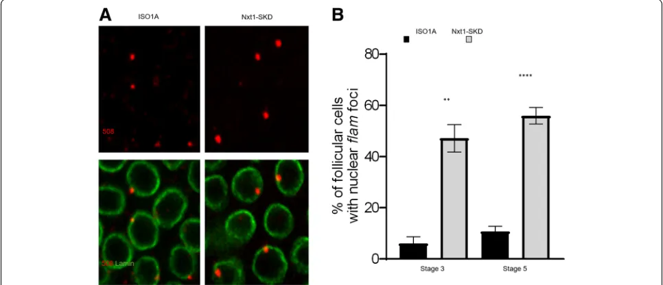
Alternatively, the nuclear accumulation could be too tran-sient before export to be detected in our immuno-RNA FISH experiments. To distinguish between these two hy-potheses, we examined the localization offlamtranscripts in follicle cells ofNxt1-SKD flies in early stages of oogen-esis (Nxt1somatic knockdown using tj-Gal4driver) when the export offlamprecursors is impeded. Nxt1 protein is a RNA export factor that is required, together with its partner Nxf1, for the export of flamprecursors [16]. We observed a clear increase in the proportion of follicle cells with a nuclear accumulation of flam transcripts in early stages 3 and 5 in Nxt1-SKD compared to WT ISO1A (Fig. 2a & b), which suggests that nuclear accumulation offlamprecursors can occur in early stages of oogenesis.
These findings indicate that, at early stages, in their traffic to the cytoplasm,flamtranscripts are shuttled to the cyto-plasm very rapidly. Export of the piRNA precursors is thus more efficient in early than in late stages of oogenesis.
At late stages of Drosophila oogenesis,flamprecursors fail to be delivered to the cytoplasm
The second issue we addressed was to understand why the delivery offlamprecursors from the membrane to the cytoplasm is steadily affected as oogenesis proceeds. We have previously shown that the export of flam precursor transcripts to the cytoplasm is the signal for the Yb-bodies to be assembled [5]. If, as suggested above,flam precur-sors do not reach the cytoplasm in late stages, then the proportion of Yb-bodies is expected to decrease.
In immunostaining experiments using Armi and Yb antibodies, we indeed observed that the size of the Yb-bodies (Fig. 3a) detected in follicle cells decreases during oogenesis together with the percentage of cells that have a discernable Yb-body (Fig. 3b). At stage 10 less than 10% of cells have Armi and Yb foci com-pared to 80% in stage 3 or 90% in OSS cells (Fig. 3b and Additional file 1: Figure S1A).
However, the decrease in Yb-body assembly in late stages of oogenesis cannot be attributed solely to the re-duction offlampiRNA precursors exported to the cyto-plasm. In the germarium, the follicle cells encapsulate the germline cyst and carry out a mitotic division pro-gram from stage 2 to stage 6 of oogenesis [17]. At stage 6, they cease to undergo mitotic cycles and start endo-cycles. This transition is regulated by the Notch pathway
[17]. It has been demonstrated that Armi protein level decreases at this switch from mitosis to endoreplication owing to Notch signaling pathway activation [18]. Immuno-histochemical experiments performed with Armi and Yb- antibodies on whole ovaries showed that not only is Armi protein level reduced in late stages of oogenesis, but also Yb staining. We observed a clear drop during oogenesis with a high level of Armi and Yb proteins only until stage 8 and a weak staining after-wards (Fig. 3b & Additional file 1: Figure S1) indicating that both proteins are presumably regulated at the tran-sition from mitosis to endoreplication.
To determine whether the reduced export of flam transcripts at late stage of oogenesis is related to the de-crease in Armi and Yb protein levels after stage 8, we analyzed whether a reduction of Armi or Yb protein level in early stages of oogenesis would have the same impact on RNA precursor export. To do so we quanti-fied the sub-cellular localization of flam transcripts in follicle cells of stage 5 depleted for Armi or Yb using RNAi or mutant lines. The localization of flam tran-scripts was found similar between WT and Armi- and
Yb-depleted lines, with a majority of follicle cells having transcripts located within the cytoplasm. However, quantifications showed a low but significant decrease in the proportion of cells exhibiting cytoplasmic flam foci (Fig. 4a & b). A reduced accumulation of flam tran-scripts in the cytoplasm may result from an increased instability of the transcripts due to the absence of Armi and Yb in the cytoplasm. Alternatively, the mild de-crease in the proportion of cells with cytoplasmic flam
Fig. 2At early stages,flamprecursors can be visualized accumulated in the nucleus. aSub-cellular localization offlamtranscripts in WT ISO1A or
Nxt1-SKD follicle cells of stage 5.flamtranscripts are labelled in red usingflam508 RNA probe and nuclear membrane is labelled in green with anti-lamin antibody.bQuantitative analysis offlamnuclear foci localization in WT ISO1A orNxt1-SKD follicle cells, at stage 3 and 5 of oogenesis. Error bars represent s.e.m. Numbers of cells and follicles counted is indicated in Additional file3: Table S1
foci is expected if the export of flam transcripts is im-peded. It has been suggested that armi is expressed in the follicle cells of the trans-heterozygous mutantarmi1/
armi72.1
[18] and we cannot exclude an incomplete de-pletion of Armi and Yb proteins in early stages in the RNAi lines used. Nevertheless, these mutants reveal a possible involvement of Armi and Yb proteins in the de-livery of piRNA precursors to the cytoplasm. It has pre-viously been shown that Armi and Yb proteins bind piRNA precursors [19–22]. It has also been reported that artificially tethering Armi or Yb protein to a heter-ologous RNA channels this RNA to the cytoplasmic pro-cessing machinery [23, 24]. Armi ATP-dependent helicase activity has also been observed in vitro [24] and been implicated in Armi specific binding to piRNAs
precursors and piRNA production [21, 22, 25], with the hypothesis that Armi binds and unpacks piRNA precur-sors while being exported to the cytoplasm. Whether this binding happens in the nucleus or at the nuclear membrane remains to be elucidated. In late stages of oo-genesis in Nxt1-SKD flies, we observed that Armi but not Yb protein can occasionally be visualized within the nucleus as a dotty pattern (Additional file 2: Figure S2). Of the 3074 follicle cells examined from 20 stage 10 egg chambers, Armi was detected in 7.9% of the nuclei, along with a great variability among the follicles (0 to 18%). This raises the possibility that Armi protein shut-tles between the cytoplasm and nucleus which is not de-tectable in WT conditions. However, we cannot exclude that this Armi pattern of staining is a consequence of a
detrimental lack of the Nxt1 export protein in the ovar-ian follicle cells.
On the basis of these findings, we propose that two in-terconnected events occur during oogenesis that lead to a drop in Yb-body assembly in late stages. On the one hand, following Notch-signaling pathway activation in stage 6–7, the levels of Armi and Yb proteins are dras-tically reduced, which not only decreases the assembly of the Yb-bodies but also impedes the export of piRNA precursors (Fig. 4). On the other hand, the export of piRNA precursors is progressively reduced along oogen-esis which further hampers the assembly of the remaining Armi and Yb into Yb-bodies.
Reduction of the assembly of Yb-bodies and of the ex-port offlampiRNA precursors is thought to cause a de-crease in the production of piRNAs in late stages of oogenesis. However, several studies indicate that the piRNA silencing is active throughout oogenesis. For ex-ample, if we consider the two retrotransposons, ZAM and gypsy, their enhancers are active at the posterior pole of the follicle cells throughout oogenesis including in late stages [26, 27]. Nevertheless, their transcripts are never detected in WT conditions owing to the silencing exerted by the piRNA pathway, which indicates that the silencing exerted on ZAM and gypsy is active even in late stages. One possible explanation of TE silencing in late stages of oogenesis when piRNA production seems impeded could be that the few and highly diminished Yb-bodies present in late stages give rise to a piRNA population that is sufficient to silence TEs. Alternatively, and not exclusively, piRNAs produced anteriorly in early stages are stable enough to remain active in late stages. Finally, it cannot be excluded that a transcriptional silen-cing exerted in early stages can still prevent transcription of the TEs in late stages with no need for piRNAs. The differential quantification of mature piRNAs produced in early versus late stages of oogenesis will provide a far better view of piRNAs produced during oogenesis.
Conclusions
From transcription to processing, multiple steps and nu-merous protein factors are required to drive piRNA pre-cursors into the cytoplasmic structure where functional piRNAs are produced. In the follicle cells of Drosophila
Fig. 4flampiRNA precursor export in early stages is slightly impeded in armi- and yb-depleted cells.aflamtranscripts (red) and nuclear membrane (green) in stages 5 of oogenesis are visualized by RNA-FISH and immunofluorescence using respectivelyflam508 RNA probe and anti-lamin antibody in follicle cells ofISO1Aline, RNAi knockdown ofYbandarmiand trans-heterozygous forarmi.b Quantitative analysis of cytoplasmicflamfoci localization at stage 5 of oogenesis in theDrosophilalines indicated. Error bars represent s.e.m. Numbers of cells and follicles counted is indicated in Additional file3: Table S1
ovaries, RNAs transcribed from piRNA clusters are first spliced, presumably concomitantly to transcription [8], before being transferred throughout the nucleoplasm in an Exon Junction complex, UAP56 protein and Nxt1-Nxf1-dependent manner, to be assembled in a single nu-clear focus [13,16]. Then, the transfer to the cytoplasm requires the export complex Nxt1-Nxf1 and our present study suggests that Armi and Yb proteins may somehow be implicated in this process (Fig. 4). Armi protein is thought to be able to bind several types of RNAs being exported - including mRNAs. In the cytoplasm, its spe-cific interaction with piRNA precursors is regulated by its ATPase hydrolysis activity and Yb protein [21, 25]. Our present study suggests that Armi could play an earl-ier role within the nucleus before RNA export. When in the cytoplasm, piRNA precursors are delivered to the Yb-bodies and mitochondria where Yb and Armi play distinct and collaborative roles to ensure the production of Zuc-dependent phased piRNAs [21,25].
Our study shows that a spatially and temporally con-trolled piRNA biogenesis exists with two critical periods taking place before and after the switch from a mitotic cycle to an endo-replication cycle. It can be anticipated that the major pool of somatic piRNAs is produced dur-ing the first period when piRNA precursors exit from the nucleus and Yb-bodies are correctly assembled.
Overall, these data show that in addition to factors specifically required for piRNA biogenesis, the spatio-temporal regulation of the whole system takes place at another level of complexity, the understanding of which will certainly help to interpret delayed and un-expected regulations.
Methods Drosophila stocks
The used fly strains were: ISO1Afrom the collection of the GReD; armi [1]/TM3 and armi [72.1]/TM6and RNAi line nxf1 (34945) from Bloomington Drosophila Stock Center; RNAi lines: armi (103589KK) and yb (110056KK) from Vienna Drosophila RNAi Centre.
Cell culture
OSS cells were grown in prepared from Shields and Sang M3 Insect Medium (Sigma) supplemented with 0.6 mg/ ml glutathione, 10 mU/ml insulin, 10% fetal bovine serum and 10% fly extract.
RNA fish
The DNA fragment to prepare the specificflam508 probe to detect flam transcripts was PCR amplified from the
ISO1A line using primers
5′-ATTCTCCTTTCTCAG-GATGC-3′ and 5′-GCATTGCTACCTTACGTTTC-3′
and cloned into pGEMT easy vector.
Riboprobe was synthesized by digestion of pGEMT easy plasmids with NcoI or SpeI enzyme, followed by in vitro transcription using Sp6 or T7 polymerase and digoxygenin labeled UTP (Roche), DNAse I treatment and purification.
RNA FISH on ovaries was performed as previously de-scribed [13]. In situ hybridization on OSS cells was car-ried out essentially described for ovaries. OSS cells were fixed in 4% formaldehyde/PBT (1X PBS, 0.1% Triton) at RT for 30 min, rinsed three times with PBT and post-fixed 10 min in 4% formaldehyde/PBT. After washes in PBT and permeabilisation (1 h in 1X PBS,0.3% Triton) prehybridization was done as follow: 10 min HYB-(50% Formamide, 5X SSC, 0.02% Tween)/PBT 1:1, 10 min HYB-, 1 h HYB+ (HYB- with yeast tRNA 0.5μg/ μl, 0.25 mg/ml heparin) at 37 °C. Ovaries were hybrid-ized overnight at 37 °C with 1μg riboprobe previously denaturated 10 min at 74 °C. Ovaries were then rinsed 20 min in HYB- and in HYB−/PBT at 37 °C then 4 times in PBT at RT before blocking 1 h at RT in TNB (Perkin-Elmer TSA kit) and immunodetection 1 h30 at RT with anti-Dig-HRP (Roche) in TNB 0.3% Tri-ton. Cells were rinsed three times in PBT, incubated 10 min with TSA-Cy5 in amplification diluent (Per-kin-Elmer) and rinsed three times in PBT.
When coupled to immunofluorescence, RNA straining was followed by incubation with mouse lamin body (ADL67–10, Hybridoma), goat Armitage anti-body (sc-34,564, Santa Cruz), Yb antianti-body (kindly provided by G. Hannon), GAPDH antibody (IMG-5143A-050, Imgenex) Secondary antibodies coupled to Cy3 or Alexa-488 were used.
Immunofluorescence
Ovaries from 2-to 4-days-old flies were dissected in PBT fixed in 4% formaldehyde/PBT at RT for 20 min, rinsed three times with PBT, incubated 1 h in PBS-0.3% Triton, rinsed three times with PBT and incubated 1 h in TNB 0,3% Triton prior staining with lamin, anti-Armitage or anti- Yb antibody. Secondary antibodies coupled to Cy3, Cy-5 or Alexa-488 were used.
Additional files
Additional file 1: Figure S1.Pattern of Armi and Yb staining in OSS cells and in whole ovariole (A)flamtranscripts (red), nuclear membrane (green) and Armi (white) (top) or Yb-foci (white) (bottom) are visualized by RNA-FISH and immunofluorescence usingflam508 RNA probe, anti-lamin antibody, anti-Armi and anti-Yb antibodies in OSS cells. (B) Armi (red) and Yb-foci (white) and nuclear membrane (green) are visualized in whole ovarioles by immunofluorescence using respectively anti-Armi, anti-Yb and anti-lamin antibodies. A clear drop in Armi and Yb staining is visualized in the surrounding follicle cells, in early versus late stages of oogenesis (TIF 142 kb)
Additional file 2: Figure S2.Unusual nuclear localization of Armi in follicle cells of late stages inNxt1-SKD flies Armi but not Yb protein can occasionally be visualized within the nucleus of follicle cells of late stages of oogenesis inNxt1-SKD flies. Armi (red), and Yb protein (white) and nuclear membrane (green) are visualized by immunofluorescence using anti-Armi, anti-Yb, and anti-lamin antibody in follicle cells ofNxt1-SKD egg chambers of stage 10 (TIF 1479 kb)
Additional file 3: Table S1.Number of cells and follicles analyzed for quantifications presented in Figs.1a-b,2b,3b,4b (XLSX 11 kb)
Acknowledgements
We are grateful to R. Pillai for useful discussions; to A. Sarkar and J. Watts for critical reading of the manuscript.
Authors’contributions
CD designed and performed experiments. CV conceived the study. CD EB and CV wrote the manuscript. All authors read and approved the final manuscript.
Funding
This work was supported by Agence Nationale pour la Recherche, project Plastisipi to CV.
Availability of data and materials
All data generated or analysed during this study are included in this published article [and its supplementary information files].
Ethics approval and consent to participate Not applicable.
Consent for publication Not applicable.
Competing interests
The authors declare that they have no competing interests.
Received: 4 April 2019 Accepted: 25 June 2019
References
1. Aravin A, Gaidatzis D, Pfeffer S, Lagos-Quintana M, Landgraf P, Iovino N, et al. A novel class of small RNAs bind to MILI protein in mouse testes. Nature. 2006; 442(7099):203–7 PubMed PMID: 16751777. Epub 2006/06/06. eng. 2. Brennecke J, Aravin AA, Stark A, Dus M, Kellis M, Sachidanandam R, et al.
Discrete small RNA-generating loci as master regulators of transposon activity in Drosophila. Cell. 2007;128(6):1089–103 PubMed PMID:17346786. Epub 2007/03/10. eng.
3. Girard A, Sachidanandam R, Hannon GJ, Carmell MA. A germline-specific class of small RNAs binds mammalian Piwi proteins. Nature. 2006;442(7099): 199–202 PubMed PMID: 16751776. Epub 2006/06/06. eng.
4. Aravin AA, Hannon GJ, Brennecke J. The Piwi-piRNA pathway provides an adaptive defense in the transposon arms race. Science. 2007;318(5851):761– 4 PubMed PMID: 17975059. Epub 2007/11/03. eng.
5. Zanni V, Eymery A, Coiffet M, Zytnicki M, Luyten I, Quesneville H, et al. Distribution, evolution, and diversity of retrotransposons at the flamenco locus reflect the regulatory properties of piRNA clusters. Proc Natl Acad Sci
U S A. 2013;110(49):19842–7 PubMed PMID: 24248389. Pubmed Central PMCID: 3856796. Epub 2013/11/20. eng.
6. Saito K, Inagaki S, Mituyama T, Kawamura Y, Ono Y, Sakota E, et al. A regulatory circuit for piwi by the large Maf gene traffic jam in Drosophila. Nature. 2009; 461(7268):1296–9 PubMed PMID: 19812547. Epub 2009/10/09. eng. 7. Goriaux C, Theron E, Brasset E, Vaury C. History of the discovery of a master
locus producing piRNAs: the flamenco/COM locus in Drosophila melanogaster. Front Genet. 2014;5:257 PubMed PMID: 25136352. Pubmed Central PMCID: 4120762. Epub 2014/08/20. eng.
8. Goriaux C, Desset S, Renaud Y, Vaury C, Brasset E. Transcriptional properties and splicing of the flamenco piRNA cluster. EMBO Rep. 2014; 15(4):411–8 PubMed PMID: 24562610. Pubmed Central PMCID: 3989672. Epub 2014/02/25. eng.
9. Qi H, Watanabe T, Ku HY, Liu N, Zhong M, Lin H. The Yb body, a major site for Piwi-associated RNA biogenesis and a gateway for Piwi expression and transport to the nucleus in somatic cells. J Biol Chem. 2011;286(5):3789–97 PubMed PMID: 21106531. Pubmed Central PMCID: 3030380. Epub 2010/11/26. eng.
10. Szakmary A, Reedy M, Qi H, Lin H. The Yb protein defines a novel organelle and regulates male germline stem cell self-renewal in Drosophila melanogaster. J Cell Biol. 2009;185(4):613–27 PubMed PMID: 19433453. Pubmed Central PMCID: 2711570. Epub 2009/05/13. eng.
11. Ishizu H, Siomi H, Siomi MC. Biology of PIWI-interacting RNAs: new insights into biogenesis and function inside and outside of germlines. Genes Dev. 2012;26(21):2361–73 PubMed PMID: 23124062. Pubmed Central PMCID: 3489994. Epub 2012/11/06. eng.
12. Siomi MC, Sato K, Pezic D, Aravin AA. PIWI-interacting small RNAs: the vanguard of genome defence. Nat Rev Mol Cell Biol. 2011;12(4):246–58 PubMed PMID: 21427766. Epub 2011/03/24. eng.
13. Dennis C, Zanni V, Brasset E, Eymery A, Zhang L, Mteirek R, et al.“Dot COM”, a nuclear transit center for the primary piRNA pathway in Drosophila. PLoS One. 2013;8(9):e72752 PubMed PMID: 24039799. Pubmed Central PMCID: 3767702. Epub 2013/09/17. eng.
14. Murota Y, Ishizu H, Nakagawa S, Iwasaki YW, Shibata S, Kamatani MK, et al. Yb integrates piRNA intermediates and processing factors into perinuclear bodies to enhance piRISC assembly. Cell Rep. 2014;8(1):103–13 PubMed PMID: 24953657. Epub 2014/06/24. eng.
15. Niki Y, Yamaguchi T, Mahowald AP. Establishment of stable cell lines of Drosophila germ-line stem cells. Proc Natl Acad Sci U S A. 2006;103(44): 16325–30 PubMed PMID: 17056713. Pubmed Central PMCID: 1637581. Epub 2006/10/24. eng.
16. Dennis C, Brasset E, Sarkar A, Vaury C. Export of piRNA precursors by EJC triggers assembly of cytoplasmic Yb-body in Drosophila. Nat Commun. 2016;7:13739 PubMed PMID: 27929060. Pubmed Central PMCID: 5155165. 17. Deng WM, Althauser C, Ruohola-Baker H. Notch-Delta signaling induces a transition from mitotic cell cycle to endocycle in Drosophila follicle cells. Development. 2001;128(23):4737–46 PubMed PMID: 11731454. 18. Olivieri D, Sykora MM, Sachidanandam R, Mechtler K, Brennecke J. An in
vivo RNAi assay identifies major genetic and cellular requirements for primary piRNA biogenesis in Drosophila. EMBO J. 2010;29(19):3301–17 PubMed PMID: 20818334. Pubmed Central PMCID: 2957214. 19. Ishizu H, Iwasaki YW, Hirakata S, Ozaki H, Iwasaki W, Siomi H, et al.
Somatic primary piRNA biogenesis driven by cis-acting RNA elements and trans-acting Yb. Cell Rep. 2015;12(3):429–40 PubMed PMID: 26166564. Epub 2015/07/15. eng.
20. Saito K, Ishizu H, Komai M, Kotani H, Kawamura Y, Nishida KM, et al. Roles for the Yb body components Armitage and Yb in primary piRNA biogenesis in Drosophila. Genes Dev. 2010;24(22):2493–8 PubMed PMID: 20966047. Pubmed Central PMCID: 2975925. Epub 2010/10/23. eng.
21. Ishizu H, Kinoshita T, Hirakata S, Komatsuzaki C, Siomi MC. Distinct and collaborative functions of Yb and Armitage in transposon-targeting piRNA biogenesis. Cell Rep. 2019;27(6):1822–35 e8. PubMed PMID: 31067466. 22. Hirakata S, Ishizu H, Fujita A, Tomoe Y, Siomi MC. Requirements for
multivalent Yb body assembly in transposon silencing in Drosophila. EMBO Rep. 2019. PubMed PMID: 31040109.
23. Rogers AK, Situ K, Perkins EM, Toth KF. Zucchini-dependent piRNA processing is triggered by recruitment to the cytoplasmic processing machinery. Genes Dev. 2017;31(18):1858–69 PubMed PMID: 29021243. Pubmed Central PMCID: 5695087.
24. Pandey RR, Homolka D, Chen KM, Sachidanandam R, Fauvarque MO, Pillai RS. Recruitment of Armitage and Yb to a transcript triggers its phased
processing into primary piRNAs in Drosophila ovaries. PLoS Genet. 2017; 13(8):e1006956 PubMed PMID: 28827804. Pubmed Central PMCID: 5578672. 25. Ge DT, Wang W, Tipping C, Gainetdinov I, Weng Z, Zamore PD. The
RNA-binding ATPase, Armitage, couples piRNA amplification in Nuage to phased piRNA production on mitochondria. Mol Cell. 2019;74(5):982–95.e6. PubMed PMID: 31076285.
26. Leblanc P, Desset S, Giorgi F, Taddei AR, Fausto AM, Mazzini M, et al. Life cycle of an endogenous retrovirus, ZAM, in Drosophila melanogaster. J Virol. 2000;74(22):10658–69 PubMed PMID: 11044110. Pubmed Central PMCID: 110940. Epub 2000/10/24. eng.
27. Pelisson A, Song SU, Prud'homme N, Smith PA, Bucheton A, Corces VG. Gypsy transposition correlates with the production of a retroviral envelope-like protein under the tissue-specific control of the Drosophila flamenco gene. EMBO J. 1994;13(18):4401–11 PubMed PMID: 7925283. Pubmed Central PMCID: 395367. Epub 1994/09/15. eng.
Publisher’s Note