P R I M A R Y R E S E A R C H
Open Access
Increase of IFN-
γ
and TNF-
α
production in
CD107a + NK-92 cells co-cultured with cervical
cancer cell lines pre-treated with the HO-1
inhibitor
Paulina Gómez-Lomelí
1,4, Alejandro Bravo-Cuellar
1,2, Georgina Hernández-Flores
1, Luis Felipe Jave-Suárez
1,
Adriana Aguilar-Lemarroy
1, José Manuel Lerma-Díaz
1,2, Jorge Ramiro Domínguez-Rodríguez
1,3,
Karina Sánchez-Reyes
1,4and Pablo Cesar Ortiz-Lazareno
1*Abstract
Background:Natural killer (NK) cells eliminate virus-infected and tumor cells through the release of perforins and granzymes; they also produce Interferon gamma (IFN-γ) and Tumor necrosis factor alpha (TNF-α), which induce apoptosis in target cells. Many tumors express Heme oxygenase 1 (HO-1), and this expression has been associated with avoiding immunosuppression and apoptosis. In this work, HO-1+ Cervical cancer cell (CCC) lines were pre-treated with HO-1 inhibitor and we assessed whether this inhibition enhanced the sensitivity of CCC to NK cell activity.
Methods:We assessed the expression of HO-1 in HeLa, SiHa, and C-33A CCC by Flow cytometry (FC). CCC were pre-treated with SnPP or ZnPP HO-1 inhibitors. After that, NK-92 cells were co-cultured with HeLa, SiHa, and C-33A CCC pre-treated or not with HO-1 inhibitors, and the expression of IFN-γ, TNF-α, CD107a, Granzyme B, NKp44, NKp46, NKp30, and NKG2D was evaluated by FC.
Results:CCC lines HeLa, SiHa, and C-33A expressed HO-1. Inhibition of HO-1 in these cells increased the expression of IFN-γand TNF-αin CD107a + NK-92 cells. We observed a reduction in the expression of NKG2D, NKp46, and NKp30 in NK cells co-cultured with HeLa and SiHa cells, and when HeLa and SiHa cells were pre-treated with the HO-1 inhibitors, the expression of NKG2D and NKp30 in NK cells was restored. We observed a similar effect in NK cells co-cultured with C-33A cells in NKp30 expression.
Conclusion:Inhibition of HO-1 in CCC induces an increase in IFN-γand TNF-αproduction in CD107a + NK-92 cells and restores NKG2D, NKp46 and NKp30 downmodulation in NK cells.
Keywords:Heme oxygenase 1, NK cells, Cervical cancer cells, IFN-γ, TNF-α, NKp30, NKG2D
Background
Cervical cancer is the third most common cancer among women worldwide [1] The main risk factor for cervical cancer is infection with the Human papillomavirus (HPV) [2,3]. HPV types 16 and 18 are responsible for 70% of cases [4]. The immune system is responsible for eliminat-ing tumor cells and infected cells with microorganisms or
foreign antigens. Natural killer (NK) cells are the major cells responsible for tumor removal and elimination of in-fected cells [5]. NK cells constitute between 2 and 18% of total lymphocytes in peripheral blood and are distributed in lymphoid as well as in non-lymphoid organs. NK cells respond to cellular signals triggered by receptor activation or inhibition; once they interact with their specific target, if these signals trigger NK cell activation, the release of per-forins and granzymes induces cellular lysis in target cells [6]. Another important function of NK cells is the produc-tion of cytokines, such as Interferon gamma (IFN-γ) and Tumor necrosis factor alpha (TNF-α); these cytokines also * Correspondence:pablolazareno@gmail.com
1División de Inmunología, Centro de Investigación Biomédica de Occidente
(CIBO), Instituto Mexicano del Seguro Social (IMSS), Sierra Mojada 800, Col. Independencia, 44340 Guadalajara, Jalisco, Mexico
Full list of author information is available at the end of the article
act as promoter agents of apoptosis in tumor cells, or they recruit and activate other cells of the immune response, such as monocytes/macrophages, dendritic cells, T cells, and B cells [7,8]. The production of these cytokines by NK cells promotes innate and adaptive immunity [9]. The major activating receptors in NK cells are NGK2D and Natural cytotoxicity receptors (NCR), that is, NKp30, NKp44, and NKp46 [10]. These activating receptors recognize ligands in tumor or infected cells [11]. These ligands for activating receptors are stress-inducible mol-ecules and include UL16-binding proteins (ULBP) and (MHC) class I-related chain A/B (MICA/B) recognized by NKG2D [12,13]. Other ligands include the poliovirus to DNAM-1, the HLA-B-associated transcript 3 (BAT-3), and the B7-H6 molecule, both recognized by NKp30 [14,15]. These ligands are absent in normal cells and their expression is increased in cancer cells. However, interaction among immune cells, tumor cells, and neighboring cells is very complex. Cells are influenced by the surrounding microenvironment and this process involves a selection mechanism of tumor cells, which initiate a number of signaling mechanisms to evade the immune response, as described in the hypothesis of immunoediting [16]. Under certain circumstances, this interaction culminates in the eradication of tumor cells, as occurs the majority of times, or instead, in suppression of the immune response and tumor formation. Within this context, tumor cells developed various escape mechanisms to avoid NK-mediated killing; tumor cells produced cyto-kines, growth factors, and enzymes that induced suppres-sion of cells of the immune response [17,18]. Heme oxygenase 1 (HO-1) is the rate-limiting enzyme in heme catabolism and leads to three products: biliverdin; free iron, and carbon monoxide [19]. It plays an important role in the modulation of inflammation, blocking the apoptotic process and antioxidant defense in the presence of any damage [20,21]. This enzyme is overexpressed in pancre-atic, colon, and lung cancer, in which it can promote tumor cell proliferation and resistance to tumor therapy [22,23]. Induction of HO-1 in tumor cells undergoing any stressor agent increases their resistance to apoptosis [24-26]. Likewise, inhibition of this enzyme leads to the re-duction of tumor growth and an increase in sensitivity to chemotherapy [19,27]. In this study, we evaluated HO-1 ex-pression in Cervical cancer cells (CCC) and whether HO-1 inhibition enhanced the sensitivity of CCC to NK cells.
Results
Cervical cancer cell lines express HO-1
We evaluated HO-1 expression in HeLa (HPV 18+),
SiHa (HPV 16+), and C-33A (HPV–) CCC (Figures 1a
and b). We can observe that the HeLa cell is the cancer cell line that expressed the highest percentage of cells positive to HO-1 (70.2% ± 4.9%) in comparison with SiHa
and C-33A CCC (54.6% ± 1.5% and 30.3% ± 6.5%, respect-ively) (p<0.01). Additionally, we determined the geometric Mean fluorescence intensity (MFI) in each cancer cell line. HeLa, SiHa, and C-33A lines have similar MFI, and we did not observe a difference for HO-1 MFI among the three CCC, suggesting that the difference it is not in the intensity of expression, but rather in the number of cells positive to HO-1. Likewise, we evaluated viability in CCC
treated with SnPP (25μM) and ZnPP (1μM) HO-1
inhibi-tors and observed that these inhibiinhibi-tors did not affect the viability in these cells (Figure 1c). In addition, we evaluated whether HO-1 inhibitors affect the expression of NK cell ligands, such as MICA and MICB. HeLa and SiHa cells express MICA, but not MICB, while C-33A expresses MICB, but not MICA, and we did not observe a change in MICA or MICB expression when cells were treated with SnPP or ZnPP inhibitors (Figure 1d). HO-1 inhibitors did not affect MICA and MICB receptors.
CD107a expression in NK-92 cells co-cultured either with cervical cancer cells pre-treated or not with the SnPP, HO-1 inhibitor
We evaluated the expression of CD107a in NK-92 cells co-cultured with HeLa, SiHa, and C-33A CCC pre-treated or not with HO-1 inhibitor (SnPP) (Figure 2). In Figure 2a, we can observe the baseline expression of CD107a in NK-92 cells and the positive-control PMA/Ionomycin increase of this expression. We did not observe differences in NK-92 cells co-cultured with HeLa cells pre-treated or not with HO-1 inhibitor in all target effector ratios (T:E) 1:5 and 1:20 (Figure 2b). In NK-92 cells co-cultured with SiHa and C-33A CCC, we observed similar behavior to that ob-served for HeLa; there were no significant differences be-tween the different T:E ranges bebe-tween pre-treated cells or not treated with the HO-1 inhibitor. When we analyzed MFI for CD107a, there was no difference in any of the ex-perimental groups; as we expected, only the positive con-trol group (PMA + Ionomycin) increased expression and MFI of CD107a in NK-92 cells.
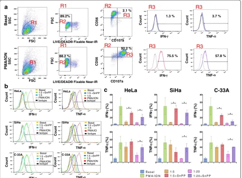
Increase of IFN-γand TNF-αproduction in NK-92 cells positive to CD107a co-cultured with cervical cancer cell lines pre-treated with the HO-1 inhibitor
In Figure 3a, we observe the baseline expression of
INF-γ and TNF-α in CD107a + NK-92 cells (Basal group)
that observed in HeLa cells. In this case, the increase in
IFN-γ expression was observed when SiHa cells were
pre-treated with SnPP (the HO-1 inhibitor) (p <0.05). When NK-92 cells were co-cultured with C-33A pre-treated with HO-1 inhibitor, we can observe a significant
increase in the production of IFN-γin comparison with
C-33A without pre-treatment with the HO-1 inhibitor (p<0.05). In general, there was an increase in the
pro-duction of IFN-γ in CD107a + NK-92 cells co-cultured
with HeLa, SiHa, and C-33A pre-treated with the HO-1 inhibitor (SnPP). In the same Figure 3b and c, we can
observe TNF-αproduction in CD107a + NK-92 cells
co-cultured with HeLa, SiHa, and C-33A pre-treated or not with the HO-1 inhibitor. In the co-culture with HeLa cells, when tumor cells were pre-treated with the HO-1 inhibitor, TNF-αproduction was significant in NK-92 cells (p <0.05). We observed a similar difference in the SiHa
co-culture. Production of TNF-α in NK-92 cells
co-cultured with C-33A pre-treated with the HO-1 inhibitor is significantly increased within the 1:20 range (p<0.05). Granzyme B expression in CD107a + NK-92 cells co-cultured with cervical cancer cell lines pre-treated with the HO-1 inhibitor
We evaluated granzyme B expression in CD107a + NK-92 cells co-cultured with HeLa, SiHa, and C-33A CCC pre-treated or not with the HO-1 inhibitor (Figure 4). We did not observe differences in granzyme B release in any T:E ratios 1:5 and (1:20 and 1:40, data not shown) in NK-92 cells co-cultured with HeLa, SiHa, and C-33A CCC pre-treated or not with the HO-1 inhibitor. It is important to stress that PMA/Ionomycin induces the release of gran-zyme B and correlates with the association between CD107a increase and granzyme B release.
Figure 1Expression of Heme oxygenase 1 (HO-1) in different Cervical cancer cell (CCC) lines.Expression of HO-1 in HeLa, SiHa, and C-33A
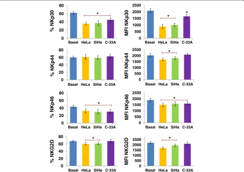
Supernatant of cervical cancer cell lines HeLa, SiHa, and C-33A induces downmodulation of NCR and NKG2D receptors in NK cells
We investigated whether the supernatant of CCC HeLa, SiHa, and C-33A induces downmodulation of NKp30, NKp44, NKp46 and NKG2D in NK-92 cell line and NK cells of healthy donors. We observed that in both cells, HeLa and SiHa cancer cells induce downmodulation of NKp30, NKp46, and NKG2D in NK cells, and in C-33A cells only NKp30 and NKp46 (p<0.05 Figures 5 and 6). Likewise, in NK-92 cells we observed downmodulation of NKp44 when the cells were treated with the super-natant of HeLa and SiHa (p<0.05).
The HO-1 inhibitor restores downmodulation in NCR and NKG2D expression in NK-92 cells co-cultured with cervical cancer cell lines
We evaluated the expression of NKp30, NKp44, NKp46, and NKG2D in NK-92 cells co-cultured with HeLa,
SiHa, and C-33A cancer cells pre-treated or not with the HO-1 inhibitors. We can observe that in NK-92 cells co-cultured with HeLa cells pre-treated with SnPP (the HO-1 inhibitor), there is an increase in NKG2D, and NKp30 ex-pression within the different T:E ratios (p<0.05) (Figure 7) in comparison with HeLa cells without pre-treatment with the HO-1 inhibitor. When NK-92 cells were co-cultured with SiHa cells, we observed a similar effect to that ob-served with HeLa cells, and an increase in NKG2D, NKp46, and NKp30 receptor when SiHa cells were pre-treated with SnPP (p<0.05), in comparison with SiHa cells without pre-treatment. When NK-92 cells were co-cultured with C-33A pre-treated with the HO-1 inhibitor, we are able to observe a significant increase in the expres-sion of NKp46 within the 1:20 ratio (p<0.05) in compari-son with cells without pre-treatment. We observed similar and comparable results when we used the ZnPP inhibitor (data not shown). Similarly, we performed transwell assays to evaluated change in NCR and NKG2D expression in
HeLa
C-33A
SiHa
CD107 a (%)
Basal
1:5 + SnPP 1:5
1:20
1:20 + SnPP CD107 a (%)
CD107 a (%)
FSC
LIVE/DEAD
CD107a
FSC
SSC SSC
CD56
SSC
a
b
3.2 % 90.3 %
CD56
CD107a
LIVE/DEAD® Fixable Near-IR 88.2 %
SSC
91.2 %
R1 R1
R1 R1
R2 R2
R2 R2
Basal
PMA/ION
® Fixable Near-IR
Figure 2Expression of CD107a in NK-92 cells co-cultured with Cervical cancer cells (CCC) pre-treated or not with Heme oxygenase 1
NK-92 and NK cells of healthy donors. We can observe, in Table 1, that the greatest change observed was in NKp30 and NKG2D receptors in HeLa and SiHa cells, and that when NK-92 cells were treated with HO-1 inhibitors (SnPP and ZnPP), this downmodulation is partially recovered (p <0.05). We observed similar results with NK primary cells of healthy donors (data not shown).
Discussion
Tumor cells acquire different capabilities that allow them to grow uncontrollably and even to evade the immune re-sponse. Among these acquired skills, we find the expres-sion of certain cytokines, chemokines, and enzymes that are not normally expressed and that can facilitate an eva-sion of this type, such as Transforming growth factor beta
(TGF-β), Interleukin (IL)-10, Prostaglandin E2 (PGE2), and indoleamine 2,3-dioxygenase [28]. In this regard, HO-1 possesses an important immune protective effect [29]. This enzyme is vital for the removal of heme, a potent pro-oxidant and inflammatory agent. HO-1 induces heme degradation and produces three metabolites: CO; ferrous iron, and biliverdin, all these metabolites with an immune protective effect [30]. We show that CCC HeLa, SiHa, and C-33A express HO-1; while each cell has a different per-centage of expression of HO-1, all possess a similar amount of HO-1 (MFI were similar in HeLa, SiHa, and C-33A cells). The expression of HO-1 has been associated with anti-apoptotic and anti-inflammatory effects [31]. In this respect, it was published that HO-1 can induce the production of IL-10 and that this promotes suppression of
a
0 20 40 60 80 * * 0 20 40 60 80 * * 0 20 40 60 80 * * 0 20 40 60 80 * 0 20 40 60 80 * 0 20 40 60 80 *B a s a l P M A /IO N
1 :5 1 :5 + S n P P
1 :2 0 1 :2 0 + S n P P
TNF -(%) TNF -( % ) TNF -( % ) IFN -(%) IFN -(%) IFN -(%)
b
c
FSC FSC CD107a CD107a IFN- TNF-Count Count Count Count CD56 CD56 FSC FSC SSC SSC Basal 1:5 +SnPP 1:5 PMA/ION Isotype Basal 1:5 Isotype 1:5 +SnPP PMA/ION Basal 1:5 Isotype 1:5 +SnPP PMA/ION Basal 1:5 Isotype 1:5 +SnPP PMA/ION Basal 1:5 Isotype PMA/ION 1:5 +SnPP Basal 1:5 Isotype 1:5 +SnPP PMA/IO Count Count Count Count Count Count IFN-TNF-HeLa SiHa C-33A
1.3 % 75.5 % 3.7 % 57.8 % 89.2% 2.1 % 88.2 % 92.2 %
LIVE/DEAD® Fixable Near-IR
LIVE/DEAD® Fixable Near-IR
R1 R1 R1 R1 R2 R2 R2 R2 R3 R3 R3 R3 R3 R3 HeLa HeLa C-33A C-33A SiHa SiHa Basal PMA/ION
Figure 3Increase of Interferon gamma (IFN-γ) and Tumor necrosis factor alpha (TNF-α) in CD107a + NK-92 cells co-cultured with
the immune response [32,33]. Overexpression of HO-1 has been reported in many cancer types, such as colorec-tal, pancreatic, and prostate [34]. Likewise, it was pub-lished that HO-1 is involved in macrophage polarization toward an M2 phenotype [35], and that HO-1+ macro-phages induce suppression of the immune response [36]. In this regard, it was also observed that HO-1 expressed by dendritic cells favors the emergence of CD4 + CD25+ regulatory T cells and inhibits the prolif-eration of T cells [37]. NK cells comprise the major cells involved in early responses against infected and tumor cells [38]. CD107a is associated with lysosomal mem-branes and has been associated with the degranulation of NK cells and CD8+ T cells, having been adopted as a marker of activation due to the association between the increased expression and increased cytotoxicity of their target cells [39,40]. In our studies, we did not observe a change in the expression of CD107a in NK cells co-cultured with HeLa, SiHa, or C-33A CCC pre-treated or not with the HO-1 inhibitor (SnPP), although we did
ob-serve an increase in the production of TNF-αand IFN-γ
in CD107a + NK-92 cells co-cultured with cancer cells pre-treated with the HO-1 inhibitor. In this regard, in our study, when NK cells were co-cultured with HeLa, SiHa, and C-33A CCC pre-treated or not with the HO-1
inhibitor (SnPP), we did not observe a difference in the ex-pression of CD107a and granzyme B in NK-92 cells. Al-though HeLa, SiHa, and C-33A cells express HO-1, this enzyme does not participate in the blocking of NK cell de-granulation, because pre-treatment with the inhibitor of the enzyme does not induce changes in CD107a expres-sion. Suppression of cytotoxicity in NK cells is associated with an important reduction in CD107a surface expression and suppression of granzyme B release [41]. It is likely that HO-1 does not interfere or intervene with the process of NK cell degranulation and cytotoxicity. It is noteworthy that CCC HeLa, SiHa, and C-33A, through other mole-cules, could interfere with the degranulation and cytotox-icity process in NK cells, as has been demonstrated with PGE2[10], although we observed a significant increase of
CD107a expression and cytotoxicity in K562 using NK-92 cells, and CD107a upregulation correlates with lysis of the target cells (data not shown). The cytotoxic action of NK cells comprises a series of steps involving combining the adhesion, activation, and secretion of lytic granules and other molecules, and NK cytotoxicity results are dependent on several changes that occur, such as phenotypical, functional, and molecular. While CD107a expression may not necessarily correlate with NK cytotox-icity, NK cells can also induce cell death by means of other
HeLa
SiHa
C-33A
1:5 + SnPP 1:5
PMA/ION Isotype Basal Granzyme B Granzyme B
Granzyme B
Count
Count
Count
Figure 4Expression of granzyme B in CD107a + NK92 cells co-cultured with Cervical cancer cell (CCC) lines pre-treated or not with
mechanisms, such as FasL and TRAIL [42,43]. Previous studies demonstrated that products of the heme catabol-ism, such as CO, induced, in T cells, a reduction in IL-2 secretion and proliferation of the cells via inhibition of the ERK MAPK pathway [44]. Similarly, the use the metabo-lites induced by the action of HO-1 on a heme group, such as CO, ferritin, or bilirubin alone or in combination, in-duces a decrease in the production of TNF-α, IL-1, IL-6, and IFN-γ, and instead promotes IL-10 production [45,46]. It was in our interest to investigate whether inhib-ition of HO-1 in tumor cells increased the production of
IFN-γ and TNF-α in the population of CD107a + NK-92
cells in our study. We observed that inhibition of the
en-zyme HO-1 increased IFN-γ and TNF-α expression in
NK-92 cells co-cultured with HeLa, SiHa, and C-33A cells pre-treated with the HO-1 inhibitor. We observed, in SiHa (HPV 16+) cells pre-treated with the HO-1 inhibitor, a
strong increase in IFN-γ and TNF-α production,
indicating that the HPV may play a role in blocking the production of these cytokines in NK cells. The effect ob-served when we employed the HO-1 inhibitor on IFN-γ
and TNF-α production in CD107a + NK-92 cells
co-cultured with CCC was important because IFN-γ and
TNF-α produced by NK cells can induce apoptosis in
tumor cells by means of interaction with localized cell-death receptors in tumor cells or through stimulation of cytotoxicity activity in CD8+ cells, in addition to helping to differentiate CD4+ T cells toward a Th1 response to promote CD8+ cell differentiation and to promote antitu-mor antibody production by B cells [47-50]. In addition, soluble E6 and E7 oncoproteins of HPV-16 inhibit NK cells to produce IFN [51]. Activating receptors are very important in NK activity, and it was reported that tumor cells can induce a decrease in the expression of activation receptors in NK cells, the latter through several mecha-nisms, such as binding competitively to the activating 0
20 40 60 80
0 20 40 60 80
0 20 40 60 80
0 20 40 60 80
0 500 1000 1500 2000 2500 0 500 1000 1500 2000 2500 0 500 1000 1500 2000 2500 0 500 1000 1500 2000 2500
*
*
*
*
*
*
*
% NKp30
% NKp44
% NKp46
% NKG2D
MFI NKp30
MFI NKp44
MFI NKp46
MFI NKG2D
Basal HeLa SiHa C-33A Basal HeLa SiHa C-33A
Basal HeLa SiHa C-33A Basal HeLa SiHa C-33A
Basal HeLa SiHa C-33A Basal HeLa SiHa C-33A
Basal HeLa SiHa C-33A Basal HeLa SiHa C-33A
*
Figure 5Downmodulation of NCR and NKG2D in NK-92 cells treated with the supernatant of HeLa, SiHa, and C-33A Cervical cancer
receptor and disrupting the binding between the activating receptor and the ligand expressed by tumor cells or by in-ducing the internalization of the activating receptor on NK cells [52,53]. In another study, we showed that CCC can induce downmodulation of the NKG2D receptor [54]. This is important because we observed that when HeLa and SiHa cells were pre-treated with the HO-1 inhibitors (SnPP and ZnPP) and subsequently co-cultured with NK-92 cells, the downmodulated expression of NKG2D and NKp30 was restored, in comparison with NK-92 cells co-cultured with HeLa and SiHa CCC without pre-treatment with HO-1 inhibitors. This same phenomenon was served for NKp46 in HeLa, SiHa and C-33A cells. We ob-served a similar response in NK cells of healthy donors (data not shown). All data strengthened the importance of HO-1 in this phenomenon because we observed these ef-fects when we employed both HO-1 inhibitors. It is note-worthy that we also observed a significant decrease in
NK-92 and primary NK cells in the expression of NKp30, NKp46, and NKG2D after 24 h of their being exposed to the culture supernatant of HeLa, SiHa, and C-33A CCC. NK cells are found in the stroma of HPV-infected Cervical intraepithelial neoplasia (CIN); however, NKp30, NKp46, and NKG2D expression is reduced in patients with pre-cancerous or pre-cancerous HPV-induced lesions and, in addition, their cytotoxicity was reduced [51,55].
We also studied the regulation of some NK cell li-gands, such as MICA and MICB, in CCC and the effect of HO-1 inhibitors on their expression. However, we did not observe any change in MICA and MICB expres-sion when we utilized HO-1 inhibitors. MICA is high expressed in HeLa and SiHa cells, but MICB is absent. In C-33A, we found higher expression of MICB in com-parison with HeLa and SiHa cells. At present, we are studying whether HO-1 inhibitors exert some effect on other NK cell ligands. As already known, cancer cells 0
20 40 60 80 100 0 2 4 6 8 0 20 40 60 80 100
0 20 40 60 80 100
0 500 1000 1500 2000
0 100 200 300 400 500
0 500 1000 1500 2000
0 500 1000 1500 2000 2500
% NKp30
% NKp44
% NKp46
% NKG2D
MFI NKp30
MFI NKp44
MFI NKp46
MFI NKG2D
Basal HeLa SiHa C-33A Basal HeLa SiHa C-33A
Basal HeLa SiHa C-33A
Basal HeLa SiHa C-33A
Basal HeLa SiHa C-33A Basal HeLa SiHa C-33A
Basal HeLa SiHa C-33A
Basal HeLa SiHa C-33A
*
*
*
*
*
*
*
Figure 6Downmodulation of NCR and NKG2D in NK cells treated with the supernatant of HeLa, SiHa, and C-33A Cervical cancer cells (CCC).
possess several mechanisms to avoid the immune re-sponse, including the production of molecules with sup-pressor activity on the immune response; tumor cells are able to induce a regulatory phenotype in immune cells, promoting immune evasion. The difference ob-served in our study among HeLa, SiHa, and C-33A cells in IFN-γ, TNF-α, NCR, and NKG2D on NK cells indi-cates that several mechanisms of resistance to cytotox-icity and to NK cell activity could be implicated, such as HPV type and their interactions with NK cells. Our findings indicate that HO-1 can interfere with IFN-γ
and TNF-α production and that are implicated in
de-regulation in the expression of activation receptors such as NKG2D, NKp30, and NKp46.
Conclusions
HO-1 inhibition in cervical cancer cells HeLa, SiHa, and
C-33A induce an increase in IFN-γand TNF-αproduction
in CD107a + NK-92 cells and restore downmodulation of NKG2D, NKp30, and NKp46 in NK cells. These re-sults demonstrate the complexity in the different inter-relations between cancer cells and immune cells. The interactions in the tumor microenvironment should be considered before designing NK cell therapies.
Methods
Antibodies and reagents
The following mAbs were used in this study: mouse-anti-human-IFN-γ-PE/Cy7 [BioLegend, San Diego, CA, USA);
-33A
0 20 40 60 80
0 20 40 60 80
0 20 40 60
*
0 20 40 60 80 0
20 40 60 80
* *
0 20 40 60
*
0 20 40 60 80
* *
0 20 40 60 80
*
0 20 40 60 80
0 20 40 60
0 20 40 60 80
* *
0 20 40 60 80
*
Basal
B a s a l 1 :5
1 :5 + S n P P
1 :2 0
1 :2 0 + S n P P 68.3 %
60.1 % 41.2 % 59.7 %
NKG2D
NKp44
NKp46
NKp30
NKG2D (%) NKG2D (%) NKG2D (%)
NKp44 (%) NKp44 (%) NKp44 (%)
NKp46 (%) NKp46 (%) NKp46 (%)
NKp30 (%) NKp30 (%) NKp30 (%)
Figure 7Downmodulation of NKG2D, NKp46, and NKp30 in NK-92 cells co-cultured with Cervical cancer cell (CCC) lines.CCC were
pre-treated with the Heme oxygenase 1 (HO-1) inhibitor (SnPP) for 48 h; after that, NK-92 cells were co-cultured with HeLa, SiHa, and C-33A CCC for 4 h. Then, the expression of NKp30, NKp44, NKp46, and NKG2D was analyzed in NK-92 cells by Flow cytometry (FC). Expression of NKp30, NKp44, NKp46, and NKG2D in NK-92 (Basal group)(a). Expression of NKp30, NKp44, NKp46, and NKG2D in NK92/HeLa cell co-culture(b). Expression of NKp30, NKp44, NKp46, and NKG2D in NK92/SiHa cell co-cultures(c). Expression of NKp30, NKp44, NKp46, and NKG2D in NK92/C-33A cell co-cultures
clone:4S.B3]; mouse-anti-human-CD56-PE [BioLegend, clone:HCD56]; mouse-anti-human-CD56-APC [BioLegend, clone:HCD56]; mouse-anti-human-CD107a-FITC [BioLe-gend, clone:H4A3]; mouse-anti-human-granzyme B-AF647 [BioLegend, clone:GB11]; mouse-anti-human-TNF-γ-PE/ Cy7 [BioLegend, clone:MAb11]; mouse-anti-human-HO1 (Abcam, Cambridge, MA, USA); goat-anti-mouse-IgG-PE (Abcam); mouse-anti-human-MICA-PE [R&D systems, Mineapolis, MN, USA; clone:159227] and mouse-anti-hu-man-MICB-APC [R&D systems, clone:236511]. LIVE/ DEAD Fixable Near-IR (Invitrogen, Life Technologies, Grand Island, NY, USA), and SYTOX Red Dead Cells Stain (Invitrogen, Life Tchnologies). Zinc and Tin-protoporphyrin IX (ZnPP and SnPP, HO-1 inhibitors) was purchased from Frontier Scientific, Inc. (Logan, UT, USA). A stock solution was prepared in DMSO and protected from light. CCC were treated with the HO-1 inhibitor (SnPP) at a final concentration of 25μM during 48 h and with Zinc protoporphyrin (ZnPP) at a final concentration of 1μM. Viability and MICA and MICB expression were evaluated using SYTOX and MICA and MICB antibodies,
respectively. The final concentration of DMSO in the cell culture medium was <0.1%.
Cell lines
HeLa (HPV-18+), SiHa (HPV-16+), and C-33A (HPV–)
CCC were kindly provided by Dr. Boukamp (DKFZH, Heidelberg, Germany). The presence of HPV type was confirmed by the Linear array genotyping test (Roche). NK-92 cells were obtained from American Type Culture Collection (ATCC CRL-2407™; Manassas, VA, USA). Blood of healthy donors was obtained with informed consent and NK primary cells were isolated using FAC-SAria I cell sorting (BD Biosciences, San Jose, CA, USA). The CCC (HeLa, SiHa, and C-33A) were maintained in vitro propagated in Dulbecco’s modified Eagle’s culture
medium (DMEM; GIBCO™ Invitrogen Corp., Carlsbad,
CA, USA) with 10% heat-inactivated Fetal bovine serum
(FBS; GIBCO™ Invitrogen Corp.), and NK-92 cells were
maintained in vitro propagated in RPMI-1640 culture
medium (GIBCO™ Invitrogen Corp.) with 100 U/mL
re-combinant human IL-2 (BioLegend) and FBS at a final
concentration of 15% (GIBCO™ Invitrogen Corp.); both
media were supplemented with 1X L-glutamine (at a
2-mM final concentration; GIBCO™ Invitrogen Corp.) and
antibiotics (penicillin/streptomycin; GIBCO™ Invitrogen Corp.). These media will be referred to as DMEM-S and RPMI-S. Cells were incubated at 37°C in a humidified at-mosphere containing 95% air and 5% CO2.
Supernatant of cervical cancer cell lines
CCC HeLa, SiHa, and C-33A were grown in flasks at
80–90% confluence and harvested with trypsin. After
that, 100,000 HeLa, SiHa, or C-33A cells were plated on 2 mL of DMEM-S on 6-well culture plates. Cells were incubated at 37°C in a humidified atmosphere contain-ing 95% air and 5% CO2for 5 days. Afterward, the
cul-tured supernatant of these cell lines was collected, and NK-92 and NK primary cells were treated or not with the supernatant of HeLa, SiHa and C-33A at a final con-centration of 30% of the total volume. Subsequently, the cells were incubated for 24 h in a humidified atmosphere containing 95% air and 5% CO2.
Evaluation of HO-1 expression on cervical cancer cell lines Expression of HO-1 in HeLa, SiHa, and C-33A CCC lines was detected by an indirect staining protocol using a PE-conjugated anti-mouse secondary antibody after incubation with mouse anti-HO-1 primary antibody (Abcam). At least 10,000 events were acquired using an EPICS XL-MCL™ Beckman Coulter model flow cytometer (Fullerton, CA, USA). Data were processed with FlowJo ver. X.0.7 software (Tree Star, Inc., Ashland, OR, USA) and results are re-ported as the % of expression or geometric Mean fluor-escence intensity (MFI).
Table 1 Downmodulation of NCR and NKG2D in NK-92 cells co-cultured with Cervical cancer cell (CCC) lines
Hela SiHa C-33A
NKG2D
Basal 2235 2235 2235
Without HO-1 Inhibitor 1690 1830 2110
SnPP 2145* 2050* 2198
ZnPP 2189* 2012* 2241
NKp30
Basal 2127 2127 2127
Without HO-1 Inhibitor 888 1609 1658
SnPP* 1848 1949 2087
ZnPP* 1929 1893 1989
NKp44
Basal 2025 2025 2025
Without HO-1 Inhibitor 1684 1801 2090
SnPP 2110* 2087* 2089
ZnPP 1989* 2046* 2105
NKp46
Basal 1900 1900 1900
Without HO-1 Inhibitor 1500 1588 1610
SnPP* 1925 2043 1892
ZnPP* 1875 1984 1931
In vitro co-culture of NK-92 cells with cervical cancer lines NK cells were co-cultured with HeLa, SiHa, and C-33A CCC pre-treated or not with HO-1 inhibitors (SnPP or ZnPP). Briefly, HeLa, SiHa, and C-33A cells (20,000) were seeded in U-bottom 96-well plates and then were placed in contact with NK-92 cells or NK primary cells at T:E (Tar-get [CCC]:Effector cells [NK cells]) at ratios of 1:5, 1:20, and 1:40; we used PMA + Ionomycin as positive control [40]. Then we added anti-CD107a-FITC. Subsequently, the cells were incubated for 1 h at 37°C in a humidified
atmos-phere containing 95% air and 5% CO2. Afterward, we
added the protein transport inhibitors Brefeldin A (BioLe-gend) at a final dilution of 1/1,000 and Monensin (Sigma Chemical Co., St. Louis, MO, USA) at a final concentration of 6μg/mL. Then, the cells were cultured for 4 h at 37°C in a humidified atmosphere containing 95% air and 5%
CO2. Afterward, we evaluated CD107a, TNF-α, IFN-γ,
granzyme B, NKp30, NKp44, NKp46, and NKG2D expres-sion in NK cells by Flow cytometry (FC).
Transwell assay
CCC HeLa, SiHa, and C-33A were cultured with NK-92 and NK primary cells in a dual-chamber transwell (0.4-μm micropores; Costar, Corning, NY, USA). NK cells were placed into the lower chamber and CCC were placed into the upper chamber and were cultured at 37°C for 24 h. After that, the NK cells were collected and we evaluated NCR and NKG2D expression.
Assessment of CD107a, TNF-α, IFN-γ, and granzyme B by flow cytometry
After the co-culture, we determined CD107a, TNF-α, IFN-γ and granzyme B (BioLegend) by FC. First, NK-92 cells were stain for viability using LIVE/DEAD Fixable Near-IR. After that NK-92 cells were harvested and resuspended in PBS, stained with CD56-PE, and incubated in the dark for 30 min at room temperature. Afterward, the cells were washed and fixed with fixation buffer (BioLegend) for 15 min. Then, the cells were washed and permeabilized with permeabilization buffer 1X (BioLegend), we added TNF-γ-PE-Cy7, granzyme B-AF647, or IFN-α-PE-Cy7, and the cells were incubated in the dark for 30 min. Then, the cells were washed with PBS, fixed with paraformaldehyde 1 %, and analyzed by FC. An appropriate isotype and Fluorescence Minus One (FMO) controls were utilized to adjust for background fluorescence, and results are reported as the % of expression. For each sample, at least 10,000 events were acquired in a FACSAria I cell sorter (BD Biosciences). Data were processed with FlowJo ver. X.0.7 software (Tree Star, Inc.).
Assessment of NKp30, NKp44, NKp46, and NKG2D by flow cytometry
After co-culture, we evaluated the expression of NKp30, NKp44, NKp46, and NKG2D in NK cells by FC. First,
NK cells were stain for viability using LIVE/DEAD Fixable Near-IR, after that NK cells were harvested, resuspended in PBS, and stained with CD56-APC, NKp30-PE, NKp44-PE, NKp46-NKp44-PE, or NKG2D-PE (BioLegend); subsequently, the cells were incubated in the dark for 30 min at room temperature. Cells were washed and fixed with parafor-maldehyde 1%. An appropriate isotype and FMO controls were utilized to adjust for background fluorescence, and results are reported as the % of expression or geometric Mean fluorescence intensity (MFI). For each sample, at least 10,000 events were acquired in a FACSAria I cell sorter (BD Bioscience). Data were processed with FlowJo ver. X.0.7 software (Tree Star, Inc.).
Statistical analysis
All experiments were carried out in triplicate and were re-peated three times. The values represent mean ± Standard deviation (SD) of the values obtained. Statistical analysis
was performed with the non-parametric Mann–Whitney
Utest, consideringp<0.05 as significant.
Competing interests
The authors declare that they have no competing interests.
Authors’contributions
PCO-L, AB-C, PG-L, and GH-F designed and performed the research, analyzed the data, and drafted the manuscript; JML-D, JRD-R, AA-L, KS-R, and LFJ-S performed NCR and NKG2D determination and analyzed the data. All of the authors read and approved the final manuscript.
Acknowledgments
This work was supported by a grant (FIS/IMSS/PROT/1011 and MD13/1257) from the Instituto Mexicano del Seguro Social (IMSS). We thank our technicians Marlin Corona-Padilla and María de Jesús Delgado-Ávila.
Author details
1División de Inmunología, Centro de Investigación Biomédica de Occidente
(CIBO), Instituto Mexicano del Seguro Social (IMSS), Sierra Mojada 800, Col. Independencia, 44340 Guadalajara, Jalisco, Mexico.2Departamento de Ciencias de la Salud, Centro Universitario de los Altos, Universidad de Guadalajara (UdeG), Tepatitlán de Morelos, Jalisco, Mexico.3Departamento de Farmacobiología, Centro Universitario de Ciencias Exactas e Ingeniería, UdeG, Guadalajara, Jalisco, Mexico.4Programa de Doctorado en Ciencias Biomédicas Orientación Inmunología, Centro Universitario de Ciencias de la Salud (CUCS), UdeG, Guadalajara, Jalisco, Mexico.
Received: 1 February 2014 Accepted: 22 September 2014
References
1. Jemal A, Bray F, Center M, Ferlay J, Ward E, Forman D:Global cancer statistics.CA Cancer J Clin2011,61(2):69–90.
2. Jayshree R, Sreenivas A, Tessy M, Krishna S:Cell intrinsic & extrinsic factors in cervical carcinogenesis.pdf.Indian J Med Res2009,130:286–295. 3. Schiffman M, Castle P, Jeronimo J, Rodriguez A, Wacholder S:Human
papillomavirus and cervical cancer.Lancet2007,370(9590):890–907. 4. Jabbour H, Sales K, Catalano R, Norman J:Inflammatory pathways in female reproductive health and disease.Reproduction (Cambridge, England)2009,138(6):903–919.
5. Levy E, Roberti M, Mordoh J:Natural killer cells in human cancer: from biological functions to clinical applications.J Biomed Biotechnol2011,
2011:676198.
6. Baier C, Fino A, Sanchez C, Farnault L, Rihet P, Kahn-Perles B, Costello RT:
7. Seeger P, Bosisio D, Parolini S, Badolato R, Gismondi A, Santoni A, Sozzani S:
Activin A as a mediator of NK-Dendritic cell functional interactions.
J Immunol2014,192(3):1241–1248.
8. Gross E, Sunwoo JB, Bui JD:Cancer immunosurveillance and immunoediting by natural killer cells.Cancer J2013,19(6):483–489. 9. Sun JC, Lanier LL:NK cell development, homeostasis and function: parallels with CD8+ T cells.Nat Rev Immunol2011,11(10):645–657. 10. Pietra G, Manzini C, Rivara S, Vitale M, Cantoni C, Petretto A, Balsamo M,
Conte R, Benelli R, Minghelli S, Solari N, Gualco M, Queirolo P, Moretta L, Mingari MC:Melanoma cells inhibit natural killer cell function by modulating the expression of activating receptors and cytolytic activity.
Cancer Res2012,72(6):1407–1415.
11. Bryceson YT, Chiang SC, Darmanin S, Fauriat C, Schlums H, Theorell J, Wood SM:Molecular mechanisms of natural killer cell activation.J Innate Immun 2011,3(3):216–226.
12. Bauer S, Groh V, Wu J, Steinle A, Phillips JH, Lanier LL, Spies T:Activation of NK cells and T cells by NKG2D, a receptor for stress-inducible MICA.
Science1999,285(5428):727–729.
13. Cosman D, Mullberg J, Sutherland CL, Chin W, Armitage R, Fanslow W, Kubin M, Chalupny NJ:ULBPs, novel MHC class I-related molecules, bind to CMV glycoprotein UL16 and stimulate NK cytotoxicity through the NKG2D receptor.Immunity2001,14(2):123–133.
14. Pogge von Strandmann E, Simhadri VR, von Tresckow B, Sasse S, Reiners KS, Hansen HP, Rothe A, Boll B, Simhadri VL, Borchmann P, McKinnon PJ, Hallek M, Engert A:Human leukocyte antigen-B-associated transcript 3 is released from tumor cells and engages the NKp30 receptor on natural killer cells.Immunity2007,27(6):965–974.
15. Brandt CS, Baratin M, Yi EC, Kennedy J, Gao Z, Fox B, Haldeman B, Ostrander CD, Kaifu T, Chabannon C, Moretta A, West R, Xu W, Vivier E, Levin SD:The B7 family member B7-H6 is a tumor cell ligand for the activating natural killer cell receptor NKp30 in humans.J Exp Med2009,
206(7):1495–1503.
16. Dunn GP, Bruce AT, Ikeda H, Old LJ, Schreiber RD:Cancer immunoediting: from immunosurveillance to tumor escape.Nat Immunol2002,3(11):991–998. 17. Teague RM, Kline J:Immune evasion in acute myeloid leukemia: current
concepts and future directions.J Immunotherapy of Cancer2013,1(13):1–13. 18. Patel S, Chiplunkar S:Host immune responses to cervical cancer.Curr Opin
Obstet Gynecol2009,21(1):54–59.
19. Chabannes D, Hill M, Merieau E, Rossignol J, Brion R, Soulillou J, Anegon I, Cuturi M:A role for heme oxygenase-1 in the immunosuppressive effect of adult rat and human mesenchymal stem cells.Blood2007,
110(10):3691–3694.
20. Piantadosi C, Withers C, Bartz R, MacGarvey N, Fu P, Sweeney T, Welty-Wolf K, Suliman H:Heme oxygenase-1 couples activation of mitochondrial biogenesis to anti-inflammatory cytokine expression.J Biol Chem2011,
286(18):16374–16385.
21. Ryter S, Alam J, Choi A:Heme oxygenase-1/carbon monoxide: from basic science to therapeutic applications.Physiol Rev2006,86(2):583–650. 22. Berberat P, Dambrauskas Z, Gulbinas A, Giese T, Giese N, Künzli B, Autschbach F,
Meuer S, Büchler M, Friess H:Inhibition of heme oxygenase-1 increases responsiveness of pancreatic cancer cells to anticancer treatment.
Clin Cancer Res2005,11(10):3790–3798.
23. Castronuovo C, Sacca P, Meiss R, Caballero F, Batlle A, Vazquez E:
Homeostatic response under carcinogen withdrawal, heme oxygenase 1 expression and cell cycle association.BMC Cancer2006,6:286.
24. Wang LH, Li Y, Yang SN, Wang FY, Hou Y, Cui W, Chen K, Cao Q, Wang S, Zhang TY, Wang ZZ, Xiao W, Yang JY, Wu CF:Gambogic acid
synergistically potentiates cisplatin-induced apoptosis in non-small-cell lung cancer through suppressing NF-kappaB and MAPK/HO-1 signalling.
Br J Cancer2013,110:341–352.
25. Rushworth SA, Bowles KM, Raninga P, MacEwan DJ:NF-kappaB-inhibited acute myeloid leukemia cells are rescued from apoptosis by heme oxygenase-1 induction.Cancer Res2010,70(7):2973–2983.
26. Goodman AI, Choudhury M, da Silva JL, Schwartzman ML, Abraham NG:
Overexpression of the heme oxygenase gene in renal cell carcinoma.
Proc Soc Exp Biol Med1997,214(1):54–61.
27. Simon T, Anegon I, Blancou P:Heme oxygenase and carbon monoxide as an immunotherapeutic approach in transplantation and cancer.
Immunotherapy2011,3(4s):15–18.
28. Hanahan D, Weinberg RA:Hallmarks of cancer: the next generation.
Cell2011,144(5):646–674.
29. Camara NO, Soares MP:Heme oxygenase-1 (HO-1), a protective gene that prevents chronic graft dysfunction.Free Radic Biol Med2005,38(4):426–435. 30. Otterbein LE, Soares MP, Yamashita K, Bach FH:Heme oxygenase-1:
unleashing the protective properties of heme.Trends Immunol2003,
24(8):449–455.
31. Yamashita K, Öllinger R, McDaid J, Sakahama H, Wang H, Tyagi S, Csizmadia E, Smith NR, Soares MP, Bach FH:Heme oxygenase-1 is essential for and promotes tolerance to transplanted organs.FASEB J2006,
20(6):776–778.
32. Tamion F, Richard V, Renet S, Thuillez C:Protective effects of heme-oxygenase expression against endotoxic shock: inhibition of tumor necrosis factor-alpha and augmentation of interleukin-10.J Trauma2006,61(5):1078–1084. 33. Lewkowicz N, Klink M, Mycko MP, Lewkowicz P:Neutrophil–CD4 + CD25+ T
regulatory cell interactions: a possible new mechanism of infectious tolerance.Immunobiology2013,218(4):455–464.
34. Was H, Dulak J, Jozkowicz A:Heme oxygenase-1 in tumor biology and therapy.Curr Drug Targets2010,11(12):1551–1570.
35. Weis N, Weigert A, von Knethen A, Brune B:Heme oxygenase-1 contributes to an alternative macrophage activation profile induced by apoptotic cell supernatants.Mol Biol Cell2009,20(5):1280–1288.
36. James N, Arnold LM, Kraman M, Fearon D:Tumoral Immune Suppression by Macrophages Expressing Fibroblast Activation Protein-Alpha and Heme Oxygenase-1.Cancer Immunology Res2014,2(2):121–126. 37. Blancou P, Anegon I:Editorial: Heme oxygenase-1 and dendritic cells:
what else?J Leukoc Biol2010,87(2):185–187.
38. Orange JS:Formation and function of the lytic NK-cell immunological synapse.Nat Rev Immunol2008,8(9):713–725.
39. Aktas E, Kucuksezer UC, Bilgic S, Erten G, Deniz G:Relationship between CD107a expression and cytotoxic activity.Cell Immunol2009,254(2):149–154. 40. Alter G, Malenfant JM, Altfeld M:CD107a as a functional marker for the
identification of natural killer cell activity.J Immunol Methods2004,
294(1):15–22.
41. Viel S, Charrier E, Marcais A, Rouzaire P, Bienvenu J, Karlin L, Salles G, Walzer T:Monitoring NK cell activity in patients with hematological malignancies.Oncoimmunology2013,2(9):e26011.
42. Kollipara PS, Kim JH, Won D, Lee SM, Sung HC, Chang HS, Lee KT, Lee KS, Park MH, Song MJ, Song HS, Hong JT:Co-culture with NK-92MI cells enhanced the anti-cancer effect of bee venom on NSCLC cells by inactivation of NF-kappaB.Arch Pharm Res2014,37(3):379–389. 43. Song DZ, Liang Y, Xiao Q, Yin J, Gong JL, Lai ZP, Zhang ZF, Gao LX, Fan XH:
TRAIL is involved in the tumoricidal activity of mouse natural killer cells stimulated by Newcastle disease virus in vitro.Anat Rec2013,
296(10):1552–1560.
44. Pae H-O, Oh G-S, Choi B-M, Chae S-C, Kim Y-M, Chung K-R, Chung H-T:
Carbon monoxide produced by heme oxygenase-1 suppresses T cell proliferation via inhibition of IL-2 production.J Immunol2004,
172(8):4744–4751.
45. Lee TS, Chau LY:Heme oxygenase-1 mediates the anti-inflammatory effect of interleukin-10 in mice.Nat Med2002,8(3):240–246. 46. Sawle P, Foresti R, Mann BE, Johnson TR, Green CJ, Motterlini R:Carbon
monoxide-releasing molecules (CO-RMs) attenuate the inflammatory response elicited by lipopolysaccharide in RAW264.7 murine macrophages.
Br J Pharmacol2005,145(6):800–810.
47. Wu J, Lanier LL:Natural killer cells and cancer.Adv Cancer Res2003,90:127–156. 48. Smyth MJ, Hayakawa Y, Takeda K, Yagita H:New aspects of natural-killer-cell
surveillance and therapy of cancer.Nat Rev Cancer2002,2(11):850–861. 49. Fluhr H, Krenzer S, Stein GM, Stork B, Deperschmidt M, Wallwiener D,
Wesselborg S, Zygmunt M, Licht P:Interferon-gamma and tumor necrosis factor-alpha sensitize primarily resistant human endometrial stromal cells to Fas-mediated apoptosis.J Cell Sci2007,120(Pt 23):4126–4133. 50. Martin-Fontecha A, Thomsen LL, Brett S, Gerard C, Lipp M, Lanzavecchia A,
Sallusto F:Induced recruitment of NK cells to lymph nodes provides I FN-gamma for T(H)1 priming.Nat Immunol2004,5(12):1260–1265. 51. Woodworth CD:HPV innate immunity.Front Biosci2002,7:d2058–d2071. 52. Clayton A, Mitchell JP, Court J, Linnane S, Mason MD, Tabi Z:Human
tumor-derived exosomes down-modulate NKG2D expression.J Immunol 2008,180(11):7249–7258.
Pereira-Suarez AL, Daneri-Navarro A, del Toro-Arreola S:Cervical cancer cell lines expressing NKG2D-ligands are able to down-modulate the NKG2D receptor on NKL cells with functional implications.BMC Immunol 2012,13:7.
55. Garcia-Iglesias T, Del Toro-Arreola A, Albarran-Somoza B, Del Toro-Arreola S, Sanchez-Hernandez PE, Ramirez-Duenas MG, Balderas-Pena LM, Bravo-Cuellar A, Ortiz-Lazareno PC, Daneri-Navarro A:Low NKp30, NKp46 and NKG2D expression and reduced cytotoxic activity on NK cells in cervical cancer and precursor lesions.BMC Cancer2009,9:186.
doi:10.1186/s12935-014-0100-1
Cite this article as:Gómez-Lomelíet al.:Increase of IFN-γand TNF-α production in CD107a + NK-92 cells co-cultured with cervical cancer cell lines pre-treated with the HO-1 inhibitor.Cancer Cell International 201414:100.
Submit your next manuscript to BioMed Central and take full advantage of:
• Convenient online submission
• Thorough peer review
• No space constraints or color figure charges
• Immediate publication on acceptance
• Inclusion in PubMed, CAS, Scopus and Google Scholar
• Research which is freely available for redistribution