R E S E A R C H
Open Access
Inhibition of PTGS1 promotes osteogenic
differentiation of adipose-derived stem
cells by suppressing NF-kB signaling
Yuejun Wang
1,2, Yunsong Liu
1,2, Min Zhang
1,2, Longwei Lv
1,2, Xiao Zhang
1,2, Ping Zhang
1,2*and
Yongsheng Zhou
1,2*Abstract
Background:Tissue inflammation is an important problem in the field of human adipose-derived stem cell
(ASC)-based therapeutic bone regeneration. Many studies indicate that inflammatory cytokines are disadvantageous for osteogenic differentiation and bone formation. Therefore, overcoming inflammation would be greatly beneficial in promoting ASC-mediated bone regeneration. The present study aims to investigate the potential anti-inflammatory role of Prostaglandin G/H synthase 1 (PTGS1) during the osteogenic differentiation of ASCs.
Methods:We performed TNFαtreatment to investigate the response of PTGS1 to inflammation. Loss- and
gain-of-function experiments were applied to investigate the gain-of-function of PTGS1 in the osteogenic differentiation of ASCs ex vivo and in vivo. Western blot and confocal analyses were used to determine the molecular mechanism of PTGS1-regulated osteogenic differentiation.
Results: Our work demonstrates that PTGS1 expression is significantly increased upon inflammatory cytokine
treatment. Both ex vivo and in vivo studies indicate that PTGS1 is required for the osteogenic differentiation of ASCs. Mechanistically, we show that PTGS1 regulates osteogenesis of ASCs via modulating the NF-κB signaling pathway.
Conclusions: Collectively, this work confirms that the PTGS1-NF-κB signaling pathway is a novel molecular
target for ASC-mediated regenerative medicine.
Keywords: PTGS1, NF-κB, Osteogenic differentiation, ASCs
Background
Traditional treatments for bone augmentation using au-tologous or allogeneic bone graft, bone substitute mater-ial, or guided bone regeneration technology often show limited clinical benefit [1]. In addition, most therapeutic bone defects or causes of bone loss, such as periodon-titis, traumatic bone injury, or insufficient bone mass around implants, are usually accompanied by tion. The above methods fail to control the inflamma-tory microenvironment. Current advances in bone tissue engineering hold significant future for regenerating bone
tissue. Considering their capabilities of self-renewal, pro-liferation, multiple differentiation, and abundant avail-ability, human adipose-derived stem cells (ASCs) are an ideal origin of adult mesenchymal stem cells (MSCs) for bone tissue regeneration [2–4]. Therefore, our focus herein is to determine how to vigorously activate the osteogenesis capability and inflammatory regulation po-tential of ASCs and to identify key parameters of a stem cell-based approach for bone tissue regeneration.
Prostaglandin G/H synthase 1 (PTGS1) is an enzyme of prostaglandin synthesis that is of great physiological sig-nificance. The promoter region of PTGS1 is GC-rich and has multiple transcription factor binding sites, but lacks functional TATA and CAAT sequences. As an inherent housekeeping enzyme, PTGS1 is mainly expressed in the stomach, kidney, and platelets, and its fundamental
* Correspondence:zhangping332@bjmu.edu.cn;kqzhouysh@hsc.pku.edu.cn 1Department of Prosthodontics, Peking University School and Hospital of
Stomatology, 22 Zhongguancun South Avenue, Haidian District, Beijing 100081, China
Full list of author information is available at the end of the article
functions include regulating the resistance of peripheral vasculature, maintenance of renal blood flow, protection of gastric mucosa, and modulation of platelet aggregation. PTGS1 is also perceived as acting critical roles on patho-physiological progress of inflammation, arthritic disease, and cancer [5–10]. Clinical studies have shown that as a specific inhibitor of PTGS1, low-dose aspirin can effect-ively exert antipyretic, analgesic, and anti-inflammatory functions; these are reported to prevent or treat numerous associated diseases [11]. Importantly, it has been reported that application of PTGS1 inhibitors or silencing of
PTGS1expression greatly attenuates the inflammatory re-sponse induced by LPS via negatively governing the nu-clear factor kappa B (NF-κB) signaling pathway [12, 13]. In addition, previous studies have indicated PTGS1 is in-volved in the differentiation of macrophages and mono-cytes [14]. Another PTGS1 inhibitor, BGJb, has been found to inhibit bone resorption [15]. However, the role of PTGS1 in the osteogenic differentiation of ASCs and its potential role in the regulation of inflammation have not been reported.
Bone remodeling is a constant homeostasis that is fre-quently disturbed by pro-inflammatory cytokines which could curb bone formation and lead to bone loss [16, 17]. NF-κB is a core transcription factor that governs osteogenesis and inflammatory response in MSCs. Sig-nificant evidence has accumulated implying the strong potential of NF-κB as a therapeutic target for treating inflammation-associated bone remodeling [18, 19]. In this study, we aimed to evaluate the role of PTGS1 in the osteogenic differentiation and inflammatory regula-tion of human ASCs. Our results demonstrate that dele-tion of PTGS1 greatly promotes the osteogenesis of ASCs ex vivo and in vivo and depletion of PTGS1 pos-sesses potential anti-inflammatory function via repres-sing NF-κB pathway, suggesting the potential utility of PTGS1 in ASC-based bone tissue engineering.
Methods
Cell cultures and osteogenic induction
Primary human ASCs from three donors (Batch number 2249, 11537, and 19382) were purchased from the ScienCell Research Laboratories (Carlsbad, CA, USA; catalogue number 7510). ASCs were cultured in a hu-midified incubator at 37 °C under 5% CO2in the DMEM alpha modified Eagle’s medium (Invitrogen, Carlsbad, CA, USA), supplemented with 10% (v/v) fetal bovine serum, 100 U/mL penicillin, 2 mmol/L glutamine, and 100μg/mL streptomycin (Invitrogen). For TNFα (R&D Systems, Minneapolis, MN, USA) treatment, ASCs were synchronized by starvation for 24 h in culture medium without fetal bovine serum, then changed to ordinary medium with TNFαtreatment.
For osteogenic differentiation induction, cells were cultured in osteogenic induction medium consisting of DMEM alpha modified Eagle’s medium with 10% (v/v) FBS, 100 U/mL penicillin, 100μg/mL streptomycin, 10 mM β-glycerophosphate, 0.2 mM L-ascorbic acid, and 100 nM dexamethasone.
Lentivirus infection
Recombinant lentivirus targeting PTGS1 was purchased from the GenePharma company. The study was per-formed as described previously [20–22]. For viral infec-tion, ASCs were cultured overnight, infected with lentivirus with 4μg/mL polybrene (Sigma-Aldrich, St. Louis, MO, USA) for 8 h, and then cultured with an ordin-ary medium. After 96 h, 1 mg/mL puromycin (Sigma-Al-drich) was added into the medium to select the infected cells. The shRNA sequences were as follows: NC, TTCT
CCGAACGTGTCACGT; PTGS1sh1, GATCCAGAA
CAGTGGCTCG; andPTGS1sh2, AAGTGCCATCCAAA
CTCTATCTT.
For gene overexpression, a lentivirus expressing PTGS1 was purchased from the Cyagen company and used for ASC infection.
ALP activity and ALP staining
For alkaline phosphatase (ALP) activity, ASCs were cul-tured in osteogenic induction medium. After 5 days, 5μL protein lysate was extracted and then tested follow-ing an ALP activity kit (Sigma-Aldrich). Signal was nor-malized by protein concentration.
After 1-week osteogenic induction, cells were fixed in 4% paraformaldehyde for 15 min. Then cells were stained with a BCIP/NBT staining kit (CWBIO, Beijing, China) at room temperature for 10 min.
Alizarin red staining and quantification
After osteogenic induction for 2 weeks, cells were fixed in 95% ethanol and then incubated with 2% Alizarin red buffer (Sigma-Aldrich). To exam the level of calcium, the coloration was destained and the absorbance of cal-cium was measured on a multiplate reader at 562 nm. The final concentration of calcium was normalized to protein concentration of each plate.
ACGACCAAATCCG-3′ and (reverse) 5′-AGCCACATC GCTCAGACACC-3′; PTGS1 (forward) 5′-CAATGCC ACCTTCATCCGAG-3′ and (reverse) 5′-GATAAGGTT GGAGCGCACTG-3′; ALP (forward) 5′-GACCTCCTC GGAAGACACTC-3′ and (reverse) 5′-TGAAGGGCT TCTTGTCTGTG-3′; OCN (forward) 5′-AGCAAAGGT GCAGCCTTTGT-3′ and (reverse) 5′-GCGCCTGGG TCTCTTCACT-3′; BSP (forward) 5′-CAGGCCACG ATATTATCTTTACA-3′ and (reverse) 5′-CTCCTCTTC TTCCTCCTCCTC-3′; RUNX2 (forward) 5′-TCTTAGA ACAAATTCTGCCCTTT-3′ and (reverse) 5′-TGCT TTGGTCTTGAAATCACA-3′; OSX (forward) 5′-CCTC CTCAGCTCACCTTCTC-3′ and (reverse) 5′-GTTG GGAGCCCAAATAGAAA-3′; IL6 (forward) 5′-CGCA ACAACTCATCTCATTCTGCG-3′and (reverse) 5′-CAT GCTACATTTGCCGAAGAGC-3′; IL8 (forward) 5′-C
GGATAAAGGGCCAAGAGAATATCCG-3′and (reverse)
5′-TCACATTCTAGCAAACCCATTCAA-3′; and SELE (forward) 5′-AGCTTCCCATGGAACACAAC-3′and (re-verse) 5′-CTGGGCTCCCATTAGTTCAA-3′. Relative gene expression is calculated by using the 2−ΔΔCtmethod.
Nuclear and cytoplasmic extraction
Cells were suspended and swollen in buffer A (10 mM HEPES, 0.1 mM EDTA, 1 mM DTT, 0.1 mM EGTA, 10 mM KCl, 0.15% NP-40, and 1:100 proteinase inhibitor cocktail) on ice for 10 min. After centrifugation, the supernatant of the sample was collected as the cytoplas-mic extract. The rest of pellet was washed with PBS and then resuspended in buffer B (20 mM HEPES, 1 mM EGTA, 1 mM EDTA, 1 mM DTT, 400 mM NaCl, 0.5% NP-40, and 1:100 proteinase inhibitor cocktail) at 4 °C for 25 min. After centrifugation, the supernatant was collected and used as the nuclear extract.
Western blot experiment
ASCs were lysed in the RIPA buffer (10 mM Tris-HCl, 1% sodium dodecyl sulfate, 50 mM sodium fluoride, 50
mM β-glycerophosphate, 1 mM EDTA, 1% NP-40, and 1:100 proteinase inhibitor cocktail). Thirty-microgram protein of each sample was used for analysis as described previously [20–22]. Primary antibodies against PTGS1, GAPDH, p-IκBα (ser32/ser36), p-p65 (Ser536), p65, tubulin, PCAF (Cell Signaling Technol-ogy), and IκBα (Abcam) were diluted 1:1500 and then incubated with the membrane overnight at 4 °C. Horseradish peroxidase-conjugated anti-mouse or anti-rabbit secondary antibodies (Cell Signaling Tech-nology) were diluted 1:10,000 and incubated with the membrane for 1 h, and the membrane was visualized. Next, band intensity was quantified by using the Image J software, and target band signal was normal-ized to the corresponding internal control (GAPDH, PCAF, or tubulin).
Immunofluorescence staining
Firstly, cells that grew on glass coverslips were fixed and then permeabilized with 0.25% Triton X-100 for 10 min, blocked with 0.8% BSA for 1 h at room temperature. Next, cells were incubated with the pri-mary antibody against p65 (Cell Signaling Technol-ogy) overnight at 4 °C and treated with appropriate secondary antibody. The nucleus was counterstained with DAPI. The experiment was performed as de-scribed previously [20, 21].
Analyses of bone formation in vivo
This study was approved by the Institutional Animal Care and Use Committee of the Peking University Health Science Center (LA2014233), and all related ex-periments were performed in accordance with the Insti-tutional Animal Guidelines. In vivo study was performed as described previously [20, 21]. The third passage of ASCs that transfected with target gene was cultured in a proliferation medium before implantation. For each im-plantation site, 2 × 10⁶ cells were mixed with 40-mg
synthograft (Bicon) at 37 °C for 1 h, then the mixture pellet was acquired and implanted on the dorsal sub-cutaneous site of homozygous nude mice (6-week-old BALB/c, n= 10). After 8 weeks, the mice were eutha-nized by CO2 asphyxiation. Next, specimens were har-vested and then fixed, followed by decalcification in 10% EDTA buffer (pH 7.4) for 1.5 weeks, then dehydrated, and embedded. Tissue section (thickness 5μm) was cut and then stained with HE, Masson’s trichrome, and im-munohistochemical staining (primary antibody against OCN (Abcam)). To quantify the bone-like tissue, 15 im-ages from each sample were randomly collected, and the area of new bone formation versus total area was calcu-lated by the SPOT 4.0 software (Diagnostic Instruments, Sterling Heights, MI, USA).
Statistics
All statistical calculations were performed by using SPSS10 statistical software. Comparisons between two groups were analyzed by independent two-tailed Student’s
ttests, and comparisons between more than two groups were analyzed by one-way ANOVA followed by Tukey’s post hoc test. Data presented are derived from three ASC strains. All data are showed as the mean ± standard devi-ation (SD) of three to ten experiments per group.P value < 0.05 was considered statistically significant.
Results
PTGS1 expression is stimulated by TNFαtreatment First, to evaluate the potential response of PTGS1 during inflammation, we investigated the expression of PTGS1
in ASCs with inflammation factor TNFα treatment. As shown in Fig. 1a, b, real-time RT-qPCR and Western blot analyses showed significantly increased expression of PTGS1 upon TNFαtreatment.
PTGS1 is required for the osteogenic differentiation of ASCs in vitro
To investigate the function of PTGS1 during the osteo-genic differentiation of ASCs, we established PTGS1 stable knockdown cells. Fluorescent staining detecting GFP showed that the majority of ASCs were infected with lentivirus (Fig.2a), and silencing efficiency was fur-ther measured by real-time RT-qPCR (Fig. 2b) and Western blot (Fig.2c). The infected cells were then cul-tured in an osteogenic induction medium to explore osteogenic differentiation capacity. The results revealed that silencing of PTGS1 strongly increased both ALP ac-tivity and staining, as shown in Fig. 2d, e. In addition,
PTGS1sh cells had elevated mineralization deposition as shown by Alizarin red staining (Fig.2f ) and quantitative calcium analysis (Fig. 2g). RT-qPCR results indicated
that knockdown of PTGS1 enhanced the expression of osteogenesis-related genes such as alkaline phosphatase (ALP), osteocalcin (OCN), bone sialoprotein (BSP), runt-related transcription factor 2 (RUNX2), and osterix (OSX) at 0, 1, and 2 weeks after induction (Fig.2h–l).
In order to avoid off-target effect, an additional se-quence targetingPTGS1was used. After transfection, re-sults indicated that the efficiency of infection was greater than 80% (Additional file1: Figure S1A-C). Con-sistently, PTGS1 knockdown promoted osteogenic dif-ferentiation as revealed by ALP activity, ALP staining, Alizarin red staining, and quantitative calcium analysis (Additional file 1: Figure S1D-G). Moreover, real-time RT-qPCR results showed upregulation of related genes (ALP, OCN, BSP, RUNX2, and OSX) (Additional file 1: Figure S1H-L) in PTGS1sh cells. Taken together, these data demonstrate that PTGS1 plays a critical function in the osteogenic differentiation of ASCs.
Next, we used ectopic PTGS1 lentivirus to infect ASCs, and PTGS1 overexpression was confirmed by RT-qPCR and Western blot (Fig. 3a, b). As shown in
Fig. 3c, PTGS1 overexpression strongly reduced ALP
staining in ASCs. Consistently, Alizarin red staining re-vealed markedly decreased mineralization in ASCs over-expressing EGFP-PTGS1 compared with control cells (Fig. 3d). Real-time RT-PCR analysis showed impaired expression of the osteogenic marker gene ALP after in-duction in EGFP-PTGS1-overexpressing ASCs (Fig. 3e). Expression of other osteogenic marker genes including
OCN, RUNX2, and OSX was also strongly decreased in these cells at 1 and 2 weeks after induction (Fig.3f–h).
PTGS1 influences osteogenesis of ASCs in vivo
To further study the function of PTGS1 in the osteogen-esis of ASCs, we tested whether knockdown or overex-pression of PTGS1 affected ASC-mediated bone regeneration in vivo. First, PTGS1-modified cells were incubated with synthograft (matrix material) and trans-planted into the dorsal space of mice. After 8-week im-plantation, the samples were harvested, processed, and subjected to histological analysis. First, HE staining showed that the PTGS1sh group generated much more
bone-like tissue compared with the NC group (Fig. 4a), and quantitative analysis verified that silencing of PTGS1 greatly promoted bone-like tissue formation in vivo (Fig.4b). Masson staining indicated increased colla-gen deposition in the PTGS1sh group compared with the NC group (Fig. 4a). The quantity and intensity of positive OCN-stained tissue was obviously increased in PTGS1-silenced cells (Fig. 4a). Consistently, overexpres-sion of PTGS1 significantly attenuated the formation of bone-like tissue, as measured by HE staining, quantita-tive measurements, and Masson staining (Fig. 4c, d). Altogether, these results demonstrate a novel role for PTGS1 in ASC-mediated bone formation.
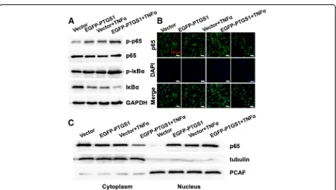
Knockdown of PTGS1 inhibits the translocation of p65 from the cytoplasm to the nucleus
In order to elucidate the molecular mechanism by which PTGS1 regulates osteogenesis in ASCs, we next investi-gated the potential effect of PTGS1 on NF-κB, the mas-ter regulator of the inflammatory response. As shown in Fig. 5a–c, expressions of the p65 targets IL6, IL8, and
SELE were impaired in PTGS1 knockdown cells, sug-gesting that PTGS1 is a positive regulator of the p65 pathway.
Next, we studied whether the classical or non-classical NF-κB signaling pathway was activated during PTGS1-mediated osteogenic differentiation of ASCs. We ob-served that silencing of PTGS1 significantly reduced the phosphorylation and degradation levels of IκBα protein in the presence of TNFα. Moreover, the phosphorylation level of p65 was also significantly inhibited in PTGS1 knockdown cells (Fig.5d). To further explore the under-lying mechanism, we studied the subcellular localization of p65 in PTGS1 knockdown cells. Confocal microscopy results showed the nuclear translocation of p65 induced
Western blot and confocal microscopy analyses. Altogether, these results demonstrate that knockdown of PTGS1 inhibits the NF-κB signal through promot-ing the nuclear exclusion of p65. (All original western membranes: Additional file 2).
Discussion
PTGS1 has been implicated in numerous pathophysio-logical processes including arthritic disease, cancer, pain, and inflammation, but its role in osteogenic differentiation and inflammatory regulation in MSCs remains fairly vague. Significant evidence has accumulated revealing that inhibition of PTGS1 yields an anti-inflammatory effect. Previous research showed that genetic deletion or pharmacological inhibition of PTGS1 could reduce the LPS-induced inflammatory response, potentially acting through the NF-κB pathway. Ptgs1-deficient mice showed a decreased level of arachidonic acid-induced inflammation [12,23].
In this study, we found that PTGS1 was activated upon TNFαtreatment in ASCs. Moreover, deletion of PTGS1 sub-stantially attenuated the expressions ofIL6, IL8, and SELE. These results indicate that inhibition of PTGS1 may have the potential to reduce inflammation in ASCs. Previously,
the role of PTGS1 in osteogenesis has rarely been docu-mented. A related study reported that the PTGS1 inhibitor BGJb can rescue bone resorption induced by IL1 [15]. Herein, we demonstrated knockdown of PTGS1 significantly enhanced the osteogenic differentiation of ASCs, and overex-pression of PTGS1 inhibited osteogenesis capacity both in vitro and in vivo. This is consistent with the above report, which indicated PTGS1 exerts a negative regulatory role in bone formation. Research about bone forming related to blocking inflammation is a meaningful part for bone tissue regeneration, which was closely coordinated by cell micro-environment. Bone injury model with inflammation is needed to carry out in our future research.
interaction with IκBa. After stimulation by TNFα, the classical pathway is triggered as follows: IKK complexes are phosphorylated by the IκB kinase, IκBa protein undergoes phosphorylation and ubiquitin-dependent degradation, and the p50-p65 dimers are released from IκBa protein, translocating to the nucleus. In addition, the nuclear p50-p65 dimer can also combine with IκBa protein to form a strong nuclear export signal. The heterodimer is crucial for the expression of genes encoding pro-inflammatory mediators and anti-osteogenesis regulators [26, 27].
Mechanistically, we found that knockdown of PTGS1 substantially repressed the expressions of NF-κB target genes (IL6, IL8, and SELE). Further-more, deletion of PTGS1 reduced the phosphorylation and degradation levels of IκBa protein and the phos-phorylation of p65. Conversely, overexpression of PTGS1 could induce the phosphorylation and degrad-ation levels of IκBa protein and the phosphoryldegrad-ation of p65. These results reveal that inhibition of PTGS1 exerts pro-osteogenic function in ASCs by regulating the classical NF-κB pathway. Furthermore, we also found that PTGS1 gene silencing can prevent nuclear translocation of p65, while its overexpression resulted in accelerated nuclear entrance of p65, as verified by confocal microscopy and Western blot analyses of nu-clear and cytoplasmic cell fractions. The new link be-tween PTGS1 and NF-κB suggests their collaboration in regulating the osteogenic differentiation and anti-inflammation of ASCs; future research is needed to further define the underlying mechanism.
In summary, our study demonstrates that knock-down of PTGS1 promotes the osteogenic differenti-ation of ASCs both in vitro and in vivo by targeting p65 cytoplasmic/nuclear translocation. More import-antly, silencing of PTGS1 may provide a potential means for modulating the inflammatory microenvir-onment during bone remodeling. Our findings dem-onstrate that PTGS1 can be a potential target for treating inflammation-associated bone defects and is a key factor to consider in stem cell-based approaches for bone tissue regeneration.
Conclusions
Our results first demonstrated that inhibition of PTGS1 promoted the osteogenic differentiation of ASCs through repressing NF-κB pathway by targeting p65 cytoplasmic/nuclear translocation. Furthermore, knock-down of PTGS1 may provide a potential means for regu-lating inflammatory microenvironment in the process of bone remodeling. Collectively, PTGS1-NF-κB signaling pathway is a novel molecular target for ASC-mediated regenerative medicine.
Additional files
Additional file 1:Figure S1.Knockdown of PTGS1 enhances the osteogenic differentiation in vitro. A Microscopic images of GFP-positive ASCs under light and fluorescence microscopy. Scale bar, 500μm. B-C Knockdown of PTGS1 was verified by real-time RT-qPCR and Western blot. D-E PTGS1 knockdown promoted ALP activity and increased ALP staining. F-G PTGS1 knockdown increased mineralization, as shown by Alizarin red staining and quantitative calcium analysis. H-L Silencing of PTGS1 increased the expressions ofALP,OCN,BSP,RUNX2, andOSX. **P< 0.01. NC negative control cells,PTGS1sh PTGS1 knockdown cells, d day, w week. (TIFF 2714 kb)
Additional file 2:Figure S2.All original western membranes. (TIFF 2886 kb)
Abbreviations
ALP:Alkaline phosphatase; ARS: Alizarin red staining; ASCs: Adipose-derived stem cells; BSP: Bone sialoprotein; GAPDH: Glyceraldehyde-3-phosphate dehydrogenase; HE: Hematoxylin and eosin; IHC: Immunohistochemistry; IL6: Interleukin 6; IL8: Interleukin 8; MSCs: Mesenchymal stem cells;
NF-κB: Nuclear factor kappa B; OCN: Osteocalcin; OSX: Osterix; PTGS1: Prostaglandin G/H synthase 1; RUNX2: Runt-related transcription factor 2; SELE: E-selectin; TNFα: Tumor necrosis factor alpha
Acknowledgements Not applicable.
Funding
This study was supported by grants from the National Natural Science Foundation of China (No. 81500822 to P.Z., No. 81570953 to Y.Z.), the Project for Culturing Leading Talents in Scientific and Technological Innovation of Beijing (Z171100001117169 to Y.Z.), the PKU School of Stomatology for Talented Young Investigators (PKUSS20140109 to P.Z.), the Beijing Nova Program (Z181100006218037).
Availability of data and materials
The authors confirm that all data underlying the findings are fully available.
Authors’contributions
YW was responsible for the collection and/or assembly of data, data analysis and interpretation, and manuscript writing; YL and MZ were responsible for the collection and/or assembly of data; LL and XZ were responsible for the provision of study material; PZ was responsible for the conception and design, collection and/or assembly of data, data analysis and interpretation, manuscript writing, and financial support; YZ was responsible for the conception and design, financial support, and manuscript writing. All authors read and approved the final manuscript.
Ethics approval and consent to participate
This study was carried out in strict accordance with the recommendations of the Guide for the Care and Use of Laboratory Animals of the National Institutes of Health. The protocol was approved by the Institutional Animal Care and Use Committee of the Peking University Health Science Center (approval no. LA2014233). All surgeries were performed under anesthesia, and all efforts were made to minimize animal suffering.
Consent for publication Not applicable.
Competing interests
The authors declare that they have no competing interests.
Publisher’s Note
Springer Nature remains neutral with regard to jurisdictional claims in published maps and institutional affiliations.
Author details
1Department of Prosthodontics, Peking University School and Hospital of
100081, China.2National Engineering Lab for Digital and Material Technology
of Stomatology, Peking University School and Hospital of Stomatology, Beijing 100081, China.
Received: 24 December 2018 Revised: 30 January 2019 Accepted: 4 February 2019
References
1. Ito K, Yamada Y, Nagasaka T, Baba S, Ueda M. Osteogenic potential of injectable tissue-engineered bone: a comparison among autogenous bone, bone substitute (Bio-Oss), platelet-rich plasma, and tissue-engineered bone with respect to their mechanical properties and histological findings. J Biomed Mater Res A. 2005;73:63–7.
2. Levi B, Longaker M. Concise review: adipose-derived stromal cells for skeletal regenerative medicine. Stem Cells. 2011;29:576–82.
3. Wilson A, Butler PE, Seifalian AM. Adipose-derived stem cells for clinical applications: a review. Cell Prolif. 2011;44:86–98.
4. Zuk PA, Zhu M, Ashjian P, De Ugarte DA, Huang JI, Mizuno H, et al. Human adipose tissue is a source of multipotent stem cells. Mol Biol Cell. 2002;13: 4279–95.
5. Smith WL, DeWitt DL, Garavito RM. Cyclooxygenases: structural, cellular, and molecular biology. Annu Rev Biochem. 2000;69:145–82.
6. Plant MH, Laneuville O. Characterization of a novel transcript of prostaglandin endoperoxide H synthase 1 with a tissue-specific profile of expression. Biochem J. 1999;3:677–85.
7. Tanabe T, Tohnai N. Cyclooxygenase isozymes and their gene structures and expression. Prostaglandins Other Lipid Mediat. 2002;68-69:95–114. 8. Wang LH, Hajibeigi A, Xu XM, Loose-Mitchell D, Wu KK. Characterization of
the promoter of human prostaglandin H synthase-1 gene. Biochem Biophys Res Commun. 1993;190:406–11.
9. Xu XM, Tang JL, Chen X, Wang LH, Wu KK. Involvement of two Sp1 elements in basal endothelial prostaglandin H synthase-1 promoter activity. J Biol Chem. 1997;272:6943–50.
10. Kargman SL, O'Neill GP, Vickers PJ, Evans JF, Mancini JA, Jothy S. Expression of Prostaglandin G/H synthase-1 and -2 protein in human colon cancer. Cancer Res. 1995;55:2556–9.
11. Fuster V, Sweeny JM. Aspirin: a historical and contemporary therapeutic overview. Circulation. 2011;123:768–78.
12. Calvello R, Lofrumento DD, Perrone MG, Cianciulli A, Salvatore R, Vitale P, et al. Highly selective cyclooxygenase-1 inhibitors P6 and Mofezolac counteract inflammatory state both in vitro and in vivo. Front Neurol. 2017; 8:251.
13. Choi SH, Langenbach R, Bosetti F. Genetic deletion or pharmacological inhibition of cyclooxygenase-1 attenuate lipopolysaccharide-induced inflammatory response and brain injury. FASEB J. 2008;22:491–501. 14. Sokolowska M, Chen LY, Eberlein M, Martinez-Anton A, Liu Y, Alsaaty S, et al.
Low molecular weight hyaluronan activates cytosolic phospholipase A2α and eicosanoid production in monocytes and macrophages. J Biol Chem. 2014;289:4470–88.
15. Allison AC, Chin RC, Cheng Y. Cyclooxygenase inhibitors vary widely in potency for preventing cytokine-induced bone resorption. Ann N Y Acad Sci. 1993;696:303–6.
16. Boyle WJ, Simonet WS, Lacey DL. Osteoclast differentiation and activation. Nature. 2003;423:337–42.
17. Khosla S, Westendorf JJ, Oursler MJ. Building bone to reverse osteoporosis and repair fractures. J Clin Invest. 2008;118:421–8.
18. Krum SA, Chang J, Miranda-Carboni G, Wang CY. Novel functions for NFkappaB: inhibition of bone formation. Nat Rev Rheumatol. 2010;6:607–11. 19. Jimi E, Ghosh S. Role of nuclear factor-κB in the immune system and bone.
Immunol Rev. 2005;208:80–7.
20. Wang Y, Liu Y, Zhang M, Lv L, Zhang X, Zhang P, et al. LRRC15 promotes osteogenic differentiation of mesenchymal stem cells by modulating p65 cytoplasmic/nuclear translocation. Stem Cell Res Ther. 2018;9:65. 21. Zhang P, Liu Y, Jin C, Zhang M, Lv L, Zhang X, et al. Histone H3K9
acetyltransferase PCAF is essential for osteogenic differentiation through bone morphogenetic protein signaling and may be involved in osteoporosis. Stem Cells. 2016;34:2332–41.
22. Wang Y, Jia Z, Diao S, Lin X, Lian X, Wang L, et al. IGFBP5 enhances osteogenic differentiation potential of periodontal ligament stem cells and Wharton’s jelly umbilical cord stem cells, via the JNK and MEK/Erk signalling pathways. Cell Prolif. 2016;49:618–27.
23. Loftin CD, Tiano HF, Langenbach R. Phenotypes of the COX-deficient mice indicate physiological and pathophysiological roles for COX-1 and COX-2. Prostaglandins Other Lipid Mediat. 2002;68-69:177–85.
24. Huang RL, Yuan Y, Tu J, Zou GM, Li Q. Opposing TNF-α/IL-1β- and activated MAPK signaling pathways converge on Runx2 to regulate BMP-2-induced osteoblastic differentiation. Cell Death Dis. 2014;5:e1187. 25. Xie J, Gu J. Identification of C-terminal Hsp70-interacting protein as a
mediator of tumour necrosis factor action in osteoblast differentiation by targeting osterix for degradation. J Cell Mol Med. 2015;19:1814–24. 26. Sun SC, Ley SC. New insights into NF-kappaB regulation and function.
Trends Immunol. 2008;29:469–78.