PRIMARY RESEARCH
miR-484 suppresses proliferation
and epithelial–mesenchymal transition
by targeting ZEB1 and SMAD2 in cervical cancer
cells
Yang Hu, Hong Xie, Yankun Liu, Weiying Liu, Min Liu and Hua Tang
*Abstract
Background: MicroRNAs (miRNAs) play important roles in cancer initiation and development. Epithelial–mesen-chymal transition (EMT) is a form of cellular plasticity that is critical for embryonic development and metastasis. The purpose of the study was to determine the function and mechanism of miR-484 in initiation and development of cervical cancer (CC).
Methods: We determined the expression levels of miR-484 in cervical cancer tissues and cell lines with RT-qPCR. Prediction algorithms and EGFP reporter assay were performed to evaluate the targets for miR-484. MTT assay, colony formation assay, flow cytometric analysis, transwell cell migration and invasion assays, and detection of EMT markers were employed to investigate the roles of miR-484 and the targets in regulation of cell proliferation and EMT process. We also used rescue experiments to confirm the effect of miR-484 on CC cells through directly regulating the expres-sion of its targets.
Results: Firstly we found miR-484 was down-regulated in cervical cancer tissues and cell lines compared with their matched non-cancerous tissues or normal cervical keratinocytes cells. Further studies revealed that overexpres-sion of miR-484 suppressed the cell proliferation, while exacerbates apoptosis. Besides, miR-484 suppressed cellular migration, invasion and EMT process of CC cells. EGFP reporter assay showed that miR-484 binds to ZEB1 and SMAD2 3′UTR region and reduced their expression. The expression of miR-484 had reverse correlation with SMAD2/ZEB1, and SMAD2/ZEB1 had positive correlation with each other in cervical cancer tissues and cell lines. Furthermore, the ectopic expression of ZEB1 or SMAD2 could rescue the malignancies suppressed by 484, suggesting that miR-484 down-regulates ZEB1 and SMAD2 to repress tumorigenic activities.
Conclusion: We found miR-484 inhibits cell proliferation and the EMT process by targeting both ZEB1 and SMAD2 genes and functions as a tumor suppressor, which may served as potential biomarkers for cervical cancer.
Keywords: miR-484, SMAD2, ZEB1, EMT, Cervical cancer
© The Author(s) 2017. This article is distributed under the terms of the Creative Commons Attribution 4.0 International License (http://creativecommons.org/licenses/by/4.0/), which permits unrestricted use, distribution, and reproduction in any medium, provided you give appropriate credit to the original author(s) and the source, provide a link to the Creative Commons license, and indicate if changes were made. The Creative Commons Public Domain Dedication waiver (http://creativecommons.org/ publicdomain/zero/1.0/) applies to the data made available in this article, unless otherwise stated.
Background
Cervical cancer (CC) is the fourth leading cause among cancer-related deaths in women, and due to delayed ini-tial screening, it mainly occurs in developing countries
and causes about 265,000 deaths every year worldwide [1]. Nowadays, advances in CC therapies have improved treatment outcomes, while the prognosis remains lim-ited and ineffective and a great number of patients died of metastasis. Although human papilloma virus (HPV) is the major risk factor for CC [2, 3], independent altera-tions in tumour suppressor genes and oncogenes are essential for the development of these cancers as well [4, 5]. Therefore, it’s crucial to identify specific molecules
Open Access
*Correspondence: tangh@tijmu.edu.cn
and markers that contribute to understanding cervical carcinogenesis and ascertaining diagnostic and treatment strategies. Recently, researchers have focused on the effect of miRNAs on CC and a lot of miRNAs were found to play great importance in the initiation and develop-ment of CC [6–8].
MicroRNAs are a class of 18-25-nucleotide, highly conserved non-coding RNAs that post-transcriptionally regulate gene expression by binding to their 3′UTRs and regulate a wide range of physiological and pathologi-cal processes including cell differentiation, proliferation, apoptosis, invasion and migration [9–12]. In addition, growing evidences indicate that miRNAs are aberrantly expressed in human cancers and may function as tumor suppressors or oncogenes [13]. miR-484 was located on chr6. The expressions and functions of miR-484 in cancers were little. Although Yang et al. [14] reported that miR-484 was overexpressed in premalignant lesions of hepatocellu-lar carcinoma (HCC), and can promote hepatocyte trans-formation and hepatoma development in two hepatocyte orthotopic transplantation models. Until now, the role and mechanism of miR-484 in CC cells are not clear.
Epithelial–mesenchymal transition (EMT) is an essen-tial requirement for cancer invasion and metastasis [15–17]. The transcription factors Snail, Slug, Twist, zinc finger E-box-binding homeobox (ZEB) play vital role in initiation of EMT process. Recent reports have showed that the miR-200 family and other miRNAs regu-late EMT through targeting these transcription factors [18–20]. ZEB family factors (ZEB1 and ZEB2) are tran-scriptional repressors that comprise two widely separated clusters of C2H2-type zinc fingers which bind to paired CAGGTA/G E-box-like promoter elements. These fac-tors promote EMT by repressing expression E-cadherin [21–23] and are important intracellular mediators of TGFβ-induced EMT. Over the past few years, ZEB1 has increasingly been considered as an important contributor to the process of malignancies including endometrioid cancer [24], breast cancer [25], lung adenocarcinomas [26] as well as cervical cancer [27]. On the one hand, it has been shown that miRNAs, such as the miR-200 family can directly bind to 3′UTR of the ZEB mRNA to down-regulate its expression and influence epithelial differentiation [28, 29]. On the other hand, it has been revealed that SMAD proteins directly act with the pro-moter of the ZEB factor and indirectly regulates estab-lishment and maintenance of EMT [30, 31].
In this report, we demonstrated that miR-484 is down-regulated in cervical cancer tissues and cell lines, and overexpression of miR-484 inhibits cell proliferation, cell viability and exacerbates apoptosis, suppresses cell migration, invasion and EMT process of CC cells as well. Moreover, miR-484 was validated to directly bind to the
3′UTR of the ZEB1 and SMAD2 transcript, inhibiting their expression in CC cells. We also found that SMAD2 is an upstream regulator of ZEB1. Therefore, miR-484 regulated EMT process through both directly and indi-rectly targeting ZEB1. Collectively, our present work provides the first evidence that miR-484 down-regulates ZEB1 and SMAD2 expression to repress malignant prop-erties in CC cells. The findings may provide insights into the mechanisms underlying carcinogenesis and potential biomarkers for cervical cancer.
Methods
Human cervical cancer tissue specimens and cell lines Fifteen CC tissues and the paired adjacent non-tumor cervical tissues were obtained from the cancer center of Sun Yat-sen University. The diagnose was evaluated by pathological analysis. Written informed consent was obtained from each patient and ethics approval for this work was granted by the Ethics Committee of Sun Yat-Sen University. The cervical samples were classified by pathologists. The human CC cell lines HeLa, Caski and ME-180 were maintained in RPMI-1640 medium. C33A, SiHa and SW756 were maintained in MEM-α medium according to Ref. [32]. Primary cultures of normal cer-vical keratinocytes (NCx) were obtained from hyster-ectomy specimens removed for non-neoplastic disease unrelated to the cervix. Cell culture and determination of growth rates were according to Ref. [33]. All the cells were maintained in a humidified incubator with 5% car-bon dioxide (CO2) at 37 °C.
Vector construction
To over-express miR-484, the primary miR-484 frag-ment was amplified from genomic DNA and cloned into pcDNA3 vector between BamHI and EcoRI sites. To block the function of miR-484, we purchased the 2′-O -methyl-modified antisense oligonucleotide of miR-484 (ASO-miR-484) and the scramble control oligonucleotides (ASO-NC) from the GenePharma (Shanghai, China).
The gene encoding ZEB1/SMAD2 was amplified from the cDNA of HeLa cells, and the product was cloned into pcDNA3-Flag vector between EcoRI and XhoI sites. The shRNA for knocking down SMAD2 and ZEB1 were synthesized from GenePharma (Shanghai, China) and were annealed and cloned into pSilencer 2.1-neo vector (Ambion) between BamHI and HindIII sites.
or pcDNA3-EGFP/SMAD2 3′UTR). All of the primers for PCR amplification and all the oligonucleotides for annealing are listed in Table 1.
Prediction of miRNA targets
miR-484 predicted targets were retrieved from TargetScan (http://genes.mit.edu/tscan/targetscanS.html), miRecords
(http://c1.accurascience.com/miRecords/) and PITA
(https://genie.weizmann.ac.il/pubs/mir07/) and the bind-ing site predictions were performed usbind-ing RNAhybrid (http://bibiserv.techfak.uni-bielefeld.de/rnahybrid/).
Cell transfection
Transient transfection was performed in antibiotic-free Opti-MEM medium (Invitrogen) with the Lipofectamine 2000 reagent (Invitrogen, Carlsbad, CA) following the manufacturer’s protocol.
RNA isolation and reverse transcription quantity (RT‑qPCR) Extraction of total RNA from cells was performed using the TRIzol reagent (Invitrogen, CA) following the manu-facturer’s instructions. Expression of mature miRNAs and mRNAs were quantified by RT-qPCR using the SYBR Premix Ex TaqTM (Promega, Madison, WI). The concen-tration of RNA were measured with a NanoDrop 2000 spectrophotometer (Thermo Fisher Scientific, Waltham, MA, USA) and stored at −80 °C for further use. Special stem-loop primers were used for the miRNA reverse tran-scription (RT) reaction, and U6 small nuclear B noncoding RNA (RNU6B) was used as the endogenous control to nor-malize the level of miRNA. The oligo (dT) primer was used for the RT reaction for gene expression. β-actin was used as the endogenous control to normalize the level of genes. All analyses were performed in triplicate and reported as 2− ΔΔCt. The primers for RT and PCR are provided in Table 1.
Table 1 The oligonucleotides and primers used in this study
Name Sequence (5′–3′)
Pri-miR-484-forward CGACGGATCCAAGCGCACCCTTCACTTC Pri-miR-484-reverse GCTCGAATTCCGCTTCAAGGTTCCTTTCG
ASO-miR-484 AUCGGGAGGGGACUGAGCCUGA
ASO-NC CAGUACUUUUGUGUAGUACAA
SMAD2-forward GCGGAATTCAACATGTCGTCCATCTTGCCATTC SMAD2-reverse CCAGCTCGAGTTATGACATGCTTGAGCAACG
ZEB1-forward GCGGATCCGCGGATGGCCCCAGGTGTAAG
ZEB1-reverse GACGCCTCGAGTTAGGCTTCATTTGTCTTTTCTTC SMAD2-3′UTR-S GATCCTTTCTAGTATCTTACAGCCTGATAAGCTTG SMAD2-3′UTR-AS AATTCAAGCTTATCAGGCTGTAAGATACTAGAAAG ZEB1-3′UTR-S GATCCTTCTGGAGAGGTCAGAGTTGACAAGCTTG ZEB1-3′UTR-AS AATTCAAGCTTGTCAACTCTGACCTCTCCAGAAG SMAD2-3′UTRmut-S GATCCTTTCTAGTATCTTACCGAGCTATAAGCTTG SMAD2-3′UTRmut-AS AATTCAAGCTTATAGCTCGGTAAGATACTAGAAAG ZEB1-3′UTRmut-S GATCCTTCTGGAGAGGTCATCTAACTACAAGCTTG ZEB1-3′UTRmut-AS AATTCAAGCTTGTAGTTAGATGACCTCTCCAGAAG
miR-484-RT primer GTCGTATCCAGTGCAGGGTCCGAGGTGCACTGGATACGACATCGGGAG
miR-484-forward TGCAGTCAGGCTCAGTCCCC
U6-RT primer GTCGTATCCAGTGCAGGGTCCGAGGTATTCGCACTGGATACGACAAAATATGGAAC
U6-forward TGCGGGTGCTCGCTTCGGCAGC
Reverse primer CCAGTGCAGGGTCCGAGGT
qPCR-actin-forward CGTGACATTAAGGAGAAGCTG qPCR-actin-reverse CTAGAAGCATTTGCGGTGGAC qPCR-ZEB1-forward TAAAGTGGCGGTAGATGGTA qPCR-ZEB1-reverse ACTGTTTGTAGCGACTGGATT
qPCR-FN1-forward CAGTGGGAGACCTCGAGAAG
qPCR-FN1-reverse TCCCTCGGAACATCAGAAAC
Fluorescent reporter assays
To identify the direct target relationship between miR-484 and the 3′UTR of ZEB1/SMAD2 mRNA, the CC cells were cotransfected with pcDNA3/pri-miR-484 or ASO-miR-484 and the 3′UTR of ZEB1/SMAD2 or the mutant 3′UTR of ZEB1/SMAD2 in 48-well plates. The vector pDsRed2-N1 (Clontech, Mountain View, CA) expressing RFP (red fluorescent protein) was transfected together with the above plasmids and used as an internal control standard. 48 h after transfection, the cells were lysed by RIPA buffer and fluorescence intensities of EGFP and RFP were detected with an F-4500 fluorescence spectro-photometer (Hitachi, Tokyo, Japan).
Cell viability assay and colony formation assay
The cell viability of CC cells was evaluated by the 3-(4,5-dimethylthiazol-2-yl)-2,5-diphenyl-tetrazolium bromide (MTT) assay. The absorbance was determined at 570 nm (A570) (Bio-Tek Instruments, Winooski, VT, USA) after CC cells transfection for 24, 48 and 72 h. The details of methods were according to Ref. [34].
For colony formation assay, the cells were seeded into 12-well plates at a density of 300 HeLa or 400 C33A per well at post-transfection 24 h. Change medium every 3 days. After 11 or 13 days, the cells were stained with crystal violet, and colonies including more than 50 cells were counted. The average number was used to evaluate the formation ability.
Cell cycle analysis and apoptosis assay by flow cytometry Cell cycle analysis and apoptosis assay was according to Refs. [6] and [35] respectively.
Cell migration and invasion assays
The migration and invasion were analyzed by 24-well Boyden chambers with an 8-μm pore size polycarbon-ate membrane (Corning, Cambridge, MA). Briefly, 8 × 10⁵ cells were resuspended in culture medium without FBS and seeded in the upper chamber. Then the chamber was placed into a 24-well plate containing 800 μL of culture media with 20% FBS. Approximately 48 h later, the cells were fixed with paraformaldehyde and stained with crystal violet. The cells did not pass through the membrane were removed with the cotton stick while the cells that passed through the membrane were counted.
Western blot analysis
The detailed procedures for western blot were described in a previous study [36]. The primary anti-bodies used in this study including ZEB1, SMAD2, E-cadherin, cytoker-atin, vimentin, N-cadherin, fibronectin and GAPDH, which were obtained from Saier Co. (Tianjin, China). The
secondary goat anti-rabbit antibodies were purchased from Sigma.
Immunohistochemistry
The tumor tissues were fixed in 4% formaldehyde for 24 h and sent to Tangshan People’s Hospital for immunohistochemistry.
Statistical analyses
All the data are presented as the mean ± SD. Each experiment was performed at least three times, and the analysis was performed using paired t test. p ≤ 0.05 was considered statistically significant (*p < 0.05, **p < 0.01, ***p < 0.001, ****p < 0.0001).
Results
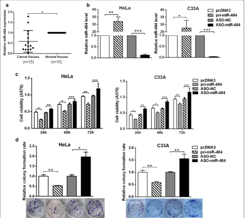
miR‑484 is down‑regulated in cervical cancer tissues We first examined the expression of miR-484 in 15 pairs of cervical cancer tissues and the adjacent noncancer-ous tissues by RT-qPCR. The results showed that miR-484 was generally down-regulated in cervical cancer tissues compared with the adjacent noncancerous tissues (Fig. 1a).
miR‑484 inhibits cell growth of cervical cancer cells, suppresses cell cycle while exacerbates apoptosis in cervical cancer cells
First, we tested the efficiency of the plasmids pcDNA3/ pri-miR-484 and ASO-miR-484 in HeLa and C33A cells with RT-qPCR. The results showed that both the plas-mid and ASO-miR-484 can change the miR-484 expres-sion efficiently (Fig. 1b). Next, MTT assays were used to test cell viability after transfecting with pcDNA3/ pri-miR-484 or ASO-miR-484 at 24, 48 and 72 h. The results showed that miR-484 decreased, whereas ASO-miR-484 increased cell viability in both HeLa and C33A cells (Fig. 1c). Meanwhile, colony formation assay was performed to test the effects of miR-484 on proliferation. The results showed that the overexpression of miR-484 suppressed, whereas ASO-miR-484 increased the colony formation rate (Fig. 1d). Taken together, these results indicate that miR-484 suppresses the proliferation of HeLa and C33A cells.
contrast, inhibition of miR-484 by antisense oligonucleo-tides in HeLa cells led to an increase in the percentage of cells in the G2/M phase from 13.85 to 21.90% and a decrease in the percentage of cells in the G1 phase from 65.32 to 56.87% (Fig. 2b). The proliferation index of the ASO-miR-484-treated HeLa cells was increased com-pared with the ASO control (Fig. 2b). In addition, flow cytometry results showed that miR-484 exacerbates apoptosis, while ASO-miR-484 inhibited apoptosis obvi-ously in CC cells (Fig. 2c). Taken together, all these results
indicated that miR-484 suppresses cell growth by both delaying the G1 to S phase and the S to G2 phase transi-tions and exacerbating apoptosis in CC cells.
miR‑484 suppresses the migration and invasion of cervical cancer cells and inhibits the EMT process
showed that overexpression of miR-484 significantly sup-pressed the migration ability by approximately 59.8 and 43.7% in HeLa and C33A cells; while blocking of miR-484 increased the migration ability by approximately 1.7- and 1.9-fold in HeLa and C33A cells respectively (Fig. 3a). The overexpression of miR-484 suppressed the invasion ability by approximately 52.1 and 44% in HeLa and C33A cells; while ASO-miR-484 increased the invasion ability by 1.6- and 1.7-fold in HeLa and C33A cells respectively (Fig. 3b).
It has been reported that EMT is an important mecha-nism correlated with migration and invasion [22]. During the transition, the expression of epithelial markers that enhance cell–cell contact decreases, while the expres-sion of mesenchymal markers increases [17]. Therefore, we tested the expression of molecular markers to clarify the effects of miR-484 on the EMT process. As shown in Fig. 3c, the overexpression of miR-484 increased epi-thelial markers (E-cadherin and cytokeratin) protein levels but decreased mesenchymal markers protein lev-els (vimentin, N-cadherin and fibronectin) in both HeLa and C33A cells. By contrast, ASO-miR-484 decreased the epithelial markers but increased the mesenchymal mark-ers protein levels. Importantly, RT-qPCR showed that miR-484 decreased the expression of transcription fac-tors Snail, Slug, Twist and ZEB1 which play vital role in initiation of EMT process (Fig. 3d). With the modulation of miR-484, the expression of ZEB1 showed the great-est alteration. In summary, these results demonstrated that miR-484 suppresses the migration and invasion and inhibits the EMT process of CC cells.
miR‑484 targets and down regulates ZEB1 and SMAD2 expression
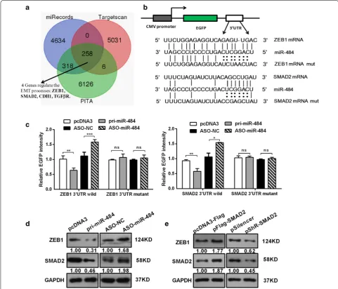
As miR-484 suppresses cervical cancer cell growth, migration, and invasion, it is important to understand which targets are directly responsible for the observed phenotypes. We used three prediction algorithms (Tar-getScan, miRecords, and PITA) to predict the targets for miR-484 in common. There were 258 candidate targets shared by all the three databases (the overlapping frac-tion), in which only four genes involved in the regulation of EMT process (Fig. 4a). Based on our data (Fig. 3d) and previous reports, we chose ZEB1 and SMAD2 for fur-ther study. ZEB1 usually acts as a key transcription factor which can induce EMT, and our data had shown ZEB1 expression was altered with the modulation of miR-484 (Fig. 3d), which suggested that ZEB1 may be a bona fide target of miR-484. Moreover, the previous work in our lab has demonstrated that SMAD2 promotes cell growth, migration, invasion, and EMT in Human CC Cell Lines [6]. Other reports have revealed that Smads interact with the ZEB promoter [30, 31]. Therefore, we chose SMAD2 as a second candidate target of miR-484.
To confirm whether SMAD2 and ZEB1 are direct tar-gets of miR-484 in human CC cells, we used an EGFP reporter system, in which we cloned the wild SMAD2/ ZEB1 3′-UTR or a mutant SMAD2/ZEB1 3′-UTR downstream of the EGFP reporter gene (Fig. 4b). Co-transfection was performed with an RFP reporter as a transfection normalizer and with either pri-miR-484 or ASO-miR-484 in HeLa cells. After 48 h, we determined the fluorescence intensity. As shown in Fig. 4c, overex-pression of miR-484 decreased the fluorescent intensity of the wild type ZEB1/SMAD2 3′UTR. In contrast, ASO-miR-484 increased the fluorescent intensity (Fig. 4c). However, neither the over-expression nor inhibition of miR-484 affected the fluorescent intensity of the mutant ZEB1/SMAD2 3′UTR (Fig. 4c).
We also explored the functions of miR-484 in the expression of endogenous ZEB1/SMAD2 protein by western blot. The results showed that the overexpression of miR-484 decreased, while ASO-miR-484 increased the expression of ZEB1/SMAD2 (Fig. 4d). Thus, the results demonstrate that miR-484 directly targets the 3′UTR of ZEB1/SMAD2 and down-regulates their protein expres-sions in CC cells.
Interestingly, we observed that overexpression of SMAD2 led to an increase in ZEB1 protein level (Fig. 4e). Conversely, decreased SMAD2 prevented up-regulation of ZEB1 protein level. This indicates that SMAD2 is an upstream regulator of ZEB1 and regulated its expression. These results demonstrate that miR-484 directly targets the 3′UTR of ZEB1 and SMAD2 mRNA and down-regu-lates their expression simultaneously.
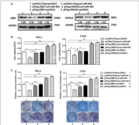
ZEB1 and SMAD2 rescue the suppression of the malignant behavior induced by miR‑484
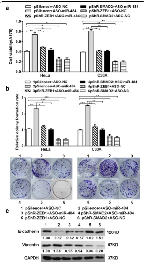
To confirm the deduce that the effect of miR-484 on CC cells is mediated by its down-regulatory effect on SMAD2 and ZEB1 expression in an inverse way, ASO-miR-484 along with pshR-ZEB1/SMAD2 was co-transfected
into HeLa and C33A cells. As anticipated, the effects of miR-484 blocking on cell growth were significantly impaired when ZEB1/SMAD2 was suppressed (Fig. 8a, b). Furthermore, the expressions of EMT markers in the (See figure on previous page.)
Fig. 3 miR-484 suppresses the migration and invasion of CC cells and down-regulates the EMT process. a, b After transfection 48 h, cell migration (a) and invasion (b) were evaluated using a transwell system with 8 μm pores in polycarbonate membranes. Representative views of migratory or invasive cells on the membrane were presented below. All pictures were photographed at ×20 magnification. c Protein levels of EMT-associated markers were assessed by western blotting after transfection 48 h. d RT-qPCR analysis for the expression of EMT transcription factors ZEB1, Snail, Slug and Twist2 in HeLa cells transfected with miR-484 or the control vector. The control was normalized to 1. All data represent mean ± SD of three independent experiments. *p < 0.05, **p < 0.01, ***p < 0.001
miR-484-blocking cells were restored to the normal lev-els after knocking-down of ZEB1/SMAD2 (Fig. 8c). In conclusion, all these data indicate that SMAD2 and ZEB1 are functional targets of miR-484 in cervical cancer cells.
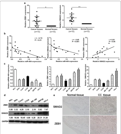
miR‑484 is inversely correlated with SMAD2 and ZEB1 expressions in cervical cancer tissues and cell lines
To further explore the expression levels of ZEB1 and SMAD2 in cervical cancer, we performed RT-qPCR analyses to examine their expression levels in 15 pairs
p < 0.05) (Fig. 9b). We also performed immunohisto-chemistry (IHC) to determine the expressions of ZEB1 and SMAD2 in clinical cervical samples. The results fur-ther demonstrated that ZEB1 and SMAD2 were both upregulated in cervical cancer tissues compared with the normal cervix (Fig. 9e).
We also determined the expressions of miR-484, ZEB1 and SMAD2 using RT-qPCR and western blotting analy-ses in a panel of cervical cancer cell lines as well as nor-mal cervical keratinocytes cells. RT-qPCR and western blotting analyses revealed a significant inverse correla-tion between miR-484 and ZEB1/SMAD2 expression
levels in different cervical cancer cell lines (Fig. 9c, d). The expression of ZEB1/SMAD2 in HeLa cells changed the most sharply while in C33A cells changed the least sharply compared with others. The expression of miR-484 and SMAD2/ZEB1 in both the tissues and cell lines indicated that miR-484 had reverse correlation with SMAD2/ZEB1, and that SMAD2 and ZEB1 had positive correlation with each other.
ZEB1 was the central molecules in this regulatory path-way. On one hand, miR-484 could inhibit cell prolifera-tion and EMT process by directly targeting ZEB1. On the
other hand, miR-484 suppresses cell growth and EMT by directly targeting SMAD2, an important upstream regulator of ZEB1 (Fig. 10). These results indicate that miR-484 exerts multiple pathways of regulation on ZEB1 expression resulting in the inhibition of cell growth and EMT.
Discussion
miRNAs have been indicated to be important regulators of a variety of biological processes, and their aberrant expressions are relevant to cancer initiation and develop-ment. In recent years, miRNAs were reported as poten-tial biomarkers as therapeutic approaches in human cancers [37]. Understanding the role of miRNAs in cer-vical cancer will provide theoretical basis for miRNA-specific personalized treatment and molecular-targeted therapy.
miR-484 was originally discovered to be associated with resistance to chemotherapeutic agents in cancer [38]. This year, an aberrant expression of miR-484 has also been described in high-fat-diet-induced tumours, with a fluctuant miR-484 overexpression during feeding [39]. Ectopic expression of miR-484 initiates tumourigen-esis and cell malignant transformation of HCC through synergistic activation of the TGF-β/Gli and NF-κB/IFN-I pathways [14]. This indicated that miR-484 may act as a significant role in tumor metastasis including EMT process. To our understanding, the functional study of miR-484 in cervical cancer is still missing. Here, we demonstrated that miR-484 was down-regulated in cer-vical cancer tissues compared to adjacent non-tumor tissues, which is different from the result in HCC. These differences may be due to the different types of cancer and the phases of the cancer, but the mechanisms need to be elucidated in the future. For example, miR-200s is downregulated and inhibit local invasion in breast can-cer but enhances spontaneous metastasis and coloniza-tion of lung adenocarcinoma [40]. Functional analyses revealed that overexpression of miR-484 suppressed the malignant behavior of cervical cancer cells. Specifi-cally, miR-484 overexpression significantly inhibited the cell proliferation, migration, invasion and EMT, whereas ASO-miR-484 enhanced these oncogenic features. In addition, western blot indicated that the expression of EMT markers including E-cadherin, cytokeratin, vimen-tin, N-cadherin and fibronectin were significantly modu-lated by miR-484, which is consistent with the formation Fig. 8 Knockdown of SMAD2/ZEB1 abrogates the effects induced
by ASO-miR-484. a After co-transfection with ASO-miR-484 and pshR-SMAD2/ZEB1 or the control vector for 48 h, MTT assay was performed to detect cell viability in CC cells. b The transfected cells were submitted to colony formation assays to test the proliferation of CC cells. c The expression of EMT markers (E-cadherin and vimentin) was detected by western blot analysis. All data represent mean ± SD of three independent experiments. *p < 0.05, **p < 0.01, ***p < 0.001 (See figure on previous page.)
Fig. 10 Schematic that describes the role of 484 in CC cells. miR-484 regulated proliferation and EMT process through both directly and indirectly targeting ZEB1 in CC cells
of EMT. Collectively, miR-484 functions as a tumor sup-pressor in cervical cancer cells.
miRNAs generally play their roles by regulating their target genes. To better understanding the role of miR-484 in CC, we applied bioinformatics analyses to predict that the 3′UTR of SMAD2/ZEB1 contains miR-484 binding sites. EGFP reporter assay demonstrated that miR-484 directly targets SMAD2 and ZEB1 transcripts. Western blot analyses showed the decreased expression of ZEB1 and SMAD2 upon overexpression of miR-484 and vise versa. Functionally, overexpression of miR-484 could inhibit cell proliferation, migration, invasion and EMT, while the ectopic expression of SMAD2/ZEB1 abrogated the inhibitory effects of miR-484 on these tumorigenic features in cervical cancer cells. Meanwhile, we also showed that knocking-down of SMAD2/ZEB1 abrogated the tumorigenic effects induced by ASO-miR-484. Thus, we concluded that miR-484 may function as tumor sup-pressor by down-regulation of ZEB1 and SMAD2 expres-sions in cervical cancer.
Upregulation of the expression of ZEB1 plays an important role in the progression and metastasis in many cancers [24–26], including cervical cancer [27]. Our pre-vious study has shown that SMAD2 promotes cell growth by facilitating the G1/S phase transition, enhances cell migration and invasion through regulated EMT in CC cell lines [6]. In addition, regulating SMAD2 by miRNAs has been identified to be involved in the TGFβ-induced EMT. For example, miR-18b targets SMAD2 and inhibits TGF-β1-induced differentiation of hair follicle stem cells into smooth muscle cells [41], and miR-27a inhibits colo-rectal carcinogenesis and progression [42]. Expectedly, ectopic expression of SMAD2 counteracts the inhibi-tion of EMT induced by miR-484 in CC cells. The resto-ration of SMAD2/ZEB1 expression mostly blocked the inhibitory influence of miR-484 on malignant behavior
(Figs. 5, 6, 7). The expression of miR-484 and SMAD2/ ZEB1 in the tissues and cell lines demonstrated that miR-484 had negative correlation with SMAD2/ZEB1, and that SMAD2 had positive correlation with ZEB1 (Fig. 9b, c). This result further supported that SMAD2 is an upstream protein of ZEB1 and they are both regu-lated by miR-484. Previous work has demonstrated that SMAD directly binds to the promoter of the ZEB factor and indirectly regulates EMT [30, 31], which is in accord-ance with our results in CC. In summary, all these results prove that miR-484 inhibits the development of CC and may partially (if not completely) through down-regulat-ing SMAD2 and ZEB1 expressions.
Except for ZEB, other transcription factors such as Snail, Slug and Twist are also the master regulators of EMT [22], which can activated by TGFβ directly and repress the transcription of E-cadherin [23]. The TGFβ/ ZEB/miR-200 double-negative feedback loop has been found and postulated to explain both the stability and interchangeability of the epithelial versus mesenchymal phenotypes in MDCK cell systems [43, 44]. In the present study, we found that miR-484 could target both SMAD2 and ZEB1 genes simultaneously, and ZEB1 is regulated by SMAD2 as well, which is different from The ZEB/ miR-200 double-negative feedback loop. Interesting, our results also showed that miR-484 affects the expression of Snail, Slug and Twist as well, which may due to miR-484 regulating ZEB1 to the EMT-related regulators, while the detailed regulatory mechanism need to be further investigated. These results indicate that miR-484 exerts multiple pathways of regulation on EMT and highlight the importance of stringently regulating transcription factors. These findings also fit the emerging concept that miRNAs fine-tune gene expression to precisely modulate essential biological processes and provide a mechanistic view of miRNA-based regulation on specific molecules and markers involved in cancer metastasis.
Despite these studies, further development of the underlying mechanisms need to be investigated. And our next work is to found out the upstream regulatory mech-anisms of miR-484 in CC cells.
Conclusions
carcinogenesis and imply its potential applications as new biomarkers in cervical cancer.
Abbreviations
miRNAs: microRNAs; CC: cervical cancer; NCx: normal cervical keratinocytes; EMT: epithelial–mesenchymal transition; ZEB: zinc finger E-box-binding homeobox; ASO-miR-484: the 2′-O-methyl-modified antisense oligonucleo-tide of miR-484; ASO-NC: the scramble control oligonucleooligonucleo-tides; MTT: the 3-(4,5-dimethylthiazol-2-yl)-2,5-diphenyl-tetrazolium bromide.
Authors’ contributions
Conceived and designed the experiments: TH, HY. Performed the experiments: HY, LY. Analyzed the data: HY. Contributed reagents/materials: LM, XH, LW. Wrote the paper: HY. Revised the paper: TH, XH. All authors read and approved the final manuscript.
Acknowledgements Not applicable.
Competing interests
The authors declare that they have no competing interests.
Availability of data and materials
The data and material of this study are included in this published article.
Ethics approval and consent to participate
Written informed consent was obtained from each patient and ethics approval for this work was granted by the Ethics Committee of Sun Yat-Sen University.
Funding
This work was supported by the National Natural Science Foundation of China (Nos. 91029714; 31270818; 81572790) and the Natural Science Foundation of Tianjin (12JCZDJC25100; 16JCYBJC42400).
Received: 9 November 2016 Accepted: 1 March 2017
References
1. Tjalma WA. Diagnostic performance of dual-staining cytology for cervical cancer screening: a systematic literature review. Eur J Obstet Gynecol Reprod Biol. 2017;210:275–80.
2. Banister CE, Liu C, Pirisi L, Creek KE, Buckhaults PJ. Identification and characterization of HPV-independent cervical cancers. Oncotarget. 2017. doi:10.18632/oncotarget.14533.
3. Tjalma WA, Kim E, Vandeweyer K. The impact on women’s health and the cervical cancer screening budget of primary HPV screening with dual-stain cytology triage in Belgium. Eur J Obstet Gynecol Reprod Biol. 2017;S0301–2115(17):30010–6.
4. Liu C, Wang J, Hu Y, Xie H, Liu M, Tang H. Upregulation of kazrin F by miR-186 suppresses apoptosis but promotes epithelial–mesenchymal transition to contribute to malignancy in human cervical cancer cells. Chin J Cancer Res. 2017. doi:10.21147/j.issn.1000-9604.2017.01.00. 5. Kim VN, Han J, Siomi MC. Biogenesis of small RNAs in animals. Nat Rev
Mol Cell Biol. 2009;10:126–39.
6. Zhao JL, Zhang L, Guo X, Wang JH, Zhou W, Liu M, Li X, Tang H. miR-212/132 downregulates SMAD2 expression to suppress the G1/S phase transition of the cell cycle and the epithelial to mesenchymal transition in cervical cancer cells. IUBMB Life. 2015;67(5):380–94.
7. Sun Y, Yang X, Liu M, Tang H. B4GALT3 up-regulation by miR-27a contrib-utes to the oncogenic activity in human cervical cancer cells. Cancer Lett. 2016;375(2):284–92.
8. Guo J, Lv J, Liu M, Tang H. miR-346 up-regulates argonaute 2 (AGO2) protein expression to augment the activity of other microRNAs (miR-NAs) and contributes to cervical cancer cell malignancy. J Biol Chem. 2015;290(51):30342–50.
9. Croce CM, Calin GA. miRNAs, cancer, and stem cell division. Cell. 2005;122:6–7.
10. Iorio MV, Ferracin M, Liu CG, Veronese A, Spizzo R, Sabbioni S, Magri E, Pedriali M, Fabbri M, Campiglio M, et al. MicroRNA gene expression deregulation in human breast cancer. Cancer Res. 2005;65:7065–70. 11. Calin GA, Liu CG, Ferracin M, Hyslop T, Spizzo R, Sevignani C, Fabbri M,
Cimmino A, Lee EJ, Wojcik SE, et al. Ultraconserved regions encoding ncRNAs are altered in human leukemias and carcinomas. Cancer Cell. 2007;12:215–29.
12. Visone R, Pallante P, Vecchione A, Cirombella R, Ferracin M, Ferraro A, Volinia S, Coluzzi S, Leone V, Borbone E, et al. Specific microRNAs are downregulated in human thyroid anaplastic carcinomas. Oncogene. 2007;26:7590–5.
13. Zhang L, Huang J, Yang N, Greshock J, Megraw MS, Giannakakis A, Liang S, Naylor TL, Barchetti A, Ward MR, et al. microRNAs exhibit high frequency genomic alterations in human cancer. Proc Natl Acad Sci USA. 2006;103:9136–41.
14. Yang Y, Lin X, Lu X, Luo G, Zeng T, Tang J, Jiang F, Li L, Cui X, Huang W, et al. Interferon-microRNA signalling drives liver precancerous lesion formation and hepatocarcinogenesis. Gut. 2016;65(7):1186–201.
15. Hu Y, Tang H. MicroRNAs regulate the epithelial to mesenchymal transi-tion (EMT) in cancer progression. MicroRNA. 2014;3(2):108–17.
16. Thiery JP, Acloque H, Huang RY, Nieto MA. Epithelial–mesenchymal transi-tions in development and disease. Cell. 2009;139:871–90.
17. Thiery JP, Sleeman JP. Complex networks orchestrate epithelial–mesen-chymal transitions. Nat Rev Mol Cell Biol. 2006;7:131–42.
18. Burk U, Schubert J, Wellner U, Schmalhofer O, Vincan E, Spaderna S, Brabletz T. A reciprocal repression between ZEB1 and members of the miR-200 family promotes EMT and invasion in cancer cells. EMBO Rep. 2008;9:582–9.
19. Gregory PA, Bert AG, Paterson EL, Barry SC, Tsykin A, Farshid G, Vadas MA, Khew-Goodall Y, Goodall GJ. The miR-200 family and miR-205 regulate epithelial to mesenchymal transition by targeting ZEB1 and SIP1. Nat Cell Biol. 2008;10:593–601.
20. Zhang LY, Liu M, Li X, Tang H. miR-490-3p modulates cell growth and epithelial to mesenchymal transition of hepatocellular carcinoma cells by targeting endoplasmic reticulum-Golgi intermediate compartment protein 3 (ERGIC3). J Biol Chem. 2013;288:4035–47.
21. Derynck R, Akhurst RJ. Differentiation plasticity regulated by TGF-β family proteins in development and disease. Nat Cell Biol. 2007;9:1000–4. 22. Kalluri R, Weinberg RA. The basics of epithelial–mesenchymal transition. J
Clin Investig. 2009;119:1420–8.
23. Vandewalle C, Van Roy F, Berx G. The role of the ZEB family of transcription factors in development and disease. Cell Mol Life Sci. 2009;66:773–87. 24. Singh M, Spoelstra NS, Jean A, Howe E, Torkko KC, Clark HR, Darling DS,
Shroyer KR, Horwitz KB, Broaddus RR, Richer JK. ZEB1 expression in type I vs type II endometrial cancers: a marker of aggressive disease. Mod Pathol. 2008;21:912–23.
25. Karihtala P, Auvinen P, Kauppila S, Haapasaari KM, Jukkola-Vuorinen A, Soini Y. Vimentin, zeb1 and Sip1 are up-regulated in triple-negative and basal-like breast cancers: association with an aggressive tumour pheno-type. Breast Cancer Res Treat. 2013;138:81–90.
26. Ohira T, Gemmill RM, Ferguson K, Kusy S, Roche J, Brambilla E, Zeng C, Baron A, Bemis L, Erickson P, Wilder E, Rustgi A, Kitajewski J, Gabrielson E, Bremnes R, Franklin W, Drabkin HA. WNT7a induces E-cadherin in lung cancer cells. Proc Natl Acad Sci USA. 2003;100:10429–34.
27. Ma Y, Zheng X, Zhou J, Zhang Y, Chen K. ZEB1 promotes the progres-sion and metastasis of cervical squamous cell carcinoma via the promotion of epithelial–mesenchymal transition. Int J Clin Exp Pathol. 2015;8(9):11258–67.
28. Hurteau GJ, Carlson JA, Spivack SD, Brock GJ. Over-expression of the microRNA hsa-miR-200c leads to reduced expression of transcription fac-tor 8 and increased expression of E-cadherin. Cancer Res. 2007;67:7972–6. 29. Korpal M, Lee ES, Hu G, Kang Y. The miR-200 family inhibits epithelial–
mesenchymal transition and cancer cell migration by direct targeting of E-cadherin transcriptional repressors ZEB1 and ZEB2. J Biol Chem. 2008;283:14910–4.
• We accept pre-submission inquiries
• Our selector tool helps you to find the most relevant journal • We provide round the clock customer support
• Convenient online submission • Thorough peer review
• Inclusion in PubMed and all major indexing services • Maximum visibility for your research
Submit your manuscript at www.biomedcentral.com/submit
Submit your next manuscript to BioMed Central
and we will help you at every step:
31. Lu J, Guo H, Treekitkarnmongkol W, Li P, Zhang J, Shi B, Ling C, Zhou X, Chen T, Chiao PJ, et al. 14-3-3zeta Cooperates with ErbB2 to promote ductal carcinoma in situ progression to invasive breast cancer by induc-ing epithelial mesenchymal transition. Cancer Cell. 2009;16:195–207. 32. Wan HY, Li QQ, Zhang Y, Tian W, Li YN, Liu M, Li X, Tang H. MiR-124
represses vasculogenic mimicry and cell motility by targeting amotL1 in cervical cancer cells. Cancer Lett. 2014;355(1):148–58.
33. Pett MR, Alazawi WO, Roberts I, Dowen S, Smith DI, Stanley MA, Coleman N. Acquisition of high-level chromosomal instability is associated with integration of human papillomavirus type 16 in cervical keratinocytes. Cancer Res. 2004;64(4):1359–68.
34. Long MJ, Wu FX, Li P, Liu M, Li X, Tang H. MicroRNA-10a targets CHL1 and promotes cell growth, migration and invasion in human cervical cancer cells. Cancer Lett. 2012;324:186–96.
35. Zhang MX, Zhang J, Zhang H, Tang H. miR-24-3p suppresses malignant behavior of lacrimal adenoid cystic carcinoma by targeting PRKCH to regulate p53/p21 pathway. PLoS ONE. 2016;11(6):e0158433. 36. Ren ZJ, Nong XY, Lv YR, Sun HH, An PP, Wang F, Li X, Liu M, Tang H.
Mir-509-5p joins the Mdm2/p53 feedback loop and regulates cancer cell growth. Cell Death Dis. 2014;5:e1387.
37. Osaki M, Takeshita F, Ochiya T. MicroRNAs as biomarkers and therapeutic drugs in human cancer. Biomarkers. 2008;13:658–70.
38. Ye FG, Song CG, Cao ZG, Xia C, Chen DN, Chen L, Li S, Qiao F, Ling H, Yao L, Hu X, Shao ZM. Cytidine deaminase axis modulated by miR-484 differ-entially regulates cell proliferation and chemoresistance in breast cancer. Cancer Res. 2015;75:1504–15.
39. Tessitore A, Cicciarelli G, Del Vecchio F, Gaggiano A, Verzella D, Fischietti M, Mastroiaco V, Vetuschi A, Sferra R, Barnabei R, Capece D, Zazzeroni F, Alesse E. MicroRNA expression analysis in high fat diet-induced NAFLD-NASH-HCC progression: study on C57BL/6J mice. BMC Cancer. 2016;16:3. 40. Korpal M, Ell BJ, Buffa FM, Ibrahim T, Blanco MA, Celià-Terrassa T, Mercatali L, Khan Z, Goodarzi H, Hua Y, Wei Y, Hu G, Garcia BA, Ragoussis J, Amadori D, Harris AL, Kang Y. Direct targeting of Sec23a by miR-200s influences cancer cell secretome and promotes metastatic colonization. Nat Med. 2011;17(9):1101–8.
41. Liu X, Song L, Liu J, Wang S, Tan X, Bai X, Bai T, Wang Y, Li M, Song Y, Li Y. MiR-18b inhibits TGF-b1-induced differentiation of hair follicle stem cells into smooth muscle cells by targeting SMAD2. Biochem Biophys Res Commun. 2013;438:551–6.
42. Bao Y, Chen Z, Guo Y, Feng Y, Li Z, Han W, Wang J, Zhao W, Jiao Y, Li K, et al. Tumor suppressor microRNA-27a in colorectal carcinogenesis and progression by targeting SGPP1 and Smad2. PLoS ONE. 2014;9:e105991. 43. Gregory PA, Bracken CP, Bert AG, Goodall GJ. MicroRNAs as regulators of
epithelial mesenchymal transition. Cell Cycle. 2008;7:3112–8. 44. Brabletz S, Brabletz T. The ZEB/miR-200 feedback loop—a motor of