R E S E A R C H
Open Access
Discordant transmission of bacteria and
viruses from mothers to babies at birth
Rabia Maqsood
1,2†, Rachel Rodgers
3†, Cynthia Rodriguez
3, Scott A. Handley
4, I. Malick Ndao
3, Phillip I. Tarr
3,5,
Barbara B. Warner
3, Efrem S. Lim
1,2*and Lori R. Holtz
3*Abstract
Background:The earliest microbial colonizers of the human gut can have life-long consequences for their hosts. Precisely how the neonatal gut bacterial microbiome and virome are initially populated is not well understood. To better understand how the maternal gut microbiome influences acquisition of the infant gut microbiome, we studied the early life bacterial microbiomes and viromes of 28 infant twin pairs and their mothers.
Results:Infant bacterial and viral communities more closely resemble those of their related co-twin than unrelated infants. We found that 63% of an infant’s bacterial microbiome can be traced to their mother’s gut microbiota. In contrast, only 15% of their viral communities are acquired from their mother. Delivery route did not determine how much of the bacterial microbiome or virome was shared from mother to infant. However, bacteria-bacteriophage interactions were altered by delivery route.
Conclusions:The maternal gut microbiome significantly influences infant gut microbiome acquisition. Vertical transmission of the bacterial microbiome is substantially higher compared to vertical transmission of the virome. However, the degree of similarity between the maternal and infant gut bacterial microbiome and virome did not vary by delivery route. The greater similarity of the bacterial microbiome and virome between twin pairs than unrelated twins may reflect a shared environmental exposure. Thus, differences of the inter-generation
transmissibility at birth between the major kingdoms of microbes indicate that the foundation of these microbial communities are shaped by different rules.
Keywords:Virome, Microbiome, Transmission
Background
The gut undergoes a profound ecological transition as the infant leaves the near sterile or sterile womb and be-comes home to diverse microbial populations. This once in a lifetime event has lasting effects on the infant by shaping growth [1], conferring resistance to infection [2], calibrating inflammation [3], and programming im-mune function [4]. Remarkably, however, we know very little about how and when the neonatal gut bacterial microbiome and virome are acquired.
Histological evidence [5] and sequencing data [6, 7] suggest that the infant might encounter microbes in the
womb, thereby starting colonization in that venue. How-ever, several recent publications find no convincing evi-dence of bacterial or viral nucleic acid in human amniotic fluid or placenta [8–11], calling into question the in utero colonization hypothesis. Several studies have demonstrated striking differences between the maternal and infant gut bacterial microbiome communities, but also provide evidence of shared bacterial species among related mothers and infants [12–15].
During the first days of life, portions of the infant gut microbiome can be traced to multiple maternal sources including the vaginal, skin, oral, and gut microbiomes. However, of these sources, colonization by bacteria that are found in the mothers’gut is the predominant driver of long-term persistence [16]. Indeed, seeding by verti-cally transmitted bacteria (such as Bifidobacterium,
Ruminococcus, Coprococcus species) causes
transcrip-tionally active colonization in the infant gut [13].
© The Author(s). 2019Open AccessThis article is distributed under the terms of the Creative Commons Attribution 4.0 International License (http://creativecommons.org/licenses/by/4.0/), which permits unrestricted use, distribution, and reproduction in any medium, provided you give appropriate credit to the original author(s) and the source, provide a link to the Creative Commons license, and indicate if changes were made. The Creative Commons Public Domain Dedication waiver (http://creativecommons.org/publicdomain/zero/1.0/) applies to the data made available in this article, unless otherwise stated.
* Correspondence:Efrem.Lim@asu.edu;loriholtz@wustl.edu
†Rabia Maqsood and Rachel Rodgers contributed equally to this work.
1School of Life Sciences, Arizona State University, Tempe, AZ 85287, USA
3Department of Pediatrics, Washington University School of Medicine, St.
Louis, MO 63110, USA
Various factors such as birth route and food source may affect transmission to her infant. The effect of birth route on the similarity of newborn and maternal bacterial gut microbiome composition is conflicted [17, 18]. While diet is known to impact bacterial microbiome composition over time, interestingly the bacterial gut microbiomes of breastfed vaginally deliv-ered newborns do not differ significantly from those receiving formula [17, 18].
Considerably less is known about early colonization of the infant gut with viruses and if vertical transmission plays a role [19]. Previously, we described a high diver-sity of bacteriophages in stools from infants during their first 96 h of life [20]. The source of these bacteriophages is unknown; our data suggest mother-to-infant transmis-sion could play a significant role. For instance, vertical transmission of bifidobacteria bacteriophages can be traced to breastmilk [21]. Further, at 1 year of age, vaginally delivered infants have increased gut viral diver-sity than infants delivered by C-section [22], suggesting that other factors may also influence infant virome acquisition.
Here, we sought to understand how microbes initially colonize the human infant gut. To do so, we ask if earli-est in life stools (obtained at 3–93 h of age) from 28 term twin pairs share their mothers’ stool virome and bacterial microbiome. This cohort additionally offers a unique opportunity to determine the consistency of vertical transmission for co-twins. Such data will greatly increase our understanding of how microbes initially occupy the infant gut.
Results
Gut bacterial microbiome of infant twin pairs and their mothers
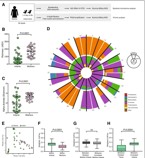
We performed 16S rRNA gene sequencing of stools from 56 infants (28 twin pairs, collected on or before the fourth day of life) and their 28 mothers (Fig. 1a). Of the 84 samples, five samples failed to amplify, four were excluded due to low (< 1000) read counts, and 18 gener-ated sequences indistinguishable from buffer control samples (see the “Methods”) and were also excluded from analysis (Additional file 6: Table S1). Sequences from the remaining 24 maternal and 33 infant samples correspond to 2615 different bacterial taxa. Both rich-ness and Shannon diversity of infant bacterial gut com-munities were significantly less than in their mothers’ stools (both p < 0.001) (Fig. 1b and c). Proteobacteria dominate several infant bacterial communities, but were much less abundant in the maternal samples (Fig. 1d). Principal coordinate analysis (PCoA) of weighted Uni-Frac distances demonstrate distinct and tight clustering of those from maternal samples and a more diffuse dis-tribution of infant samples (Fig. 1e). Group significance
testing with ADONIS confirmed that the clustering of samples by source (mother or infant) was statistically significant and accounts for approximately 23% of the variation in weighted UniFrac distances (p< 0.001,R2= 0.2347). To determine if other factors might be associ-ated with specific microbial communities, we performed a multivariate analyses and random forest classification; both methods demonstrate that mothers and infants have significant differences between their bacterial microbiome (Additional file 1: Figure S1B–E). Race was identified as a significant determinant of bacterial com-munity composition by multivariate analysis, but not by random forest (Additional file1: Figure S1B). Therefore, age of host (i.e., adult vs. infant) was the only factor that separated the bacterial communities in this cohort.
Infant bacterial communities and the relationship to maternal communities were further characterized by pairwise weighted UniFrac distances. Similar to the PCoA data (Fig.1e), the pairwise distance between all in-fant bacterial communities was significantly greater (p< 0.001) than the mean distance between all maternal communities (Fig. 1f). To test if the infant gut bacterial communities more closely resembled those of their own mother than all unrelated mothers, we measured the distances of infant bacterial community structure to their cognate mother versus the distances to all mothers in the study. Interestingly, the distance of an infant to their own mother was no different than to an unrelated mother (Fig. 1g). However, the mean distance between co-twin bacterial communities was significantly less (p
< 0.001) than to unrelated infants (Fig. 1h). The dis-tance between co-twins did not differ with zygosity (Additional file 1: Figure S1F), suggesting that age and environmental exposures are the more important determinants of the bacterial communities.
B
Infants Mothers
0 100 200 300 400
Ri
chness
(AS
V
)
P<0.0001
C
Infants Mothers
0 2 4 6
Alpha
diversity
(Shannon
)
P<0.0001
D
G
H
37
4
8
12
14
17
19
21
23
24 25
26 35 36
39 45
46
47
Acidobacteria Actinobacteria Bacteroidetes Firmicutes Proteobacteria Verrucomicrobia Other
Family group #
Mother Infant I1 I2
E
−0.1 0.0 0.1 0.2
−0.2 −0.1 0.0 0.1 0.2
PCoA 1 (37.9%)
PCoA 2 (18.1%)
Mother Infant
F
Within Infants
Within Mothers 0.0
0.2 0.4 0.6 0.8 1.0
W
e
ighte
d
UniF
rac
P<0.0001
Between Related Mother-Infants
Between Unrelated Mother-Infants 0.0
0.2 0.4 0.6 0.8 1.0
W
e
ighte
d
UniFra
c
Between Related Twins
Between Unrelated Infants 0.0
0.2 0.4 0.6 0.8 1.0
We
ig
h
te
d
U
ni
F
rac
ns P<0.0004
Mother Infant twins 1-4 day of life
28 dyads
Beadbeating DNA extraction
0.45µM filtration Total nucleic acid extraction
16S rRNA V4 PCR
Phi29 amplification
Illumina MiSeq NGS
Illumina MiSeq NGS
Bacterial microbiome analysis
Virome analysis
A
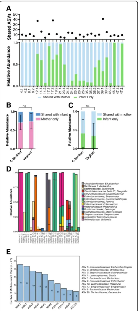
both twins had a high relative abundance in the infant, they represented only a small fraction of the maternal bacterial community (Fig.2d and Additional file7: Table S2). We additionally examined if there were ASVs which were frequently shared by mother-infant pairs across all the families (Fig.2e). One ASV belonging to the Escheri-chia/Shigella genus was shared in 23 of the 27 mother-infant pairs (Additional file 8: Table S3). The relative abundance of these 10 commonly shared ASVs was again generally less in the maternal samples than the in-fant samples (Additional file 2: Figure S2). These data collectively indicate that the mother shares a relatively small proportion of bacterial community with her in-fants, that these shared taxa make significant contribu-tions to the infant’s bacterial microbiome, and that the amount either shared (mother) or received (infant) does not vary by delivery route.
Gut virome of infant twin pairs and their mothers
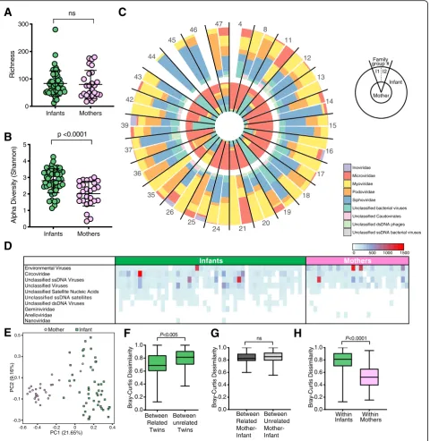
To assess early life mother-infant virome transmission, we performed metagenomic sequencing of the same fecal specimens from 56 healthy infants (28 twin pairs, collected on or before the fourth day of life) and their 28 healthy mothers. Seven of the 84 samples were not entered into analysis: five did not amplify, one had insuf-ficient volume after filtering, and one had low sequen-cing reads (3768) (Additional file 6: Table S1). This resulted in a virome dataset for 50 infants and 27 mothers, i.e., 26 families with mother and at least one infant. There was no significant difference between the sequence depth of infants (mean 131,846 ± 105,022) and maternal samples (mean 103,040 ± 39,986) (p = 0.271) (Additional file3: Figure S3A). The mean infant and ma-ternal gut virome species richness were similar (83 vs 78 species, respectively) (P = 0.6091) (Fig. 3a), but the gut virome Shannon diversity was significantly greater in in-fants than in mothers (p < 0.0001) (Fig. 3b). The most abundant bacteriophages in the maternal gut virome were from the Microviridae family, whereas infant gut viromes consisted predominantly of bacteriophages from
the Myoviridae, Podoviridae, and Siphoviridae families
(Fig.3c). Circoviruses (Circoviridaefamily) and unclassi-fied viruses (within the “ssDNA viruses” and “environ-mental viruses” taxonomic group classifications) were frequently detected in both mother and infant samples (Fig. 3d). Taken together, these data suggest consider-able age-dependent virome differences (i.e., whether the specimen is from an adult or infant) that might con-found assessments of familial relationships between re-lated mother-infant pairs.
To resolve this, we first compared virome diversity by measuring the unweighted Bray-Curtis distance between gut virome communities. PCoA indicated that the ma-ternal gut viromes differ from infant gut viromes 4.1 4.2 8.1 8.2 12.1 14.1 14.2 17.1 17.2 19.2 21.1 23.1 24.1 24.2 25.1 26.1 26.2 35.1 35.2 36.2 37.1 39.1 39.2 45.2 46.1 47.1 47.2
0.0 0.5 1.0 Relative Abundanc e Infant Only Shared With Mother
10 20 30 40 50 Share d ASV s C-Section Vaginal 0.0 0.5 1.0 Relative Abundan c e Infant only Shared with mother
C-Section Vaginal 0.0 0.5 1.0 Relative Abundanc e Mother only Shared with infant
A
B
nsC
ns0.00 0.25 0.50 0.75 1.00 inf ant 4.1 inf ant 4.2
mother 4 inf
ant 8.1
in
fant 8.2
mother 8 inf
ant 14.1
in
fant 14.2
mother 14infant 17.1 infant 17.2 mother 17 inf
ant 24.1
inf
ant 24.2
mother 24 infant 26.1 inf
ant 26.2
mother 26 infant 35.1 inf
ant 35.2
mother 35infant 39.1 inf
ant 39.2
mother 39 inf
ant 47.1 inf ant 47.2 mother 47 Relativ e Ab undance Alicyclobacillaceae; Effusibacillus Bacillaceae 1; Aeribacillus Bacteroidaceae; Bacteroides Clostridiales Incertae Sedis XI; Finegoldia Corynebacteriaceae; Corynebacterium Enterobacteriaceae; Enterobacter Enterobacteriaceae; Escherichia/Shigella Enterobacteriaceae; Pantoea Enterococcaceae; Enterococcus Peptoniphilaceae; Peptoniphilus Ruminococcaceae; Gemmiger Staphylococcaceae; Staphylococcus Streptococcaceae; Streptococcus unclassified Enterobacteriaceae Veillonellaceae; Veillonella
D
23 22 19 18 1713 11 8 7 6 0 5 10 15 20 25
ASV1 ASV3 ASV2 ASV7 ASV5 ASV4 ASV13 ASV17 ASV9 ASV20
Number of Mother−Inf
ant
P
airs (n = 27)
E
ASV 1: Enterobacteriaceae; Escherichia/Shigella
ASV 3: Streptococcaceae; Streptococcus
ASV 2: Staphylococcaceae; Staphylococcus
ASV 7: Lachnospiraceae; Blautia
ASV 5: Bacteroidaceae; Bacteroides
ASV 4: Enterobacteriaceae; Enterobacter
ASV 13: Lachnospiraceae; Roseburia
ASV 17: Streptococcaceae; Streptococcus
ASV 9: Bacteroidaceae; Bacteroides
ASV 20: Bacteroidaceae; Bacteroides
Fig. 2Bacterial ASV transmission analysis.aThe number of ASVs shared between mother and infant and the relative abundance of the infant bacterial microbiome that is shared with mother.b
(Fig. 3e). To further understand additional factors that might influence the composition of the infant gut vir-ome, we performed multivariate analyses of the cohort metadata (e.g., delivery route, feeding type, delivery site)
and found that age (i.e., maternal or infant origin of stool) was significantly associated with differences in the virome (Additional file3: Figures S3B–D). This was cor-roborated by building classifier models with random
A
B
D
0 500 1000 1500
Inoviridae
Microviridae
Myoviridae
Podoviridae
Siphoviridae
Unclassified bacterial viruses
Unclassified Caudovirales
Unclassified dsDNA phages
Unclassified ssDNA bacterial viruses
Family group #
Mother Infant I1 I2
C
4 8
11
12
13
14
15
16
17
18
19 20 21 24 25 26 35 36 37 39
42 43
44 45
46 47
Environmental Viruses Circoviridae
Unclassified ssDNA Viruses Unclassified Viruses
Unclassified Satellite Nucleic Acids Unclassified ssDNA satellites Unclassified dsDNA Viruses Geminiviridae Anelloviridae Nanoviridae
Infants Mothers
Between Related
Twins
Between unrelated Twins 0.0
0.2 0.4 0.6 0.8 1.0
B
ray-C
urtis
D
issim
ila
rit
y
P<0.005
E
-0.6 -0.4 -0.2 0 0.2 0.4
PC1 (21.65%)
-0.3 -0.1 0.1 0.3 0.5
PC2 (9.16%)
Mother Infant
F
H
Between Related Mother-Infant
Between Unrelated Mother-Infant 0.0
0.2 0.4 0.6 0.8 1.0
Bra
y
-Curt
is Diss
imilari
ty
G
nsWithin
Infants MothersWithin
0.0 0.2 0.4 0.6 0.8 1.0
B
ra
y-Cur
tis Diss
imilarit
y
P<0.0001
Infants Mothers
0 100 200 300
Richness
ns
Infants Mothers
0 1 2 3 4 5
Alpha
Diversity (Shannon
)
p <0.0001
Fig. 3Virome analysis of mothers and infants.aRichness of viral species in infants and mothers. Statistical significance assessed by Mann-Whitney test.bAlpha diversity of viral species in mothers and infants using Shannon index. Statistical significance assessed by Mann-Whitney test.
cRelative abundance of phages at family level for mothers and related infants within each family. I1 (infant twin 1) and I2 (infant twin 2).d
forest that accurately distinguished the virome of mothers from infants (AUC = 0.996) (Additional file 3: Figure S3E). Maternal BMI (pre-pregnancy) was also identified as a determinant by the multivariate analyses but was not a significant factor in random forest (Add-itional file3: Figure S3B).
Because mother-infant dyads shared a substantial subset of their bacterial microbiome, we hypothesized that they would also harbor relatively similar viromes. Consistent with this hypothesis, we found that related infants shared a more similar gut virome to their co-twin than to unrelated infants (Fig. 3f, p < 0.005). This co-twin virome similarity was not influenced by their zygosity (Additional file 3: Figure S3F). However, infant gut viromes were highly dissimilar from their own mother. In fact, the ecological distance between an in-fant and their own mother was the same as it was to un-related mothers (Fig. 3g, p = 0.1400). This might be explained in part by the significantly lower virome inter-personal variation among mothers as compared to in-fants (Fig.3h,p< 0.0001).
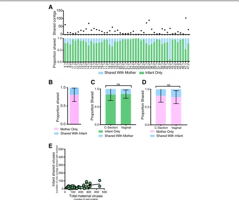
These diversity measurements do not account for differences in virome abundance (i.e., unweighted dis-tances). Hence, we assessed virome transmission in terms of the shared viral abundance. For example, a large proportion of the infant virome might be estab-lished from transmission of a relatively small number of viruses from the mother. To quantify the proportion of the infant virome shared with their mother, we mapped the infants’sequencing reads to viral contigs assembled from the infant(s) and their mothers’ virome. On aver-age, only 15.0% of the viruses present in the infant stool could be mapped back to the sequences in their mother’s stool (Fig. 4a). Conversely, 19.8% of the mother’s virome was shared with her infant (Fig. 4b). The proportion of the viruses shared between mothers and their infants was not influenced by delivery route (Fig. 4c and d). Further, the number of viruses transmit-ted from mother-to-infant was independent of the num-ber of viruses in the mother’s virome (Fig.4e, slopem= 0.127). Thus, unlike the bacterial microbiome, infants shared only a limited proportion (approximately 15%) of their gut virome with their mother. These results were also validated by analyses of bacteriophage contigs iden-tified by VirSorter (Additional file4: Figure S4).
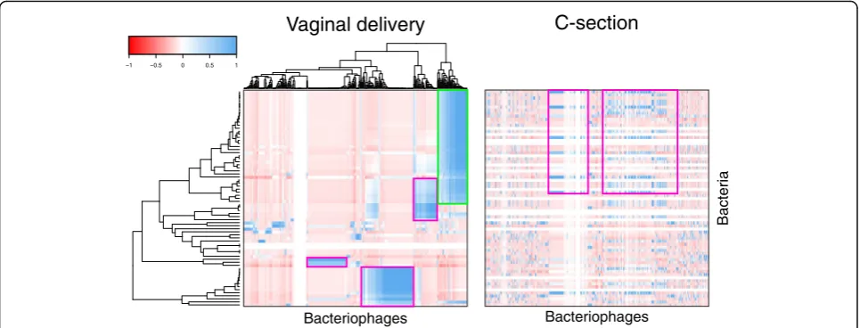
Route of delivery influences transkingdom interactions between bacteria and bacteriophage
After demonstrating that only a subset of the virome and bacterial microbiome is shared between mothers and infants, we sought clinical factors that might influence their transkingdom interactions. Hierarchical clustering indicated that the gut bacterial microbiomes of infants born by vaginal delivery (Ruminococcaceae,
Lachnospiraceae, Bacteroidales, and Lactobacillales
amplicon sequence variants (ASV)) were strongly corre-lated with bacteriophages of the Microviridae,
Myoviri-dae, Siphoviridae, and Podoviridae families (Fig. 5). In
contrast, microbiome interactions of infants born by C-section delivery were discordant from infants born by vaginal delivery. First, bacteriophages also detected in the stools of infants born by C-section correlated with different bacteria than the same bacteriophages from in-fants delivered vaginally. In children born by C-section,
Microviridae, Myoviridae, Siphoviridae, and Podoviridae
bacteriophages were strongly correlated with
Staphylo-coccaceae, Bacteroidaceae, Lachnospiraceae, and other
Ruminococcaceae ASVs. For example, Parabacteroides
phage YZ-2015b from Microviridae family correlated
with Bacteroidales ASVs (Bacteroides uniformis and
Parabacteroides distasonis) in infants born through
vagi-nal delivery, but correlated with other Bacteriodales
ASVs (Bacteriodes ovatus) in infants born through C-section (Fig. 5, compare pink outline; Additional file 5: Figure S5A). Second, microbiome interactions in stool were also absent from infants born by C-section com-pared to those born vaginally (Fig.5, compare green out-line), indicating a transkingdom signature which was unique to each delivery route. This was corrobo-rated by Pearson’s correlations between bacterial ASVs-and VirSorter-identified contigs (Additional file5: Figure S5B). Therefore, this suggests that delivery route might influence interactions between the bacterial microbiome and virome.
Discussion
were indistinguishable from buffer background, and if we interpret these as having no maternal transmission, 63% may be an overestimate. Addtionally, 16S rRNA gene sequencing may not always have sufficient reso-lution to distinguish different strains which would also lead to an overestimation of shared bacteria. However, together, these studies emphasize that the maternal gut microbiome can be a substantial, though not the exclu-sive, source of an infant’s early bacterial microbiome.
Our virome data are unique, and offer substantial con-trast to the bacterial microbiome. Our finding of only 15% of the infant’s virome in the cognate newborn’s mother’s stool strongly suggest that infant gut viruses are likely from other habitats (e.g., mother’s skin,
breastmilk) or the inanimate environment. Second, it is possible that the early window in this study (1–4 days of life) is when bacterial colonization is still underway. This builds on the longitudinal study of Ferretti et al., which demonstrated that maternal gut-sourced bacteria in-creasingly dominate the infant microbiome by 4 months [16]. In this model, it is plausible that “maternally sourced” viruses that can replicate in “maternally sourced” bacteria would become enriched weeks to months after bacterial colonization. This would be con-sistent with the shift in bacteriophage community previ-ously reported to occur from 0 to 2 years [20].
This twin study also offers unique data to compare vertical transmission and co-twin relationships. The
0 50 100 150
Shared contig
s
4.1 8.1 8.2
1
1.1
1
1.2
12.1 12.2 13.1 13.2 14.1 14.2 15.1 15.2 16.1 16.2 17.1 17.2 18.1 18.2 19.1 19.2 20.1 20.2 21.1 21.2 24.1 24.2 25.1 25.2 26.1 26.2 35.1 35.2 36.1 36.2 37.1 37.2 39.1 39.2 42.1 42.2 43.1 43.2 44.2 45.2 46.1 47.1 47.2
0.0 0.5 1.0
Infant Only Shared With Mother
Propor
tion shared
A
C-Section Vaginal
0.0 0.5 1.0
Proportion Shared
Infant Only Shared With Mother
ns
C-Section Vaginal
0.0 0.5 1.0
Proportion Shared
Mother Only Shared With Infant
ns
0 100 200 300 400 500
0 100 200 300 400 500
Infan
t shared
viruse
s
(number
of viral
c
ontigs
shared
w
ith
m
other)
E
D
C
Total maternal viruses
(number of viral contigs)
0.0 0.5 1.0
Proportion shared
Mother Only Shared With Infant
B
greater similarity of the bacterial microbiome and vir-ome between twin pairs than unrelated twins may reflect commonality of a shared environmental exposure. Shared exposure from a maternal site other than stool, such as maternal breastmilk, might be another source. Although breastfeeding did not significantly influence mother-to-infant bacterial transmission, the viromes of breastfed infants are more similar to their mother than were those of formula fed infants (Additional file1: Fig-ure S1G and Additional file 3: Figure S3G). Given the limitations of this cohort, future studies will be needed to clarify the role of diet. Nonetheless, this highlights that the dynamics of the acquisition of the human bac-terial microbiome and virome clearly differ.
The neonatal gut virome consists of largely bacterio-phage with only a small portion being eukaryotic viruses. Of note, we focused on the DNA virome as our previous study of the infant virome [20] (those four twin pairs are also included here) found a sparse RNA virus compo-nent early in life. As with the neonatal bacterial micro-biome, the virome varies across individuals. In contrast to what has been described in adult twins, bacteriophage diversity does not correlate with bacterial diversity [27]. Additionally, the discrepancy in proportion of overlap between the maternal and infant virome and bacterial microbiome suggests that bacterial microbiome and vir-ome are acquired from different sources.
Previous studies of the effect of birth route on the newborn gut bacterial microbiome have offered conflict-ing findconflict-ings. While we found bacterial communities dif-fered significantly between infants born vaginally versus those born by C-section, we were unable to find
discriminating ASVs for delivery route. The shared pro-portion of the infant and maternal bacterial microbiome is not significantly different by delivery route. Further-more, we found no difference between the Bray-Curtis dissimilarity of infant gut virome by delivery route, and we found no viral contig that discriminated between in-fants born vaginally versus by C-Section. Additionally, the proportion of infant and maternal virome that is shared is not significantly different between infants born vaginally versus by C-section. However, when we exam-ined transkingdom interactions, we found that this rela-tionship did differ by delivery route. This transkingdom interaction might reconcile why some studies show de-livery route is an important factor, while others do not.
Conclusions
In summary, our data demonstrates that while mothers and infants do share some of their gut microbial com-munities, the degree of sharing is more substantial for bacteria than for virus. Additionally, the proportion and specific taxa shared varies between and even within fam-ilies. Lastly, bacterial-bacteriophage interactions differed depending on delivery route. Therefore, our data depict that the inter-generation transmissibility at birth di-verges between the major kingdoms of microbes sug-gesting that the founding sources of these communities may differ.
Methods
Subjects
This study was approved by the Human Research Protection Office of Washington University School of
−1 −0.5 0 0.5 1
Vaginal delivery
C-section
Bacteriophages Bacteriophages
Bacteria
Medicine in St. Louis and Missouri-Baptist Medical Center institutional review board. We obtained written informed consent from women pregnant with twin ges-tations were consented to collect stool specimens around the time of birth from the mother and her twins. Stools from the mothers were collected at home or in the hospital; infant stools were collected in the hospital. Home-produced samples were couriered to the labora-tory in insulated envelopes containing frozen (−20 °C) freezer packs and stored at −80 °C until analysis. Twenty-eight families were selected in which maternal stools were collected peripartum and there was adequate sample from early in life from the twins. Metadata collected included mode of delivery, feeding content, maternal age, maternal weight and weight gain, and ma-ternal race (Table 1 and Additional file 6: Table S1). This birth cohort has been described in various detail in prior publications [20,23,28–33].
Bacterial 16S rRNA gene sequencing
One hundred milligrams of stool was disrupted by bead beating and DNA was extracted using QIAamp DNA Stool Mini Kit on a QIAcube automated DNA extraction unit. In parallel, four buffer-only controls were disrupted by bead beating and similarly extracted to serve as ex-traction negative controls. PCR was performed using Golay-barcoded primers specific for the V4 region (F515/R806). Reactions were held at 94 °C for 2 min to denature the DNA, with amplification proceeding for 40 cycles at 94 °C for 15 s, 50 °C for 30 s, and 68 °C for 30 s; a final extension of 2 min at 68 °C. Stool and buffer sam-ples, as well as 4 water negative controls, were amplified in triplicate, combined, and cleaned using Agencourt Ampure XP beads (Beckman-Coulter). Equimolar librar-ies were pooled and sequenced using an Illumina MiSeq sequencer (2 × 250 v2 kit) at the Center for Genome Sciences & Systems Biology at Washington University.
Bacterial 16S rRNA gene analysis
16S rRNA amplicon sequences were demultiplexed using QIIME (Quantitative Insights Into Microbial Ecology, v 1.8.0) [34]. Read quality control and the reso-lution of amplicon sequence variants (ASVs) using the forward-reads only was performed with the DADA2 R package [35]. Taxonomy was assigned to the ASVs from the DADA2-formatted training files derived from the Ribosomal Database Project’s Training Set 16 [36]. ASVs were aligned using an R implementation of MSA [37] and arranged into a maximum likelihood phylogeny (GTR model with optimization of the proportion of in-variable sites and the gamma rate parameter) using the phangorn package [38]. The ASV counts, taxonomy assignments, phylogeny, and sample metadata were combined to generate a PhyloSeq object for further analyses [39]. Five samples failed to amplify. Four sam-ples with less than 1000 reads were excluded from ana-lysis. To compare the bacterial community structure between the stool samples and controls we generated a multidimensional scaling (MDS) plot of the unweighted UniFrac distance with normal confidence ellipses. Eighteen samples encapsulated by the confidence ellipse of the buffer control samples were removed from further analysis (Additional file1: Figure S1A). The Decontam R package “prevalence”method at a threshold of 0.25 was used to identify and remove remaining contaminating sequences [40]. Further ecological analyses were per-formed in R.
If a mother and one of her related infants had an iden-tical ASV, then it was assumed to be shared between mother and infant; otherwise, it was exclusive to mothers or infants. Only families that had a sample from mother and at least one of her infants were used to de-termine potential mother to infant transmission.
Virome sequencing
Fecal specimens (approximately 100 mg) were diluted 1:6 with phosphate-buffered saline (PBS) and filtered through a 0.45-μm-pore-size membrane. Total nucleic acid was ex-tracted from the filtrate using COBAS Ampliprep (Roche). In parallel, PBS was filtered and extracted to serve as ex-traction reagent only control. Total nucleic acid from stool and negative controls was amplified with Phi29 polymer-ase (GenomiPhi V2 kit, GE Healthcare) according to the manufacturer’s instructions and used for Nextera DNA library construction (Illumina). Additionally, to evaluate the level of specimen cross-contamination that might occur after library construction [41], a uniquely indexed li-brary of cDNA derived from the nematode Orsay virus RNA1 segment [42] was included in the pool for each se-quencing run. Libraries were purified and size-selected using Agencourt Ampure XP beads (Beckman-Coulter), Table 1Cohort demographics
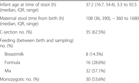
Infant age at time of stool (h) (median, IQR, range)
37.2 (16.7, 54.4), 3.3 to 92.5
Maternal stool time from birth (h) (median, IQR, range)
108 (36, 390),−360 to 1680
C-section no. (%) 35 (62.5%)
Feeding (between birth and sampling) no. (%)
Breastmilk 8 (14.3%)
Formula 16 (28.6%)
Mix 32 (57.1%)
followed by quantification on a 2100 Bioanalyzer (Agilent Technologies).
Virome analysis (read-based)
To control for sample extraction and cross-sample con-tamination, we included two types of negative controls: buffer-only controls that underwent the same extraction process, and PBS spiked with Orsay virus (a nodavirus of nematodes). Illumina MiSeq sequencing reads (2 × 250 bp) were processed through the VirusSeeker workflow (version 0.063) [43]. In brief, VirusSeeker follows a hier-archical workflow to perform a quality filter, subtract host sequences (human), identify candidate viral sequences through curated virus-only databases, then search sequen-tially against comprehensive databases to remove potential false positives (i.e., candidate viral sequences that have higher similarity to non-viral sequences). VirusSeeker takes a multi-step approach to remove bacterial se-quences. Candidate viral sequences are queried against NCBI bacteria reference genomes to remove bacterial se-quences. Second, VirusSeeker also performs three sequen-tial BLAST searches (MegaBLAST against the complete NCBI NT database, BLASTn against the complete NT database, and BLASTx against the complete NCBI NR database) to remove potential falpositive viral se-quences that have higher similarity to non-viral sese-quences (bacterial, fungal, etc.).
Contaminants were identified by decontam (version 1.0.0) [40] in a 2-step process, which uses a prevalence-based method to determine contaminant taxa. First, samples were compared to“Orsay”controls in decontam at a threshold of 0.1, and identified contaminant taxa were removed. Next, buffer controls were compared to samples using decontam at a stricter threshold of 0.5, and contaminants removed likewise. The resulting quality-controlled sequencing reads of each sample were then normalized to 31,000. Virus species with fewer than three reads were masked.
Ecological analyses (richness, alpha diversity—Shannon Index) were performed using Vegan R package (version 2.5-2) [44].The beta diversity and PCoA plots were ob-tained by using QIIME [34] using the Bray-Curtis dis-tance on a presence-absence species data matrix. Results were plotted in GraphPad Prism (version 8) and the stat-istical significance was assessed using a non-parametric Mann-Whitney U test. For the familial relative abun-dance plot, only paired data of one mother and at least one infant per family were used.
Metadata
MaAslin
MaAslin, a multivariate linear modeling tool, was uti-lized in finding the association between study metadata
and relative abundance [45]. The significance of each as-sociation is given via a q-value; any relationship with a
q-value of less than 0.05 is shown. For the metadata cat-egories exclusive to infants such as, delivery route, feed-ing type, and zygosity, only the infant data was used. All sample data was used for the other general categories such as, mother/infant, delivery site, pre-pregnancy BMI [category], and race.
Random forest
RandomForest was used to classify metadata categories (version 4.6-14) [46]. Five hundred trees were built using randomForest and the Out-Of-Bag samples were used to test the classifier and obtain their error rates. The ROC curves and AUC measures for the classifiers were made by a R package, pROC, which plots pseudo-probabilities from classification tree votes for OOB samples (version 1.13.0) [47]. Similar to MaAslin metadata analysis, for the metadata categories exclusive to infants, such as, de-livery route, feeding type, and zygosity, only the infant data was used. For the other general categories such as mother/infant, delivery site, pre-pregnancy BMI [cat-egory], and race, all sample data was used.
Virome contig analyses
Contigs databases
Using IDBA_UD, the QC reads of each sample were used to create initial raw contigs (version 1.1.0) [48]. These contigs were then filtered using bbtools [31] to re-move contigs with length 500 bp and de-duplicated at minidentity ≥99. The contigs created from buffer sam-ples were queried against NT database using megablast and any contig with a coverage≥95 and percent identity
≥97, was designated as a true contaminant and used to create a database of contaminant contigs. Megablast was used to query sample contigs against contig contaminant database and any hit to this database resulted in the re-moval of such contig from sample contigs. After con-taminated contig removal, for each family, the contigs were concatenated into one file and then merged to cre-ate longer contigs using minimus2 [49]. These familial contigs were queried against viral NR database using blastx and the contigs with a hit against this database were used to create the familial viral contig database. We also corroborated these contig analyses by identify-ing bacteriophage contigs with VirSorter (parameters— virome, -db 1) (Additional file4: Figure S4) [50].
Contig transmission
The mapped reads were used to measure shared contigs between mother and infant and used to determine po-tential transmission of virome from mother to infant. If a mother and one of her related infants had a read mapped to the same contig, then it was assumed to be shared between mother and infant; otherwise, it was ex-clusive to mothers or infants. Only families that had a sample from mother and at least one of her infants were used to determine potential mother to infant transmis-sion. VirSorter-identified contigs were also analyzed in an identical manner, by generating a VirSorter contig database and mapping reads to the database to deter-mine potential mother to infant transmission (Add-itional file4: Figure S4).
Transkingdom analysis
To characterize transkingdom interactions in mother-to-infant transmission, Pearson’s correlations were per-formed between bacterial ASVs and bacteriophage contigs that were shared between mothers and at least one of her infants. Bacterial ASVs and bacteriophage contigs that were present in only one family were excluded. Correla-tions were performed separately by delivery route and clustered on the vaginal delivery dataset using the R pack-age gplots. To contrast differences in the C-section profile, the same clustering order (vaginal delivery) was used to visualize correlations in the C-section dataset. Pearson’s correlation was also performed between the ASVs and VirSorter identified bacteriophages.
Statistics
Mann-WhitneyUtest was used to test significance when there were two independent groups, for example, related twins versus unrelated twins. Kruskal-Wallis test and Dunn’s multiple comparison were used to test signifi-cance between three or more groups, for example the feeding type of breastmilk, formula, or mixed. Pearson’s correlation was used obtain correlation between trans-mitted viruses and bacteria. Allpvalues were two-tailed.
Code availability
A fully reproducible workflow of the analysis presented in this manuscript can be found at https://github.com/ RachelRodgers/Holtz-Lim_MotherTwinInfant_Virome_ BacterialMicrobiome
Supplementary information
Supplementary informationaccompanies this paper athttps://doi.org/10. 1186/s40168-019-0766-7.
Additional file 1: Figure S1.Metadata analysis of bacterial ASVs. (A) Multidimensional scaling (MDS) plot of the unweighted UniFrac distance with normal confidence ellipses. Expanded version shows eighteen samples encapsulated by the confidence ellipse of the buffer control
samples (B) Output of MaAslin and random forest analysis. (C) Important ASVs and their statistical significance as assessed by MaAslin and random forest. (D) Heatmap of MaAslin identified ASVs for mothers and infants. (E) ROC curves and AUC measures for delivery route and mother/infant classification using random forest and pROC packages in R. Pseudo-probabilities are plotted on graph using Out-Of-Bag (OOB) sample tree classification votes. Only infant data used in vaginal vs. C-section classifi-cation while both mother and infant data used for mother vs. infant clas-sification. (F) Weighted UniFrac pairwise comparisons. Statistical significance assessed by Mann-Whitney, and Kruskal-Wallis with Dunn’s multiple correction (feeding type: breastmilk vs. formula; breastmilk vs. mix; formula vs. mix). (G) Weighted UniFrac pairwise comparisons for re-lated mother infant pairs. Statistical significance assessed by Kruskal-Wallis and Mann-Whitney.
Additional file 2: Figure S2.Relative abundance of the 10 most frequently shared (mother-infant) ASVs. Taxonomy shown at the genus level.
Additional file 3: Figure S3.Metadata analysis of viral species. (A) Number of sequencing reads in infant and maternal samples. Statistical significance assessed by Mann-Whitney. (B) Output of MaAslin and ran-dom forest analysis. (C) Important viral species and their statistical signifi-cance as assessed by MaAslin and random forest. (D) Heatmap of MaAslin identified viral species for mothers and infants. (E) ROC curves and AUC measures for delivery route and mother/infant classification using ran-dom forest and pROC packages in R. Pseudo-probabilities are plotted on graph using Out-Of-Bag (OOB) sample tree classification votes. Only infant data used in vaginal vs. C-section classification while both mother and in-fant data used for mother vs. inin-fant classification. (F) Bray-Curtis Dissimilar-ity pairwise comparisons. Statistical significance assessed by Mann-Whitney. Kruskal-Wallis with Dunn’s multiple correction was used to as-sess feeding type (breastmilk vs. formula; breastmilk vs. mix; formula vs. mix) and Pre-pregnancy BMI (obese vs. overweight; obese vs. normal; overweight vs. normal). (G) Bray-Curtis Dissimilarity pairwise comparisons for related mother infant pairs. Statistical significance assessed by Mann-Whitney. Kruskal-Wallis with Dunn’s multiple correction was used to as-sess feeding type (breastmilk vs. formula; breastmilk vs. mix; formula vs. mix) and Pre-pregnancy BMI (obese vs. overweight; obese vs. normal; overweight vs. normal).
Additional file 4: Figure S4.VirSorter viral contig transmission analysis. Analyses of VirSorter contigs were performed as in Fig.4. (A) The number of infant VirSorter viral contigs shared with their mother (top), and their proportion out of the total infant virome (VirSorter identified) is shown (bottom). Percent of infant VirSorter contigs that are shared with their mother is shown in blue and infant only VirSorter contigs are shown in green. (B) Average VirSorter contig proportion of mothers’virome that is shared with infant or unique to the mother. (C) Average VirSorter contig proportion of infant virome that is shared with mother by delivery route. Statistical significance assessed by Mann-Whitney test. (D) Average VirSorter contig proportion of maternal virome that is shared with infant by delivery route. Statistical significance assessed by Mann-Whitney test. (E) Plot of the number of infant shared VirSorter viral contigs vs. the total number of ma-ternal VirSorter contigs. Linear regression line fit to data is shown.
Additional file 5: Figure S5.Transkingdom interaction between bacteria and bacteriophage. Correlation of shared bacterial ASVs and viral contigs. (A) Heatmap shows the Pearson correlation between bacterial ASVs and bacteriophage contigs in infants from C-section delivery (left) clustered by hierarchical clustering. Second heatmap shows correlations from infants delivered vaginally clustered in the same order as correla-tions from infants delivered by C-section. B) Heatmap shows the Pearson correlation between bacterial ASVs and VirSorter identified bacteriophage contigs in infants delivered vaginally (left) clustered by hierarchical clus-tering. Second heatmap shows correlations from infants delivered by C-section clustered in the same order as correlations from infants delivered vagi-nally. These VirSorter analyses corroborate the contig analyses shown in Fig.5.
Additional file 6: Table S1.Cohort metadata and sample inclusion status.
Additional file 7: Table S2.Read counts and taxonomy of ASVs shared by both twins.
Acknowledgements
We thank the families and their physicians for their participation in, and cooperation with, the study.
Authors’contributions
ESL and LRH conceived and designed the experiments. CR prepared samples for sequencing. CR performed PCR experiments. RR, RM, ESL, and SH processed and analyzed the sequencing data. PIT, BBW, and MN recruited the study participants and managed the metadata. ESL and LRH wrote and edited the manuscript. All authors read and approved the final manuscript.
Funding
This work was supported in part by the Children’s Discovery Institute (MD-FR-2013-292), Doris Duke Charitable Foundation (2017076), March of Dimes (BOC 388999), NIH grant R00 DK107923, Arizona State University startup funding (ESL), the National Institutes of Health (5P30 DK052574 (Biobank, DDRCC) to P.I.T. and UH3AI083265 to P.I.T. and B.B.W.). P.I.T. and B.B.W. received funding from the Eunice Kennedy Shriver National Institute of Child Health and Human Development, and the Foundation for the National Institutes of Health (made possible by support from the Gerber Foundation).
Availability of data and materials
Sequence data has been deposited to the European Nucleotide Archive under accession number PRJEB33578. Reads mapping to human have been removed from the submitted metagenomic sequence data. A fully reproducible workflow of the analysis presented in this manuscript can be found athttps://github.com/RachelRodgers/Holtz-Lim_MotherTwinInfant_ Virome_BacterialMicrobiome
Ethics approval and consent to participate
The institutional review board at Washington University School of Medicine and Missouri-Baptist Medical Center approved the study. Subjects provided informed consent to be included.
Consent for publication
Not applicable
Competing interests
P.I.T. is a consultant to Takeda Pharmaceuticals on childhood gastrointestinal diseases. The other authors declare that they have no competing interests.
Author details 1
School of Life Sciences, Arizona State University, Tempe, AZ 85287, USA.
2Center for Fundamental and Applied Microbiomics, The Biodesign Institute,
Tempe, AZ 85287, USA.3Department of Pediatrics, Washington University
School of Medicine, St. Louis, MO 63110, USA.4Department of Pathology &
Immunology, Washington University School of Medicine, St. Louis, MO
63110, USA.5Department of Molecular Microbiology, Washington University
School of Medicine, St. Louis, MO 63110, USA.
Received: 4 September 2019 Accepted: 8 November 2019
References
1. White RA, Bjornholt JV, Baird DD, Midtvedt T, Harris JR, Pagano M, Hide W, Rudi K, Moen B, Iszatt N, et al. Novel developmental analyses identify longitudinal patterns of early gut microbiota that affect infant growth. PLoS Comput Biol. 2013;9:e1003042.
2. Britton RA, Young VB. Interaction between the intestinal microbiota and host in Clostridium difficile colonization resistance. Trends Microbiol. 2012;20:313–9. 3. Frank DN, Robertson CE, Hamm CM, Kpadeh Z, Zhang T, Chen H, Zhu W,
Sartor RB, Boedeker EC, Harpaz N, et al. Disease phenotype and genotype are associated with shifts in intestinal-associated microbiota in inflammatory bowel diseases. Inflamm Bowel Dis. 2011;17:179–84.
4. Olszak T, An D, Zeissig S, Vera MP, Richter J, Franke A, Glickman JN, Siebert R, Baron RM, Kasper DL, Blumberg RS. Microbial exposure during early life has persistent effects on natural killer T cell function. Science. 2012;336:489–93. 5. Stout MJ, Conlon B, Landeau M, Lee I, Bower C, Zhao Q, Roehl KA, Nelson
DM, Macones GA, Mysorekar IU. Identification of intracellular bacteria in the basal plate of the human placenta in term and preterm gestations. Am J Obstet Gynecol. 2013;208:226 e221–7.
6. Aagaard K, Ma J, Antony KM, Ganu R, Petrosino J, Versalovic J. The placenta harbors a unique microbiome. Sci Transl Med. 2014;6:237ra265.
7. Collado MC, Rautava S, Aakko J, Isolauri E, Salminen S. Human gut colonisation may be initiated in utero by distinct microbial communities in the placenta and amniotic fluid. Sci Rep. 2016;6:23129.
8. Lauder AP, Roche AM, Sherrill-Mix S, Bailey A, Laughlin AL, Bittinger K, Leite R, Elovitz MA, Parry S, Bushman FD. Comparison of placenta samples with contamination controls does not provide evidence for a distinct placenta microbiota. Microbiome. 2016;4:29.
9. Perez-Munoz ME, Arrieta MC, Ramer-Tait AE, Walter J. A critical assessment of the“sterile womb”and“in utero colonization”hypotheses: implications for research on the pioneer infant microbiome. Microbiome. 2017;5:48. 10. Lim ES, Rodriguez C, Holtz LR. Amniotic fluid from healthy term pregnancies
does not harbor a detectable microbial community. Microbiome. 2018;6:87. 11. de Goffau MC, Lager S, Sovio U, Gaccioli F, Cook E, Peacock SJ, Parkhill J,
Charnock-Jones DS, Smith GCS. Human placenta has no microbiome but can contain potential pathogens. Nature. 2019;572:329.
12. Yassour M, Jason E, Hogstrom LJ, Arthur TD, Tripathi S, Siljander H, Selvenius J, Oikarinen S, Hyoty H, Virtanen SM, et al. Strain-Level Analysis of Mother-to-Child Bacterial Transmission during the First Few Months of Life. Cell Host Microbe. 2018;24:146–154 e144.
13. Asnicar F, Manara S, Zolfo M, Truong DT, Scholz M, Armanini F, Ferretti P, Gorfer V, Pedrotti A, Tett A, Segata N. Studying Vertical Microbiome Transmission from Mothers to Infants by Strain-Level Metagenomic Profiling. mSystems. 2017;2:e00164.
14. Nayfach S, Rodriguez-Mueller B, Garud N, Pollard KS. An integrated metagenomics pipeline for strain profiling reveals novel patterns of bacterial transmission and biogeography. Genome Res. 2016;26:1612–25. 15. Milani C, Mancabelli L, Lugli GA, Duranti S, Turroni F, Ferrario C, Mangifesta M,
Viappiani A, Ferretti P, Gorfer V, et al. Exploring Vertical Transmission of Bifidobacteria from Mother to Child. Appl Environ Microbiol. 2015;81:7078–87. 16. Ferretti P, Pasolli E, Tett A, Asnicar F, Gorfer V, Fedi S, Armanini F, Truong DT,
Manara S, Zolfo M, et al. Mother-to-Infant Microbial Transmission from Different Body Sites Shapes the Developing Infant Gut Microbiome. Cell Host Microbe. 2018;24:133–145 e135.
17. Chu DM, Ma J, Prince AL, Antony KM, Seferovic MD, Aagaard KM. Maturation of the infant microbiome community structure and function across multiple body sites and in relation to mode of delivery. Nat Med. 2017;23:314–26.
18. Backhed F, Roswall J, Peng Y, Feng Q, Jia H, Kovatcheva-Datchary P, Li Y, Xia Y, Xie H, Zhong H, et al. Dynamics and Stabilization of the Human Gut Microbiome during the First Year of Life. Cell Host Microbe. 2015;17:690–703. 19. Lim ES, Wang D, Holtz LR. The Bacterial Microbiome and Virome Milestones
of Infant Development. Trends Microbiol. 2016;24:801–10.
20. Lim ES, Zhou Y, Zhao G, Bauer IK, Droit L, Ndao IM, Warner BB, Tarr PI, Wang D, Holtz LR. Early life dynamics of the human gut virome and bacterial microbiome in infants. Nat Med. 2015;21:1228–34.
21. Duranti S, Lugli GA, Mancabelli L, Armanini F, Turroni F, James K, Ferretti P, Gorfer V, Ferrario C, Milani C, et al. Maternal inheritance of bifidobacterial communities and bifidophages in infants through vertical transmission. Microbiome. 2017;5:66. 22. McCann A, Ryan FJ, Stockdale SR, Dalmasso M, Blake T, Ryan CA, Stanton C,
Mills S, Ross PR, Hill C. Viromes of one year old infants reveal the impact of birth mode on microbiome diversity. PeerJ. 2018;6:e4694.
23. Yatsunenko T, Rey FE, Manary MJ, Trehan I, Dominguez-Bello MG, Contreras M, Magris M, Hidalgo G, Baldassano RN, Anokhin AP, et al. Human gut microbiome viewed across age and geography. Nature. 2012;486:222–7. 24. Zhao G, Vatanen T, Droit L, Park A, Kostic AD, Poon TW, Vlamakis H, Siljander
H, Harkonen T, Hamalainen AM, et al. Intestinal virome changes precede autoimmunity in type I diabetes-susceptible children. Proc Natl Acad Sci U S A. 2017;114:E6166–75.
25. Drell T, Stsepetova J, Simm J, Rull K, Aleksejeva A, Antson A, Tillmann V, Metsis M, Sepp E, Salumets A, Mandar R. The Influence of Different Maternal Microbial Communities on the Development of Infant Gut and Oral Microbiota. Sci Rep. 2017;7:9940.
26. Avershina E, Lundgard K, Sekelja M, Dotterud C, Storro O, Oien T, Johnsen R, Rudi K. Transition from infant- to adult-like gut microbiota. Environ Microbiol. 2016;18:2226–36.
28. Baumann-Dudenhoeffer AM, D'Souza AW, Tarr PI, Warner BB, Dantas G. Infant diet and maternal gestational weight gain predict early metabolic maturation of gut microbiomes. Nat Med. 2018;24:1822–9.
29. Gibson MK, Wang B, Ahmadi S, Burnham CA, Tarr PI, Warner BB, Dantas G. Developmental dynamics of the preterm infant gut microbiota and antibiotic resistome. Nat Microbiol. 2016;1:16024.
30. Planer JD, Peng Y, Kau AL, Blanton LV, Ndao IM, Tarr PI, Warner BB, Gordon JI. Development of the gut microbiota and mucosal IgA responses in twins and gnotobiotic mice. Nature. 2016;534:263–6.
31. Moore AM, Ahmadi S, Patel S, Gibson MK, Wang B, Ndao MI, Deych E, Shannon W, Tarr PI, Warner BB, Dantas G. Gut resistome development in healthy twin pairs in the first year of life. Microbiome. 2015;3:27. 32. Gurnee EA, Ndao IM, Johnson JR, Johnston BD, Gonzalez MD, Burnham CA,
Hall-Moore CM, McGhee JE, Mellmann A, Warner BB, Tarr PI. Gut Colonization of Healthy Children and Their Mothers With Pathogenic Ciprofloxacin-Resistant Escherichia coli. J Infect Dis. 2015;212:1862–8. 33. Gurnee EA, Ndao IM, McGhee JE, Warner BB, Tarr PI, Fritz SA, Burnham CA.
Fecal carriage of methicillin-resistant Staphylococcus aureus and vancomycin-resistant Enterococcus in healthy children. Antimicrob Agents Chemother. 2014;58:1261–2.
34. Caporaso JG, Kuczynski J, Stombaugh J, Bittinger K, Bushman FD, Costello EK, Fierer N, Pena AG, Goodrich JK, Gordon JI, et al. QIIME allows analysis of high-throughput community sequencing data. Nat Methods. 2010;7:335–6. 35. Callahan BJ, McMurdie PJ, Rosen MJ, Han AW, Johnson AJ, Holmes SP.
DADA2: High-resolution sample inference from Illumina amplicon data. Nat Methods. 2016;13:581–3.
36. Cole JR, Wang Q, Fish JA, Chai B, McGarrell DM, Sun Y, Brown CT, Porras-Alfaro A, Kuske CR, Tiedje JM. Ribosomal Database Project: data and tools for high throughput rRNA analysis. Nucleic Acids Res. 2014;42:D633–42. 37. Bodenhofer U, Bonatesta E, Horejs-Kainrath C. Hochreiter S: msa: an R
package for multiple sequence alignment. Bioinformatics. 2015;31:3997–9. 38. Schliep KP. phangorn: phylogenetic analysis in R. Bioinformatics. 2011;27:592–3. 39. McMurdie PJ. Holmes S: phyloseq: an R package for reproducible interactive analysis and graphics of microbiome census data. PLoS One. 2013;8:e61217. 40. Davis NM, Proctor DM, Holmes SP, Relman DA, Callahan BJ. Simple statistical
identification and removal of contaminant sequences in marker-gene and metagenomics data. Microbiome. 2018;6(1):226.
41. Kircher M, Heyn P, Kelso J. Addressing challenges in the production and analysis of illumina sequencing data. BMC Genomics. 2011;12:382. 42. Felix MA, Ashe A, Piffaretti J, Wu G, Nuez I, Belicard T, Jiang Y, Zhao G,
Franz CJ, Goldstein LD, et al. Natural and experimental infection of Caenorhabditis nematodes by novel viruses related to nodaviruses. PLoS Biol. 2011;9:e1000586.
43. Zhao G, Wu G, Lim ES, Droit L, Krishnamurthy S, Barouch DH, Virgin HW, Wang D. VirusSeeker, a computational pipeline for virus discovery and virome composition analysis. Virology. 2017;503:21–30.
44. Oksanen J, Blanchet FG, Friendly M, Kindt R, Legendre P, McGlinn D, Minchin PR, O'Hara RB, Simpson GL, Solymos P, Stevens MHH, Szoecs E, Wagner H. vegan:Community Ecology Package. R package version 2.5-2; 2018. 45. Morgan XC, Tickle TL, Sokol H, Gevers D, Devaney KL, Ward DV, Reyes JA, Shah
SA, LeLeiko N, Snapper SB, et al. Dysfunction of the intestinal microbiome in inflammatory bowel disease and treatment. Genome Biol. 2012;13:R79. 46. Liaw A, Wiener M. Classification and Regression by randomForest. R News.
2002;2:18–22.
47. Robin X, Turck N, Hainard A, Tiberti N, Lisacek F, Sanchez JC, Muller M. pROC: an open-source package for R and S+ to analyze and compare ROC curves. BMC Bioinformatics. 2011;12:77.
48. Peng Y, Leung HC, Yiu SM, Chin FY. IDBA-UD: a de novo assembler for single-cell and metagenomic sequencing data with highly uneven depth. Bioinformatics. 2012;28:1420–8.
49. Sommer DD, Delcher AL, Salzberg SL, Pop M. Minimus: a fast, lightweight genome assembler. BMC Bioinformatics. 2007;8:64.
50. Roux S, Enault F, Hurwitz BL, Sullivan MB. VirSorter: mining viral signal from microbial genomic data. PeerJ. 2015;3:e985.
51. Li H. Aligning sequence reads, clone sequences and assembly contigs with BWA-MEM. arXiv. 2013;1303:3397.
Publisher’s Note