R E S E A R C H A R T I C L E
Open Access
Outcomes after the surgery for acquired
nonaccommodative esotropia
Eunbi Kim and Dong Gyu Choi
*Abstract
Background:To analyze the surgical outcomes for patients diagnosed with acquired nonaccommodative
esotropia (ANAET).
Methods:In this retrospective study, the medical records of 35 patients who had undergone the surgery for ANAET with a postoperative follow-up period of 6 months or more were reviewed. The main outcome measures were postoperative esodeviation angle, final success rate, and factors affecting surgical outcome. Surgical success was considered to be an alignment within 8 prism diopters (PD) at distance and near.
Results:The preoperative mean esodeviation angles were 37.3 ± 13.7 PD at distance and 38.6 ± 16.6 PD at near. The postoperative mean esodeviation angles at distance were as follows: 4.2 PD at day 1, 4.0 PD at month 1, 3.9 PD at month 3, 4.9 PD at month 6, 4.7 PD at year 1, and 4.8 PD at final follow-up. There was no statistically significant difference in angle of esodeviation between the initial postoperative period (day 1 to month 6) and the final follow-up day (p> 0.05). The surgical success rate at final follow-up was 65.7% (23/35). Among the 12 patients for whom the surgery failed, 9 (24.3%) showed esotropia and 3 (8.1%) exotropia of more than 8 PD. Six patients (16.2%) underwent reoperation (4 for esotropia and 2 for exotropia). There was no factor influencing surgical outcome (p> 0.05).
Conclusions:The surgical outcome in patients with ANAET was relatively favorable: the final success rate was 65.7% and the reoperation rate was 17.1%.
Keywords:Acquired nonaccommodative esotropia, Surgical outcome, Reoperation, Influencing factor
Background
Acquired nonaccommodative esotropia (ANAET) is a type of strabismus characterized by a constant nonac-commodative esodeviation that develops after 6 months of age, in the absence of any significant refractive error and in an otherwise healthy child or adult [1, 2]. Many studies have identified factors affecting surgical outcome in infantile esotropia and accommodative esotropia. However, the literature regarding surgical outcomes or the factors influencing them in ANAET is thin, notwith-standing the research demonstrating older age at onset as a factor associated with postoperative stereopsis.
The purpose of the present study was to analyze the surgical outcomes in patients who had undergone
surgery for ANAET and to determine the factors associ-ated with favorable outcome.
Methods
Design and patients
We retrospectively reviewed the medical records of 35 patients who had undergone surgery for ANAET with a postoperative follow-up period of 6 months or more. ANAET was diagnosed if esotropia developed after 6 months of age in an otherwise healthy child and eso-deviation angles at distance and near were unchanged even after the full correction of hyperopia of +2.0 diop-ters (D) or more, if revealed in cycloplegic refraction. Patients with any history of accommodative or partial accommodative esotropia, infantile esotropia, paralytic or restrictive strabismus, previous extraocular muscle surgery were excluded. And we included only the pa-tients whose difference between the preoperative * Correspondence:[email protected]
Department of Ophthalmology, Hallym University College of Medicine, Kangnam Sacred Heart Hospital, Shingil-ro 1, Youngdeungpo-gu, Seoul 07441, South Korea
esodeviation angles at distance and near were less than 5 prism diopters (PD). Patients with associated findings of dissociated vertical deviation (DVD), latent nystag-mus or inferior oblique overaction (IOOA) were ex-cluded regardless of history, due to concern for unrecognized infantile esotropia. In the very young age group, we obtained the information about onset of strabismus which was acute, subacute, acquired or con-genital from their parents or previous photography. This study was approved as a retrospective study by the Institutional Review Board of Hallym University Medical Center (IRB No. 2015-10-117) before data col-lection in order to review patient records and use the data, and adheres to the tenets of the Declaration of Helsinki. Informed written consent was obtained from all participants or their parents.
We noted the following preoperative characteristics: sex, age at onset, age at surgery, refractive error, symptom duration, amblyopia, stereopsis, dominance of fixation, accompanying strabismus (e.g. vertical strabis-mus [≥ 5 PD in the primary position]), preoperative follow-up period, and type of surgery.
Cycloplegic refraction (with 1% cyclopentolate and 1% tropicamide) and fundus examination were performed on all of the patients. One operating surgeon measured all deviations using the alternate prism cover test at near and at distance (0.3 m and 6 m) (with spectacle correction based on cycloplegic refraction if necessary). Preoperative measurements were made no more than 3 days prior to surgery. The presence of amblyopia was defined as a dif-ference of two or more lines between the best-corrected visual acuities of the right and left eyes or a best-corrected visual acuity lower than 20/30 on the Snellen visual acuity chart. The sensory status was evaluated by Titmus stereot-est (Stereo Optical Co., Chicago, II, USA) at 33 cm and by Worth-4-dot test (Worth-4-dot Attachment; Richmond Products, Albuquerque, NM, USA) at 6 m.
Surgical techniques
All of the surgeries were performed under general anesthesia by a single surgeon (D.G.C.). The selection of the surgical procedure was determined by the preopera-tive angle of esodeviation, the presence of dominance of fixation and the patient’s and surgeon’s preference. Four-teen patients underwent bilateral medial rectus recession (BMR), 17 unilateral medial rectus muscle recession and lateral rectus muscle resection (RR), and the remaining 4, with esotropia of 20 PD or less, unilateral medial rectus recession (UMR).
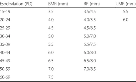
The extent of recession and resection was measured from the original muscle insertion. No hang-back or ad-justable sutures were used. Surgical dosages were determined, based on the formulas suggested by Parks [3] and Wang [4] (Table 1).
Main outcome measures
Postoperative alignment was measured on postoperative day 1, month 1, 3, 6, year 1, and at final follow-up. The main outcome measures included the postoperative esode-viation angles at each follow-up day, the final success rate, and the factors affecting surgical outcomes (i.e. sex, age at onset, age at surgery, refractive error, symptom duration, amblyopia, stereopsis, fusion by Worth-4-dot, alternate fix-ation, accompanying strabismus, preoperative follow-up period, and type of surgery). Surgical success was defined as esotropia or exotropia of 8 PD or less at distance and near.
Statistics
SPSS software V.12.0 K (SPSS Inc., Chicago, Illinois, USA) was employed for the statistical analysis. The Wilcoxon signed rank test was used to compare the preoperative and postoperative angles of deviation. The Mann-Whitney U test and Fisher’s exact test were applied to analyze the demographic data. P-values less than 0.05 were considered significant.
Results
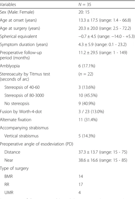
The baseline characteristics of the total 35 patients are summarized in Table 2. The mean age at onset was 13.3 ± 17.5 years (range: 1.4-66.8 years), and the mean age at sur-gery was 20.3 ± 20.0 years (range: 2.5-72.2 years). The mean spherical equivalent was −0.7 ± 4.5, and the mean symptom duration was 4.3 ± 5.9 years. The mean pre-operative follow-up period was 11.2 ± 29.5 months (range: 3-149 months), and the mean postoperative follow-up period was 57.8 ± 60.0 months (range: 6-201 months). Seven patients had amblyopia, which was defined as a two-line difference in acuity between eyes. For 10 patients, alternate fixation was possible. Five patients presented with vertical strabismus. The mean preoperative angle of esodeviation was 37.3 ± 13.7 PD (range: 15-75 PD) at distance and 38.6 ± 16.6 PD (range: 15-85 PD) at near.
Table 1Surgical dosages for acquired nonaccommodative esotropia (ANAET)
Esodeviation (PD) BMR (mm) RR (mm) UMR (mm)
15-19 3.5 3.5/4.5 5.5
20-24 4.0 4.0/5.5 6.0
25-29 4.5 4.5/6.5
30-34 5.0 5.0/7.0
35-39 5.5 5.5/7.5
40-44 6.0 6.0/8.0
45-49 6.5 6.5/8.0
50-59 7.0 7.0/8.5
60-69 7.5
BMRbilateral medial rectus recession,RRunilateral medial rectus recession and lateral rectus resection,UMRunilateral medial rectus recession,
Surgical outcomes
The mean esodeviation angles at distance and at near were 4.2 and 3.8 PD at postoperative day 1 and 4.8 and 4.8 PD at final follow-up, respectively. There was no statistically significant difference in angle of esode-viation between the initial postoperative period (day 1 to month 6) and the final follow-up day (p> 0.05, Wilcoxon signed rank test, Tables 3 and 4). The initial postoperative angles of deviation were stably maintained during the follow-up period.
The surgical success rates at postoperative day 1 were 88.6%: the final success rates were 65.7% (Table 5).
Reoperation
Six patients underwent reoperation, 4 for recurrent or residual esotropia and 2 for consecutive exotropia. The mean interval period between the 1st and 2nd op-eration was 71.8 ± 37.2 months (range: 25-120 months) (Table 6).
Factors affecting surgical outcome
We analyzed the factors affecting surgical success (i.e. sex, age at onset, age at surgery, refractive error, symp-tom duration, amblyopia, stereopsis, fusion by Worth-4-dot, alternate fixation, accompanying strabismus [e.g. vertical strabismus], preoperative follow-up period, and type of surgery). There was no factor influencing surgical outcome (P> 0.05, Table 7).
Discussion
Conventionally, ANAET is considered to occur infre-quently, but is sometimes associated with intracranial tumor or other central nervous system (CNS) lesions [5–14]. However, Jacobs et al. [1] reported that with this form of esotropia, neurologic problems are only rarely present. In this study we excluded any patient with known neurological disorder.
According to Jacobs et al. [1]’s report, approximately three-quarters of those who underwent surgery for ANAET had good alignment after a mean duration of 1 decade, and two-thirds of them were within 10 PD of orthotropia. Chan et al. [2] found that 64.7% of patients had successful outcomes after surgery for ANAET. Sturm et al. [15] reported a 92% surgical success rate (within 8PD or less of orthotropia) among acute ac-quired concomitant esotropia patients. Our results indi-cated the final success rates (an alignment within 8 PD)
Table 2Demographic data on ANAET patients
Variables N= 35
Sex (Male: Female) 20: 15
Age at onset (years) 13.3 ± 17.5 (range: 1.4 - 66.8)
Age at surgery (years) 20.3 ± 20.0 (range: 2.5 - 72.2)
Spherical equivalent −0.7 ± 4.5 (range:−14.0 - +5.3)
Symptom duration (years) 4.3 ± 5.9 (range: 0.1 - 23.2)
Preoperative follow-up period (months)
11.2 ± 29.5 (range: 1 - 149)
Amblyopia 6 (17.1%)
Stereoacuity by Titmus test (seconds of arc)
(n= 22)
Stereopsis of 40-60 3 (13.6%)
Stereopsis of 80-3000 10 (45.5%)
No stereopsis 9 (40.9%)
Fusion by Worth-4-dot 3 / 23 (13.0%)
Alternate fixation 11 (31.4%)
Accompanying strabismus
Vertical strabismus 5 (14.3%)
Preoperative angle of esodeviation (PD)
Distance 37.3 ± 13.7 (range: 15 - 75)
Near 38.6 ± 16.6 (range: 15 - 85)
Type of surgery
BMR 14
RR 17
UMR 4
Postoperative follow-up period (months) 57.8 ± 60.0 (range: 6-201)
ANAETacquired nonaccommodative esotropia, DVD dissociated vertical deviation,PDprism diopters,BMRbilateral medial rectus recession,
RRunilateral medial rectus recession and lateral rectus resection,
UMRunilateral medial rectus recession
Table 3Mean angle (PD) of esodeviation at distance
Angle of esodeviation P-valuea
Preoperative 37.3 ± 13.7 0.000
Postoperative day 1 4.2 ± 8.4 0.772
Postoperative month 1 4.0 ± 5.3 0.751
Postoperative month 3 3.9 ± 7.0 0.432
Postoperative month 6 4.9 ± 7.0 0.497
Postoperative year 1 4.7 ± 6.4 0.916
Final follow-up 4.8 ± 15.1
PDprism diopters
a
Wilcoxon signed rank test (comparison with final follow-up day)
Table 4Mean angle (PD) of esodeviation at near
Angle of esodeviation P-valuea
Preoperative 38.6 ± 16.6 0.000
Postoperative day 1 3.8 ± 6.7 0.446
Postoperative month 1 4.2 ± 5.2 0.581
Postoperative month 3 4.6 ± 6.5 0.700
Postoperative month 6 4.1 ± 6.6 0.672
Postoperative year 1 5.5 ± 6.5 0.934
Final follow-up 4.8 ± 16.1
PDprism diopters
a
of 65.7%, which are in line with the findings of the relevant previous studies.
However, surgical failures for persistent esotropia were 3 times more numerous than for consecutive exotropia in this study. This would mean that the correction of the surgical tables might be considered. Because the study population was too small, the true difference between the groups could be hidden. Further prospective study involving more data would be needed.
In the present study, 6 patients (17.1%) underwent re-operation compared with 26.7 and 5.9% in the Jacobs et al. [1] and Chan et al. [2] investigations, respectively. These differences were thought to be owed to the sig-nificantly varying mean postoperative follow-up periods: 57.8 months (present study), 10.9 years (Jacobs et al.), and 17.8 months (Chan et al.).
Several studies about the surgical outcome of ANAET including Jacobs et al. [1] and Chan et al. [2] had the age limit of the childhood at onset or surgery. However, there was no limitation in the age of onset or surgery, so in this respect, there is some limitation in comparing the surgical result for ANAET between our study and the other studies mentions above. Because of this, we ana-lyzed the success rates after separating the two groups by age, more than 18 years old or not. The surgical suc-cess rate of less than 18 years old group(1.4 ~ 17.5 years
old, mean 10.8 years old) was 66.7% and that of more than 18 years old group(18.9 ~ 66.8 years old, mean 42.5 years old) was 54.5%. It was a little difference be-tween the two groups. However, there was no statistical difference between two groups (p= 0.374, Fisher’s exact test).
The main limitation of our study is the non-standardized and retrospective data collection. The other limitation was that the sample was so diverse and in-cluded 6 patients with amblyopia and 3 with perfect stereopsis. And moreover, the range of refractive errors was from−14.0 to +5.3 D even though all of the patients were eligible for according to the diagnostic criteria of
Table 5Surgical success ratesafor ANAET
Surgical success rates
Postoperative day 1 88.6%
Postoperative month 1 80.0%
Postoperative month 3 80.0%
Postoperative month 6 80.0%
Postoperative year 1 72.7%
Final follow-up 65.7%
ANAETacquired nonaccommodative esotropia
a
Surgical success was defined as esotropia or exotropia of 8 PD or less at distance and near
Table 6Reoperation after surgery for ANAET
N= 6 Reason for reoperation
Recurrent or residual esotropia 4
Consecutive exotropia 2
Preoperative angle of esodeviation before 1stoperation (PD)
At distance: 40.0 ± 12.2 At near: 48.3 ± 17.6
Preoperative angle of esodeviation
before 2ndoperation (PD) At distance: 6.7 ± 29.8At near: 10.8 ± 31.5
Mean interval period between 1st
and 2ndoperation (months)
71.8 ± 37.2 (range: 25 - 120)
Surgical success rate after reoperation 4/6 (66.6%)
ANAETacquired nonaccommodative esotropia,PDprism diopters
Table 7Patient characteristics in Successaand Non-success ANAET Groups
Variable Success Group
(n= 26)
Non-success Group (n= 9)
P-value
Sex (Male: Female) 15: 11 5: 4
Age at onset (years) 10.0 ± 12.1 19.3 ± 24.7 0.392b
Age at surgery (years) 18.8 ± 17.7 22.7 ± 23.9 0.674b
Spherical equivalent −1.3 ± 5.6 0.5 ± 3.2 0.628b
Duration of symptom (years) 3.9 ± 4.7 5.0 ± 8.4 0.837b
Preoperative follow-up period (months)
12.1 ± 32.1 9.8 ± 25.9 0.420b
Amblyopia 5 (19.2%) 1 (11.1%) 0.891c
Stereopsis by Titmus test (seconds of arc)
(n= 16) (n= 6)
Stereopsis of 40-60 3 (18.8%) 0 (0%) 0.235c
Stereopsis of 80-3000 7 (43.8%) 3 (50.0%) 0.456c
No stereopsis 6 (37.5%) 3 (50.0%) 0.443c
Worth-4-dot (n= 17) (n= 6)
Fusion 2 (11.8%) 1 (16.7%) 0.306c
Alternate fixation 5 0 0.374c
Accompanying strabismus
Vertical strabismus 2 3 0.125c
Preoperative angle of esodeviation (PD)
Distance 36.6 ± 10.3 38.5 ± 18.0 0.940b
Near 37.7 ± 13.9 40.5 ± 22.2 1.000b
Type of surgery
BMR 11 3 0.581c
RR 13 4 0.285c
UMR 2 2 0.134c
Postoperative follow-up period (months)
45.8 ± 51.5 78.2 ± 68.3 0.203b
ANAETacquired nonaccommodative esotropia,DVDdissociated vertical deviation,PDprism diopters,BMRbilateral medial rectus recession,
RRunilateral medial rectus recession and lateral rectus resection,
UMRunilateral medial rectus recession
a
Surgical success was defined as esotropia or exotropia of 8 PD or less at final follow-up(at both distance and near)
b
Mann-Whitney U test;c
ANAET. Because the patients in whom esotropia had developed after 6 months of age were included in this study, even though patients with DVD, latent nystagmus or IOOA were excluded, the possibility infantile esotro-pia patients to be included in this study group were not completely ruled out. Further, a large prospective study looking at only individuals with childhood onset ANAET will be needed.
Conclusions
In conclusion, this paper provides data on the clinical characteristics of, and surgical outcomes for, ANAET. The surgical outcome at final follow-up was favorable. And there was no factor influencing surgical outcome.
Abbreviations
ANAET:Acquired nonaccommodative esotropia; BMR: Bilateral medial rectus recession; CNS: Central nervous system; DVD: Dissociated vertical deviation; IOOA: Inferior oblique overaction; IRB: Institutional Review Board; PD: Prism diopters; RR: Unilateral medial rectus muscle recession and lateral rectus muscle resection; UMR: Unilateral medial rectus recession
Acknowledgements None.
Funding None.
Availability of data and materials
The datasets used and/or analyzed during the current study are available from the corresponding author on reasonable request.
Authors’contributions
DGC Designed and supervised the study; EK drafted the manuscript; DGC and EK collected the data; DGC and EK analyzed the data and helped to draft the manuscript. All authors read and approved the final manuscript.
Ethics approval and consent to participate
This study followed the tenets of the Declaration of Helsinki and was approved by the Institutional Review Board of Hallym University Medical Center (IRB No. 2015-10-117). Informed written consent was obtained from all participants or their parents.
Consent for publication Not applicable.
Competing interests
The authors declare that they have no competing interest.
Publisher’s Note
Springer Nature remains neutral with regard to jurisdictional claims in published maps and institutional affiliations.
Received: 27 September 2016 Accepted: 18 July 2017
References
1. Jacobs SM, Green-Simms A, Diehl NN, Mohney BG. Long-term follow-up of acquired nonaccommodative esotropia in a population-based cohort. Ophthalmology. 2011;118(6):1170–4.
2. Chan TY, Mao AJ, Piggott JR, Makar I. Factors affecting postoperative stereopsis in acquired nonaccommodative esotropia. Can J Ophthalmol. 2012;47(6):479–83.
3. Parks MM. Pediatric ophthalmology. 2ndedition. Chapter 67:
strabismus surgery. 1997. p. 976.
4. Wang L, Wang X. Comparison between graded unilateral and bilateral medial rectus recession for esotropia. Br J Ophthalmol. 2012;96(4):540–3. 5. Von Noorden GF. Esodeviations. In: Burian-von Noorden’s binocular vision
and ocular motility: theory and management of strabismus. 3rd ed. St Louis: Mosby; 1985. p. 320–9.
6. Astle WF, Miller SJ. Acute comitant esotropia: a sign of intracranial disease. Can J Ophthalmol. 1994;29(3):151–4.
7. Hoyt CS, Good WV. Acute onset concomitant esotropia: when is it a sign of serious neurological disease? Br J Ophthalmol. 1995;79(5):498–501. 8. Macpherson H, De Becker I, MacNeill JR. Beware: armed and
dangerous-acquired non-accommodative esotropia. Am Orthopt J. 1996;46:44–56. 9. Williams AS, Hoyt CS. Acute comitant esotropia in children with brain
tumors. Arch Ophthalmol. 1989;107(3):376–8.
10. Greenberg AE, Mohney BG, Diehl NN, Burke JP. Incidence and types of childhood esotropia: a population-based study. Opthalmology. 2007;114:170–4. 11. Mohney BG. Acquired nonaccommodative esotropia in childhood.
J AAPOS. 2001;5:85–9.
12. Simon JW, Waldman JB, Couture KC. Cerebellar astrocytoma manifesting as isolated, comitant esotropia in childhood. Am J Ophthalmol. 1996;121:584–6. 13. Harada T, Ohasi T, Ohki K, Sawamura Y, Yoshida K, Ito T, et al. Clival
chordoma presenting as acute esotropia due to bilateral abducens palsy. Opthalmologica. 1997;211:109–11.
14. Fukuo Y, Abe T, Hayasaka S. Acute comitant esotropia in a boy with head trauma and convulsions receiving carbamazepine. Ophthalmologica. 1998;212:61–2.
15. Sturm V, Menke MN, Knecht PB, Schoffler C. Long-term follow-up of children with acute acquired concomitant esotropia. J AAPOS. 2011;15:317–20.
• We accept pre-submission inquiries
• Our selector tool helps you to find the most relevant journal
• We provide round the clock customer support
• Convenient online submission
• Thorough peer review
• Inclusion in PubMed and all major indexing services
• Maximum visibility for your research
Submit your manuscript at www.biomedcentral.com/submit