C A S E R E P O R T
Open Access
Molecular monitoring of minimal residual disease
in two patients with
MLL-rearranged acute
myeloid leukemia and haploidentical
transplantation after relapse
Thomas Burmeister
1*, Mara Molkentin
1, Claus Meyer
2, Nils Lachmann
3, Stefan Schwartz
1, Birte Friedrichs
1,
Jörg Beyer
4, Igor Wolfgang Blau
1, Gunnar Lohm
5, Carola Tietze-Bürger
6, Rolf Marschalek
2,7and Lutz Uharek
6Abstract
This report describes the clinical courses of two acute myeloid leukemia patients. Both hadMLLtranslocations, the first a t(10;11)(p11.2;q23) withMLL-AF10and the second a t(11;19)(q23;p13.1) withMLL-ELLfusion. They achieved a clinical remission under conventional chemotherapy but relapsed shortly after end of therapy. Both had a history of invasive mycoses (one had possible pulmonary mycosis, one systemic candidiasis). Because no HLA-identical donor was available, a haploidentical transplantation was performed in both cases. Using a specially designed PCR
method for the assessment of minimal residual disease (MRD), based on the quantitative detection of the individual chromosomal breakpoint in theMLLgene, both patients achieved complete and persistent molecular remission after transplantation. The immune reconstitution after transplantation is described in terms of total CD3+/CD4+, CD3+/CD8+, CD19+, and CD16+/CD56+cell numbers over time. The KIR and HLA genotypes of donors and recipients are reported and the possibility of a KIR-mediated alloreactivity is discussed. This report illustrates that haploidentical transplantation may offer a chance of cure without chronic graft-versus-host disease in situations where no suitable HLA-identical donor is available even in a high-risk setting and shows the value of MRD monitoring in the pre- and posttransplant setting.
Keywords:Minimal residual disease, Haploidentical stem cell transplantation,MLL, KIR, Acute myeloid leukemia
Background
Acute myeloid leukemia (AML) is a molecularly
hetero-geneous disease. Around 5–10% of affected patients
show MLL translocations and an additional 8% have a
MLLpartial internal duplication. WhileMLLaberrations
have historically and generally been associated with an adverse prognosis, this view has become more differen-tiated during recent years and at least for some rarer
MLL aberrations this verdict appears to be unfounded
[1]. The 5 most commonMLLaberrations in adult AML
are t(9;11)/MLL-AF9, t(6;11)/MLL-AF6, t(11;19)/
MLL-ELL, t(11;19)/MLL-ENL, and t(10;11)/MLL-AF10,
to-gether accounting for around 80% of cases [2,3]. The
general recommendation for younger and medically fit AML patients is allogeneic transplantation. The only ex-ception are the core-binding factor leukemias with either
CBFA2-ETOor CBFB-MYH11, and acute promyelocytic
leukemia withPML-RARA, if no additional high-risk
fea-tures (e.g. FLT3-ITD) are present. Around 15–30% of
patients referred for transplantation have a suitable HLA-identical sibling donor. Large donor registries can be accessed for those patients without a HLA-identical sibling donor, but the likelihood of finding a suitable donor depends strongly on the ethnic background of the patient [4]. Currently it is possible to identify a suitable HLA-identical donor in around 70% of patients of Euro-pean ancestry [5]. Different options exist for those patients without a fully (10/10) HLA antigen-matched donor. Clinical trials with donors showing only a 9/10,
8/10 or even 7/10 HLA match have shown a
* Correspondence:thomas.burmeister@charite.de 1
Klinik für Hämatologie, Charité CBF, Hindenburgdamm 30, 12200 Berlin, Germany
Full list of author information is available at the end of the article
significantly higher transplant-related mortality, mainly due to the higher incidence of severe graft-versus-host disease (GvHD) [6-8]. The Perugia group of Martelli and coworkers first introduced the concept of transplanting
“megadoses” of highly purified CD34+ cells of a
HLA-haploidentical donor in the clinical setting to overcome the HLA barrier and prevent graft failure [9,10]. To achieve an alloreactive antileukemic effect in the absence of alloreactive T-cells, donor-derived natural killer (NK) cells were infused [11].
This report describes the clinical courses of two
patients withMLL-rearranged AML and particular high
risk features (early relapse, invasive mycoses) with par-ticular emphasis on the MRD monitoring method which was based on the quantitative detection of the individual
chromosomal breakpoints in the MLL gene. Both
patients had no suitable HLA-identical donor and thus haploidentical transplantations were performed. The im-mune reconstitution after transplantation in terms of B-cells, CD4+and CD8+T-cells and NK cells is described.
Case presentation Case 1
Features at presentation
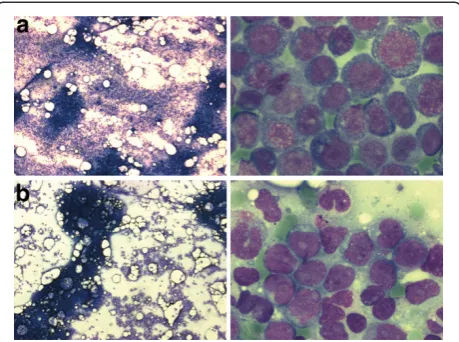
A 33-year-old male patient was diagnosed with acute mye-loid leukemia. The first clinical symptoms were dyspnoea on exertion, a generalized maculopapular rash, gingival hyperplasia, and general weakness. The blood count at diagnosis revealed 52/nl leukocytes (79% blasts) a hemoglobin of 11.1 g/dl (hematocrit 0.33), and platelets of 110/nl. The LDH was 1306 U/l (normal<248 U/l), cre-atinine 1.27 mg/dl, C-reactive protein 16.1 mg/dl (normal <0.5 mg/dl). Bone marrow cytology revealed an increased overall cellularity and an 80% infiltration by medium-sized blasts with basophilic cytoplasm, partial cytoplasmic vacuolisation, prominent nucleoli, and positive esterase staining, compatible with acute myeloid leukemia of the FAB M5a subtype (Figure 1). Flow cytometric analysis of the bone marrow revealed an expression of myeloid anti-gens and HLA-DR with aberrant CD4 and CD56 expres-sion. Cytogenetic analysis revealed a 46,XY,t(10;11)(p11.2; q23) karyotype and molecular analysis by RT-PCR an MLL-AF10fusion transcript.
Initial treatment
The patient was randomized into arm C of the German AML2003 study after informed consent. The study
protocol comprises two cycles DA (100 mg/m2= 200 mg
cytarabine continuously, d1-7 + 60 mg/m2= 120 mg
dau-norubicine, d3-5) followed by 3 cycles post-remission
therapy (MAC-MAMAC-MAC) with MAC = 2 × 1 g/m2
cytarabine d1-6 + 10 mg/m2 mitoxantrone d4-6 and
MAMAC = 2 × 1 g/m2cytarabine d1-5 + 100 mg/m2
amsa-crine d1-5. An allogeneic transplant is recommended after
completion of the first two cycles, if a donor is available. However, no HLA-identical sibling or unrelated donor could be identified in this case.
The inital clinical course was complicated by severe adverse events. During the first days of DA induction the patient suffered from acute dyspnea which led to his admission to the intensive care unit where he first received CPAP (Continuous Positive Airway Pressure) and then mechanical ventilation. A thoracic CT scan showed bilateral pleural effusions and atypical infiltrates. During these first days right-sided hemiplegia and deso-rientation developed. A cranial CT scan showed a left frontal and right pareto-occipital parenchymal bleeding. The renal function deteriorated and hemodialysis had to be performed. The clinical situation improved under broad-spectrum antibiotic treatment, the mechanical ventilation and hemodialysis could be discontinued and after 24 days on the ICU the patient was readmitted to the ward. The patient made a full recovery under inten-sive physical therapy with no residual neuro-psychiatric deficits. A bone marrow biopsy on day 15 after start of therapy showed a partial remission with around 10 to 20% blasts, and a second bone marrow biopsy on day 27, i.e. after leukocyte and platelet recovery, showed cytologically a suspicious residual blast population; how-ever, flow cytometry did not confirm a significant blast population. The second induction cycle started on day 34 and had less adverse events. However, the patient developed febrile neutropenia and a methicillin-resistant
Staphylococcus aureus (MRSA) was isolated from a
blood culture. Despite receiving antibiotic treatment according to the resistogram, the patient developed pneumonic infiltrates and increasing C-reactive protein levels which only improved after a combination of vori-conazole and liposomal amphotericin was added to the antibiotic regimen. After leukocyte recovery all symp-toms rapidly resolved and the patient could be dis-charged with a prophylactic posaconazole medication. The therapy was continued with 3 consolidation cycles (MAC-MAMAC-MAC). Stem cells apheresis was per-formed after the second consolidation cycle. Complica-tions during consolidation included reactivation of the pulmonary mycosis and a marked cervical lymphaden-itis. Bone marrow cytology after each cycle showed a complete remission.
Assessment of minimal residual disease (MRD)
A patient-specific PCR for detecting minimal residual disease (MRD) was established as described previously
[12]. The chromosomal breakpoint in theMLLgene was
identified by long distance inverse PCR [13] and a highly sensitive quantitative PCR using a hydrolysis probe for detecting this breakpoint sequence was established [12]
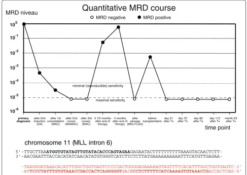
reproducible sensitivitywas 10-4in each case (terminology according to van der Velden [14]). This PCR showed con-tinuously decreasing MRD which was below the detection limit after the second and third consolidation cycle (Figure 1).
Relapse and salvage therapy
Three and a half months after end of the therapy the pa-tient presented with an inguinal swelling. A biopsy and a bone marrow investigation was undertaken. The inguinal biopsy revealed an extramedullary AML relapse. While bone marrow cytology appeared normal, MRD investiga-tion showed MRD positivity (Figure 2). A second bone marrow investigation two weeks later confirmed the re-lapse. In addition, patchy skin infiltrates appeared. The patient was treated according to the Mito-FlAG scheme
(2×15 mg/m2fludarabine; 2×1000 mg/m2cytarabine
d1-5; 7 mg/m2 mitoxantrone d1,3,5). The treatment was
relatively well-tolerated and besides a venous catheter-associatedS. epidermidisbacteriaemia, no major compli-cations occurred. After therapy the patient achieved complete cytomorphologic remission, the extramedullary manifestations had disappeared and no MRD was detect-able in the bone marrow.
Haploidentical transplantation
Since no HLA-identical donor was available, the haploi-dentical mother of the patient was chosen as stem cell donor. After successful eligibility assessment she
under-went stimulation with filgrastim (2×10 μg/kg body
weight) on days −5 to +1 and CD34+ stems cells were
apheresed via an inguinal shaldon catheter on days 0 and +1. The patient was conditioned in parallel with
5 mg/kg thiotepa on day −9 and −8, 40 mg/m2
fludara-bine on day−7 to−4 and fractionized 12 Gy total body irratiation on days−3 to−1 (Figure 3). Muromonab was
given daily in a dose of 5 mg on days −9 to +3. The
apheresed stem cells were T-cell depleted and 9.6 × 10⁶
CD34+ donor cells/kg recipient body weight were
infused on days 0 and +1. The infusion of donor NK cells which had been scheduled for day +1 was
post-poned because of a relatively high CD3+ cell count
(19.6 × 10³/kg = 1.5 × 10⁶in total) and concern regarding severe graft-versus-host disease. The collected NK cells were archived in liquid nitrogen. The conditioning re-gime was tolerated relatively well. The clinical course after transplantation was characterized by infectious complications which could however be controlled with adequate antibiotic treatments, e.g. a CMV reactivation on day +27, which was successfully treated with
valgan-ciclovir. The leukocyte count was >1/nl since day +8
and the platelet count reached values >50/nl after
day +13. No signs of GvHD grade >1 were noted after
transplantation and thus it was decided to infuse the with-held donor NK cells on day +27. No GvHD occurred after this infusion.
Long-term follow-up and immune reconstituion
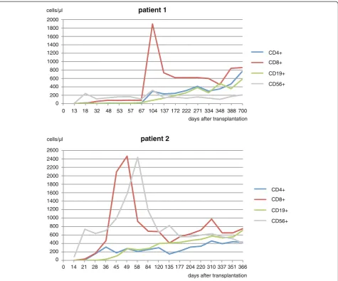
The further clinical course was uneventful and required no hospitalizations. MRD follow-ups for almost three years showed no persistent leukemia and the patient returned to normal life working in his former profession and is now, 4 years after transplantation, clinically free of disease. Lymphocyte subpopulations were measured in the peripheral blood at different time points after transplantation (Figure 4). The T helper cell count was>200/μl after day +104. Around day +104, a sharp in-crease in CD8+cells was observed, however this could not be related to any clinical event. All cell subsets increased to normal values over time.
Case 2
Features at presentation (relapse)
A 28-year-old female patient was admitted to our hos-pital after diagnosis of relapsed acute myeloid leukemia. She had been diagnosed with AML FAB M2 16 months earlier and had been treated elsewhere according to the AML2003 protocol (see above) arm B with two cycles of DA induction therapy and 3 cycles of high-dose
Figure 1A. Bone marrow smear at diagnosis from the first patient at two different magnifications.The marrow shows increased cellularity by round-shaped medium-sized blasts with basophilic occasionally vacuolated sparsely granulated cytoplasm and perinuclear halos. The nuclei show 1-3 nucleoli. Peroxidase stain: 10% positive, esterase stain: 100% markedly positive, PAS stain: approx. 50% granular positive. The cytomorphology is compatible with the diagnosis of AML FAB M5a.B.Bone marrow smear at the time of relapse from the second patient at two different
cytarabine (2 × 3 g/m2 d1,3,5) consolidation therapy. Cytological bone marrow examinations revealed a delayed blast clearance (persistence on day 15 of first in-duction, blast clearance after second induction). No HLA-identical sibling or unrelated donor was available. On presentation in our hospital she was in good overall condition. The blood count showed 3.3/nl leukocytes (30% blasts), 10.1 g/dl hemoglobin (0.29 hematocrit) and 79/nl platelets. The clinical chemical parameters and LDH were otherwise normal. Bone marrow examination confirmed the relapse (Figure 5). Cytogenetic analysis showed a t(11;19)(q23;q13.1) which had also been
detected at the time of primary diagnosis. A MLL-ENL
fusion transcript was detected by RT-PCR.
Salvage treatment and disseminated candidiasis
The patient was treated according to a modified FlAmsa
protocol (30 mg/m2 fludarabine d1-4, 100 mg/m2
amsa-crine d1-4, 2000 mg/m2cytarabine). During the subsequent
aplasia she developed an increase in C-reactive protein to
around 30 mg/l (normal<0.5 mg/l) despite
broad-spectrum antibiotic treatment. A thoracic CT scan showed small bilateral pulmonary and hepatic lesions suggestive of atypical or fungal pneumonia. An MR scan of the abdomen showed hepatomegaly (22 cm craniocaudal diameter) with multiple lesions in liver, spleen and kidneys, measuring up to 8 mm in diameter. At the same time, an elevated Can-dida antigen level was detected in the serum. The patient was initially treated with liposomal amphotericin B and later with voriconazole. A bronchoalveolar lavage revealed 100–1.000 colony-forming units/ml Candida dubliniensis and a liver biopsy showed neither leukemic infiltrates nor fungi. Candida dubliniensis was later also isolated from a stool specimen. Systemic candidosis was diagnosed, the antimycotic treatment was changed to caspofungin and after a further increase in Candida antigen titer to micafun-gin with liposomal amphotericin B. The manifestation in lungs, liver, spleen and kidneys regressed and the C-reactive
Quantitative MRD course
10
10
10
10
10 10 10
-2
-3
-4
-5
-6 -1 0
MRD niveau
MRD negative MRD positive
••
primary diagnosis
••
after 1st consolidation
(MAC)
••
after 2nd induction (DA)
after 2nd consol. (MAMAC)
minimal (reproducible) sensitivity
after 3rd consol. (MAC)
••
3 1/2 months after end of
therapy
4 months after end of
therapy
••
after salvage (Mito-FLAG)
••
before transplantation
day 21 after Tx
day 70 after Tx
day 85 after Tx
day 113 after Tx
month 23 after Tx
maximal sensitivity
time point
••
5’-TTGCTTAAATGGTGTATAGTTGTATACACCCAGTAGAA
GAGAATACTTTTTTTTTTAAAGTACAACTCTT-
3’-AACGAATTTACCACATATCAACATATGTGGGTCATCTTCTCTTATGAAAAAAAAAATTTCATGTTGAGAA-chromosome 11 (
MLL
intron 6)
chromosome 10 (
AF10
intron 13)
-TAAGGGATAAACACATTTGGCTGGTGAGTCCCCCACTGGGGAGAAAAAGTAGTTTTCACATTTGGCTGGTGAGTC-3’ -ATTCCCTATTTGTGTAAACCGACCACTCAGGGGGTGACCCCTCTTTTTCATCAAAAGTGTAAACCGACCACTCAG-5’
protein and the Candida antigen titer normalized after leukocytic regeneration. Liposomal amphotericin was dis-continued 11 days before transplantation and micafungin was continued until day +31 after transplantation after which it was replaced by posaconazole. The candidiasis delayed the scheduled transplantation by about 4 weeks.
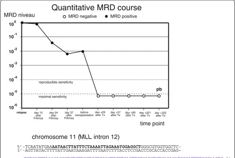
Minimal residual disease was assessed by quantifying the patient-specific chromosomal breakpoint sequence in the MLL gene as described in the first case. A real-time quantitative PCR was established and every bone marrow specimen was investigated for MRD. The MRD level be-fore transplantation was 10-2relative to the time of relapse (Figure 5).
Haploidentical transplantation
The younger brother of the patient was chosen as a suitable haploidentical stem cell donor. The conditioning regimen for haploidentical was similar to that described above but the 12 Gy total body irradiation was applied before the chemotherapy for logistical reasons (Figure 1). A bone mar-row investigation shortly before start of conditioning revealed an MRD level of 10-2, which was only slightly higher compared to that achieved one month ago after FlAmsa therapy (Figure 1). The patient received a high stem cell dose (15.87 × 10⁶/kg body weight) from the donor (Tables 1, 2, and 3). On day +1 after CD34+cell transplant-ation donor NK cells (5.46 × 10⁶/kg) were infused. The clinical course after transplantation was relatively uncom-plicated. In particular, there were no signs of recurrence of invasive fungal infection under continuous daily micafungin medication. The patient showed a mild graft-versus-host reaction (grade 1) of the skin and intestine (both not histo-logically verified) which regressed without immunosuppres-sion. Clinical engraftment of leukocytes (>1/nl) and platelets (>50/nl) occurred on day +10 and +13.
Immune reconstitution and MRD after transplantation
The MRD level was continuously negative in all investi-gations after transplantation during the follow-up period of more than one year (Figure 5). Immune reconstitution occurred relatively fast, and T helper cells first reached a level of >200/nl on day +36. A peak in CD8+ cells was
observed on day +49 followed by a peak in CD8+ cells
on day +58.
Conclusions
Haploidentical transplantation and the possible role of KIR mismatches
The difficulties in finding a suitable HLA-identical donor for every patient in need for it led to the idea of considering the“always present but overlooked”[6] haploidentical rela-tives as potential donors. The patient’s parents and children are always HLA-haploidentical and the probability of a patient’s sibling being HLA-haploidentical is 50% in con-trast to 25% probability for full HLA identity. Thus in practical reality the probability of finding a HLA-haploidentical relative is nearly 100%.
Two major problems have to be overcome in haploiden-tical transplantation: the first is graft rejection and the sec-ond is severe graft-versus-host disease. The group of Martelli et al.first demonstrated clinically that this is pos-sible by using large amounts (“megadoses”, i.e.> 107/kg) of highly purified CD34+ hematopoietic stem cells. These purified stem cells are largely devoid of T-cells which are responsible for the graft-versus-host effect.
To achieve some kind of alloreactivity against residual recipient’s blood cells, donor-derived natural killer (NK) cells have been infused. The basis of this NK alloreactiv-ity is not fully understood. NK cells are able to attack other cells that show downregulation of HLA class I molecules on their surface which is often the case in
-11 -10 -9 -8 -7 -6 -5 -4 -3 -2 -1
patient 2 patient 1
-12 0 +1 +2 +3
5 mg/kg thiotepa 2x2 Gy total body irradiation
40 mg/m fludarabine2 5 mg muromonab (OKT3)
CD34
day
CD56
+4 +5 ... +55 CD56 +
+
+
virus-infected cells or tumor cells. However, HLA class I downregulation is neither necessary nor sufficient for NK cell activation as is illustrated by the fact that nor-mal cells or tissues lacking HLA class I expression (e.g., erythrocytes) or showing low HLA class I ex-pression (e.g., neural cells) are not attacked by autologous NK cells. Cytotoxic NK cell activity is believed to be mainly determined by the presence of inhibitory or activating receptors on the NK cell surface. Two major types of
in-hibitory receptors have been identified: killer cell
immunoglobulin-like (KIR) receptors and the CD94/ NKG2A heterodimer, a C-type lectin, which interacts with the non-classical class Ib HLA-E molecule (reviewed in [15]). While the ligands for many KIR receptors are un-known, the inhibitory receptors KIR2DL1, KIR2DL2 and
KIR3DL1 bind epitopes of HLA-C (’HLA-C2, -C1’groups)
and HLA-A and HLA-B molecules with ’Bw4”motif,
respectively [16]. Upon encountering self-class HLA
anti-gen, NK cell–mediated lysis and cytokine release are
inhibited.
While HLA genes are located in the MHC complex on chromosome 6p21.3, KIR genes are clustered in the leukocyte receptor complex (LRC) on chromosome 19q13.4. Thus they segregate independently in human pedigrees. The LRC is highly polymorphic and contains seven to 15 KIR gene loci dispersed within a region of 65 to 200 kb [17]. It is not yet fully understood how NK cells are“educated”not to attack other autologous cells lacking inhibitory HLA ligands for their KIR receptors [18,19]. Various models have been proposed to explain
0 200 400 600 800 1200 1400 1600 1800 2000
1000
0 13 18 32 48 53 57 67 104 137 172 222 271 334 348 388 700
CD4+
CD8+
CD19+
CD56+
days after transplantation cells/µl
CD4+
CD8+
CD19+
CD56+
0 200 400 600 800 1200 1400 1600 1800 2000
1000 cells/µl
2200 2400 2600
14 21 28 36 45 49 58 84 120 135 177 204 220 310 351 366 days after transplantation
337 0
patient 1
patient 2
the experimentally observed alloreactivity of NK cells. The“licensing model”predicts that NK cells that do not receive a signal through an inhibitory receptor become
hyporesponsive instead of alloreactive. The “Ruggieri
model”assumes that NK cells with inhibitory KIR recep-tors for ligands absent on both donor and recipient are
nonalloreactive. The effect of NK cell alloreactivity in the setting of a fully HLA-matched transplantation has been controversially discussed (synopsis in [16]). Some groups have found a benefit in overall survival in case of a KIR-HLA class I mismatch [20], but others could not confirm this and found no effect [21] or even a worse
Table 1 Immunogenetic characteristics (HLA and KIR genotypes)
Recipient HLA Donor HLA Recipient KIR Donor KIR
case 1
A*0101, A*0301,B*1801, B*4101, Cw*0102, Cw*1701, DRB1*0301, DRB1*1301, DQB1*0201, DQB1*0603
A*0101, A*2902,B*1801, B*4403, Cw*0102, Cw*1601, DRB1*0301, DRB1*0701, DQB1*0201, DQB1*0202
2DL1+, 2DL2+, 2DL3-, 2DL4+, 2DL5+, 2DP1 +, 2DS1 +, 2DS2+, 2DS3+, 2DS4-, 2DS5-,
3DL1+, 3DL2+, 3DL3+, 3DP1+, 3DS1+ (2DS4 could not be excluded).
2DL1+, 2DL2+, 2DL3+, 2DL4+, 2DL5-, 2DP1+, 2DS1-, 2DS2+, 2DS3-, 2DS4+, 2DS5-, 3DL1+, 3DL2+, 3DL3+, 3DP1+,
3DS1+
case 2
A*0201, A*2601,B*4403, B*5701, Cw*0401, Cw*0602, DRB1*0402, DRB1*0701, DQB1*0201, DQB1*0302
A*0201, A*6801,B*1302, B*4402,Cw*0501, Cw*0602, DRB1*0401, DRB1*0701, DQB1*0201, DQB1*0301
2DL1+, 2DL2-, 2DL3+, 2DL4+, 2DL5+, 2DP1 +, 2DS1+, 2DS2-, 2DS3-, 2DS4+, 2DS5+, 3DL1+, 3DL2+, 3DL3+, 3DP1+, 3DS1+
2DL1+, 2DL2-, 2DL3+, 2DL4+, 2DL5+, 2DP1+, 2DS1+, 2DS2-, 2DS3-, 2DS4+, 2DS5+, 3DL1+, 3DL2+, 3DL3+, 3DP1 +, 3DS1+
Patient 2 was serologically HLA-haploidentical to her donor. However, high resolution DNA typing and HLA typing of the patient’s father revealed that donor and patient had inherited different HLA haplotypes from their parents and were coincidentially nearly haploidentical (haploidentity apart from one molecular B44 mismatch).
relapse
Quantitative MRD course
MRD niveau
MRD negative MRD positivemaximal sensitivity
•
•
•
day 15 after FlAmsa
day 24 after FlAmsa
day 37 after FlAmsa
•
before transplantation
•
day +29
after Tx day +57after Tx day +99after Tx day +221after Tx
reproducible sensitivity
•
day +325 after Tx
time point
1010
10
10
10 10 10
-2
-3
-4
-5
-6 -1 0
5’-TCAATATGAAAATAACTTATTTCTAAAATTAGAAATGGAGGCT
GGGCGTGGTGGCTC-3’-AGTTATACTTTTATTGAATAAAGATTTTAATCTTTACCTCCGACCCGCACCACCGAG
-chromosome 11 (MLL intron 12)
chromosome 19 (
ELL
intron 1)
pb
-TCTCATTTAAACACCGTCTCTTCCTCCTCGGCTTCTTCAGTTGTCATCTCTCAGAGATGGGATTTTGGGAAGTC-3’ -AGAGTAAATTTGTGGCAGAGAAGGAGGAGCCGAAGAAGTCAACAGTAGAGAGTCTCTACCCTAAAACCCTTCAG-5’
outcome [22]. In haploidentical transplantation the Perugia group has published evidence that NK cells with a mismatch between donor KIR and recipient's HLA molecules in graft-versus-host direction positively cor-relate with outcome at least in patients with myeloid leukemias [10,11].
Both our patients and their donors showed inhibitory and activating KIR genes corresponding to KIR haplo-type group B. KIR ligands and mismatches were calcu-lated using the EMBL IPD-KIR database release 2.4.0 [23]. Both donors were KIR3DL1-positive but only the second patient had the corresponding inhibitory recep-tor Bw4 (B44 and B57 with Bw4). The donor of patient 1 expressed KIR2DL2, 2DL3, 2DS2, 2DL1 and 2DS2, all of which are ligands for Cw1 und Cw17, which were expressed by patient 1. The first patient showed mis-matches in both directions, a mismatch in GvH direc-tion (donor KIR3DL1-based) and a mismatch in HvG direction (recipient KIR2DL1-based). The second patient showed neither a mismatch in GvH nor HvG direction. Since no functional tests were performed it was not pos-sible to assess any NK cell-based alloreactivity in both cases.
Minimal residual disease before and after transplantation
Due to the extraordinary genetic heterogeneity of AML, it is difficult to set up general recommendations for mo-lecular monitoring of residual disease (recently reviewed by [24-26]). Astonishingly few studies have addressed the issue of MRD monitoring before and after allogeneic transplantation in AML. Although the same MRD levels
may have a different prognostic impact in “good risk”
and“bad risk”AML, there is little doubt that the level of molecular remission is in principle of critical importance for the probability of long-term remission and the risk of relapse. The issue how often and when to assess MRD in AML is still controversial, but attempts have been made to provide general recommendations for MRD monitoring [24]. Basically two different methods have been used for MRD assessment, polymerase chain reac-tion (PCR) and flow cytometry. The main advantage of flow cytometry is the speed of getting results; however,
disadvantages of the method are the lack and principal difficulties in standardization. The theoretical maximum achievable sensitivity is higher for flow cytometry than PCR, but in reality it is often significantly lower. To ob-tain a higher sensitivity, multiparameter flow has to be used, but this again makes the procedure more compli-cated and investigator-dependent. PCR is based on the quantitative detection of clonal markers which are often, but not necessarily, also disease markers, such as leukemogenic fusion genes. In lymphoid neoplasms the clonally rearranged immune genes of the malignant clone can often be used as an MRD target. They are no disease but surrogate markers and may sometimes get lost during the course of disease due to further immune gene rearrangements. Detection of MRD by PCR can be based on RNA or DNA targets. RNA targets, such as chimeric transcripts originating from leukemogenic fusion genes, can often be assessed more easily, but MRD detec-tion based on such RNA molecules has the disadvantage that no absolute quantification is possible. Instead the quantification is calculated relative to a “housekeeping gene” or “control gene” which is supposed to be stably expressed; of course this can only be an approximation. Be-sides that, RNA is relatively unstable and degrades easily if not stored at low temperatures. In contrast, DNA is re-markably stable, usually over several days at room temperature. PCR methods based on the quantification of DNA targets allow a precise absolute quantification of the target. It is not trivial to find such a clonal PCR target in
every case of AML. However, in acute leukemia withMLL
translocation it is possible and feasible to use the
chromo-somal breakpoint in theMLLgene as a molecular marker
for PCR [12]. The resulting PCR usually has a very high sensitivity and stability. TheMLLbreakpoint can relatively easily and elegantly be identified using a generic inverse PCR method [13].
Sixteen AML patients at our institution received a haploi-dentical stem cell transplant in the time period between 2004 and 2011, mostly because no suitable HLA-identical donor was available, but in a few cases also because of relapse after HLA-identical stem cell transplantation. Fifteen out of the sixteen patients were transplanted with
Table 3 NK cell doses
Total WBC (106) CD56+count (106), total and per kg CD56+content CD3+count (103), total and per kg
case 1 659 337 (4.32/kg) 51% 1530 (19.6/kg)
case 2 375 284.5 (5.46/kg) 91.6% 740 (14.0/kg)
Table 2 CD34+cells transplant characteristics
Total WBC (106) CD34+count (106), total and per kg CD34+purity CD3+count (103), total and per kg CFU (105/kg)
case 1 770 753 (9.6/kg) 97.8% 548 (7.0/kg) 13.7
active disease/relapse at transplantation. The overall sur-vival was 24% with a median follow-up of 3.7 years (detailed data soon to be published). Several factors may have con-tributed to the favourable outcome despite several risk fac-tors in our two patients. First, both were relatively young and had virtually no comorbidities. Second, in spite of early relapses, the leukemias were still chemotherapy-sensitive and responded relatively well to salvage therapy. Third, the MRD niveau before transplantation was not very high. A high pre-HCT MRD level has been associated with an increased risk of relapse after myeloablative HCT for AML [27]. Last but not least the regular MRD assessments were certainly helpful to guide therapeutical decisions during the course of disease.
In short summary, the two cases presented here illus-trate the value of regular MRD monitoring in the pre-and post-transplant setting for guiding clinical decisions. They show exemplarily the immune reconstitution after haploidentical transplantation and illustrate that under certain circumstances haploidentical transplantation may be an option even for patients with a history of invasive fungal infections.
Consent
Written informed consent was obtained from the patients for publication of this case report and any ac-companying images. A copy of the written consent is available for review by the Editor-in-Chief of this journal.
Competing interests
The authors declare that they have no competing interests.
Acknowledgements
TB was supported by the Deutsche José Carreras Leukämiestiftung e.V. We thank Michael H Notter, Olaf Hopfer, Johannes Lakner and Klaus Axel Nogai for their contributions in the treatment of the patients and Constanze Schönemann for supervising the HLA typing. The authors thank the laboratory technicians of the flow cytometry and bone marrow morphology lab and all involved nursing staff for their professional and skilful work.
Author details 1
Klinik für Hämatologie, Charité CBF, Hindenburgdamm 30, 12200 Berlin Germany.2Diagnostikzentrum für akute Leukämie (DCAL), Zentrum für
Kinder- und Jugendmedizin, Goethe-Universität, Theodor Stern Kai 7, 60590, Frankfurt, Germany.3Labor für Gewebetypisierung/HLA-Labor, Charité CVK,
Augustenburger Platz 1, 13353 Berlin, Germany.4Vivantes Klinik Am Urban, Klinik für Hämatologie, Dieffenbachstraße 1, 10967 Berlin, Germany.5Klinik für
Radioonkologie und Strahlentherapie, Charité CBF, Hindenburgdamm 30, 12200 Berlin, Germany.6Stem Cell Facility, Charité CBF, Hindenburgdamm 30,
12200 Berlin, Germany.7Institut für Pharmazeutische Biologie, Biocenter, Goethe-Universität, Marie-Curie-Str. 9, 60439 Frankfurt, Germany.
Authors' contributions
TB: wrote the manuscript, designed MRD method, treated patients. MM: performed technical PCR work. CM and RM: obtained and analyzed genomic chromosomal breakpoint sequences. NL: performed HLA and KIR typing. SS: performed bone marrow and flow cytometry analysis. BF, IWB, JB, LU (head of stem cell facility): treated patients, analyzed data. GL: planned and supervised radiation therapy. CTB: obtained and processed stem cells and NK cells. All authors read and approved the final manuscript.
Received: 16 March 2012 Accepted: 18 April 2012 Published: 18 April 2012
References
1. Balgobind BV, Raimondi SC, Harbott J, Zimmermann M, Alonzo TA, Auvrignon A, Beverloo HB, Chang M, Creutzig U, Dworzak MN, Forestier E, Gibson B, Hasle H, Harrison CJ, Heerema NA, Kaspers GJ, Leszl A, Litvinko N, Nigro LL, Morimoto A, Perot C, Pieters R, Reinhardt D, Rubnitz JE, Smith FO, Stary J, Stasevich I, Strehl S, Taga T, Tomizawa D, Webb D, Zemanova Z, Zwaan CM, van den Heuvel-Eibrink MM:Novel prognostic subgroups in childhood 11q23/MLL-rearranged acute myeloid leukemia: results of an international retrospective study.Blood2009,114:2489–2496.
2. Meyer C, Kowarz E, Hofmann J, Renneville A, Zuna J, Trka J, Ben Abdelali R, Macintyre E, De Braekeleer E, De Braekeleer M, Delabesse E, de Oliveira MP, Cave H, Clappier E, van Dongen JJ, Balgobind BV, van den Heuvel-Eibrink MM, Beverloo HB, Panzer-Grumayer R, Teigler-Schlegel A, Harbott J, Kjeldsen E, Schnittger S, Koehl U, Gruhn B, Heidenreich O, Chan LC, Yip SF, Krzywinski M, Eckert C, Moricke A, Schrappe M, Alonso CN, Schafer BW, Krauter J, Lee DA, Zur Stadt U, Te Kronnie G, Sutton R, Izraeli S, Trakhtenbrot L, Lo Nigro L, Tsaur G, Fechina L, Szczepanski T, Strehl S, Ilencikova D, Molkentin M, Burmeister T, Dingermann T, Klingebiel T, Marschalek R:New insights to the MLL recombinome of acute leukemias.Leukemia2009,23:1490–1499. 3. Shih LY, Liang DC, Fu JF, Wu JH, Wang PN, Lin TL, Dunn P, Kuo MC, Tang
TC, Lin TH, Lai CL:Characterization of fusion partner genes in 114 patients with de novo acute myeloid leukemia and MLL rearrangement.
Leukemia2006,20:218–223.
4. Joshua TV, Rizzo JD, Zhang MJ, Hari PN, Kurian S, Pasquini M, Majhail NS, Lee SJ, Horowitz MM:Access to hematopoietic stem cell transplantation: effect of race and sex.Cancer2010,116:3469–3476.
5. Tiercy JM, Nicoloso G, Passweg J, Schanz U, Seger R, Chalandon Y, Heim D, Gungor T, Schneider P, Schwabe R, Gratwohl A:The probability of identifying a 10/10 HLA allele-matched unrelated donor is highly predictable.Bone Marrow Transplant2007,40:515–522.
6. Spitzer TR:Haploidentical stem cell transplantation: the always present but overlooked donor.Hematology Am Soc Hematol Educ Program2005,
2005:390–395.
7. Reisner Y, Hagin D, Martelli MF:Haploidentical hematopoietic transplantation: current status and future perspectives.Blood2011,
118:6006–6017.
8. Huang XJ:Current status of haploidentical stem cell transplantation for leukemia.J Hematol Oncol2008,1:27.
9. Aversa F, Martelli MM, Reisner Y:Use of stem cells from mismatched related donors.Curr Opin Hematol1997,4:419–422.
10. Aversa F, Tabilio A, Velardi A, Cunningham I, Terenzi A, Falzetti F, Ruggeri L, Barbabietola G, Aristei C, Latini P, Reisner Y, Martelli MF:Treatment of high-risk acute leukemia with T-cell-depleted stem cells from related donors with one fully mismatched HLA haplotype.N Engl J Med1998,
339:1186–1193.
11. Ruggeri L, Capanni M, Urbani E, Perruccio K, Shlomchik WD, Tosti A, Posati S, Rogaia D, Frassoni F, Aversa F, Martelli MF, Velardi A:Effectiveness of donor natural killer cell alloreactivity in mismatched hematopoietic transplants.Science2002,295:2097–2100.
12. Burmeister T, Marschalek R, Schneider B, Meyer C, Gökbuget N, Schwartz S, Hoelzer D, Thiel E:Monitoring minimal residual disease by quantification of genomic chromosomal breakpoint sequences in acute leukemias with MLL aberrations.Leukemia2006,20:451–457.
13. Meyer C, Schneider B, Reichel M, Angermueller S, Strehl S, Schnittger S, Schoch C, Jansen MW, van Dongen JJ, Pieters R, Haas OA, Dingermann T, Klingebiel T, Marschalek R:Diagnostic tool for the identification of MLL rearrangements including unknown partner genes.Proc Natl Acad Sci U S A2005,102:449–454.
14. van der Velden VH, Hochhaus A, Cazzaniga G, Szczepanski T, Gabert J, van Dongen JJ:Detection of minimal residual disease in hematologic malignancies by real-time quantitative PCR: principles, approaches, and laboratory aspects.Leukemia2003,17:1013–1034.
15. Moretta L, Locatelli F, Pende D, Marcenaro E, Mingari MC, Moretta A:Killer Ig-like receptor-mediated control of natural killer cell alloreactivity in haploidentical hematopoietic stem cell transplantation.Blood2011,
117:764–771.
16. Nguyen S, Beziat V, Roos-Weil D, Vieillard V:Role of natural killer cells in hematopoietic stem cell transplantation: myth or reality?J Innate Immun
17. Norman PJ, Parham P:Complex interactions: the immunogenetics of human leukocyte antigen and killer cell immunoglobulin-like receptors.
Semin Hematol2005,42:65–75.
18. Elliott JM, Yokoyama WM:Unifying concepts of MHC-dependent natural killer cell education.Trends Immunol2011,32:364–372.
19. Held W, Kijima M, Angelov G, Bessoles S:The function of natural killer cells: education, reminders and some good memories.Curr Opin Immunol
2011,23:228–233.
20. Scquizzato E, Zambello R, Teramo A, Baesso I, Varotto S, Albergoni MP, Boscaro E, Cesaro S, Pillon M, Calore E, Gazzola MV, Semenzato G, Messina C, Trentin L:KIR/HLA-I mismatching and risk of relapse in paediatric patients undergoing non-haploidentical allogeneic haematopoietic stem cell transplantation.Pediatr Transplant2011,15:198–204.
21. Björklund AT, Schaffer M, Fauriat C, Ringdén O, Remberger M, Hammarstedt C, Barrett AJ, Ljungman P, Ljunggren HG, Malmberg KJ:NK cells expressing inhibitory KIR for non-self-ligands remain tolerant in HLA-matched sibling stem cell transplantation.Blood2010,115:2686–2694.
22. Sun JY, Dagis A, Gaidulis L, Miller MM, Rodriguez R, Parker P, Nademanee A, Falk P, Rosenthal J, Forman SJ, Senitzer D:Detrimental effect of natural killer cell alloreactivity in T-replete hematopoietic cell transplantation (HCT) for leukemia patients.Biol Blood Marrow Transplant2007,
13:197–205.
23. Robinson J, Mistry K, McWilliam H, Ropez R, Marsh SGE:IPD - the immuno polymorphism database.Nucleic Acids Res2010,38:D863–D869. 24. Hokland P, Ommen HB:Towards individualized follow-up in adult acute
myeloid leukemia in remission.Blood2011,117:2577–2584. 25. Bacher U, Haferlach T, Fehse B, Schnittger S, Kröger N:Minimal residual
disease diagnostics and chimerism in the post-transplant period in acute myeloid leukemia.ScientificWorldJournal2011,11:310–319.
26. Dominietto A:Minimal residual disease markers before and after allogeneic hematopoietic stem cell transplantation in acute myeloid leukemia.Curr Opin Hematol2011,18:381–387.
27. Walter RB, Gooley TA, Wood BL, Milano F, Fang M, Sorror ML, Estey EH, Salter AI, Lansverk E, Chien JW, Gopal AK, Appelbaum FR, Pagel JM:Impact of pretransplantation minimal residual disease, as detected by multiparametric flow cytometry, on outcome of myeloablative hematopoietic cell transplantation for acute myeloid leukemia.J Clin Oncol2011,29:1190–1197.
doi:10.1186/2162-3619-1-6
Cite this article as:Burmeisteret al.:Molecular monitoring of minimal residual disease in two patients withMLL-rearranged acute myeloid leukemia and haploidentical transplantation after relapse.Experimental Hematology & Oncology20121:6.
Submit your next manuscript to BioMed Central and take full advantage of:
• Convenient online submission
• Thorough peer review
• No space constraints or color figure charges
• Immediate publication on acceptance
• Inclusion in PubMed, CAS, Scopus and Google Scholar
• Research which is freely available for redistribution