R E V I E W
Open Access
Excimer laser 6
th
generation: state of the art
and refractive surgical outcomes
Mohamed El Bahrawy
3and Jorge L Alió
1,2,4*Abstract
After nearly three decades of innovation in excimer laser, today we are presented with a state of the art generation targeting minimally invasive refractive surgery with high speed laser, faster trackers, pupil monitoring systems and better customization profiles. These systems are capable of delivering better treatments with less induced postoperative high order aberrations. The results reported by many authors had confirmed the superiority in efficiency and safety profiles of this generation compared to previous generations. Still, current technology is facing major challenges in the correction of high hyperopic errors and in presbyopic treatments, with upgrades in ablation centration and thermal control needed, which will ensure better biomechanical results, as a step closer to
perfection in refractive surgery.
Keywords:Refractive surgery, Excimer laser, 6thGeneration
Introduction
The birth and evolution of excimer laser refractive surgery
The concepts of modern refractive surgery witnessed its breakthrough when Professor Jose I. Barraquer described in 1949 his coined technique of keratomeliosis, setting the foundation for all following innovation in this field. The nameexcimer lasercame as an abbreviation of “ ex-cited dimer”, introduced by the Russian, Nikolay Basov, in 1970 using a xenon dimer gas [1]. Few years later, the argon-fluoride excimer laser was developed and was first tried on an organic tissue by IBM scientists. The intro-duction of excimer laser to be used in the human eye was done by Stephen Trokel as a precise and safe tool of corneal shaping,these concepts later defined the refract-ive techniques widely used now, when Marguerite McDonald under the supervision of Steve Kaufmann, performed the most commonly used epithelium removal technique photorefractive keratectomy (PRK). Peyman, presented the first patency of using excimer laser as a corneal refractive tool, and it was accepted in Jun 1989 (personal correspondence Gholam Peyman). Following Ioannis Pallikaris, among others, introduced the most
widely used and commonly accepted technique of laser in situ keratomeliosis(LASIK) in 1990 [1].
Several ophthalmic authorities had set the benchmark for laser keratorefractive surgery; The Food and Drug Administration (FDA) based on data presented by sev-eral evidence based reviews, defined the correction limi-tation of excimer laser (Table 1) [2]. The American Academy of Ophthalmologist (AAO) reports stated that the substantial level II and III evidence proved that excimer laser refractive surgery whether LASIK or PRK, is a safe and effective tool of correcting the full spectrum of refractive errors but with some limitations in high hyperopic refractive errors [2,3].
The previous lasers were presented with a number of limitations, where treatment of hyperopia and to a greater extent presbyopia, represented the fundamental challenge as the biomorphological and biomechanical corneal structure and architecture seem to render the planned correction [4]. Early broad beam lasers were associated with Laser-induced deviations from the intended uniform corneal profiles, increasing depth abla-tion and therefore decreased the predictability of the re-fractive outcomes [5]. The variations in energy of the laser beam that could happen during a refractive surgical procedure also reduced refractive predictability, fluctua-tions noted between two series of pulses averaged 11.02%, tending to decrease progressively till the end of * Correspondence:jlalio@vissum.com
1
Vissum Corporación Alicante, Alicante, Spain
2Division of Ophthalmology, Universidad Miguel Hernández, Alicante, Spain
Full list of author information is available at the end of the article
the treatment and the total loss of energy was 45.16% [6]. Another major limitation is biological interactions, as wound healing responses are thought to be a key fac-tor limiting the predictability of refractive surgery in some patients and may contribute to some of the com-plications, including haze formation [7]. Also a central island is a type of astigmatism that occurs after laser re-fractive surgery. It is generally defined as a central area of steeper corneal tissue having increased refractive power [8]. This evolutional technology that passed by several generations demonstrated in (Table 2).
Review
Sixth generation excimer lasers
This generation of excimer laser platforms can be de-fined as an excimer laser delivery system that targets the goal of minimally invasive laser refractive surgery by re-ducing the amount of time and tissue ablated with a fas-ter laser system, delivering more laser spots per second,
with a faster treatment time, through the ability of ablat-ing more corneal tissue in a given time (Table 3) [9-11]. The 6th generation lasers speed varies from 400 to 1050 Hz, being 400 Hz in Wavelight Eye-Q up to 1050 Hz in Schwind Amaris. On average, a 500 Hz plat-form will reduce the time needed per diopter ablation in a 6.5 mm optical zone from 7–10 seconds using older generation laser platforms to an effective 4 seconds [12]. Another feature to reduce treatment time is the ad-vanced fluence level adjustment system, in which a mix of high and low fluence levels are used. High fluence level will perform 80% of corneal ablation, while low flu-ence will be used for fine correction, improving reso-lution, with remarkable precision in high refractive errors (Figure 1).
The reduction of induced aberrations, is a critical trend in modern laser refractive surgery, 6th generation platforms feature advanced ablation profiles, with the re-duction of spot size as a key factor of control of the in-duced aberration; which is 0.54 mm and 0.68 mm for Schwind Amaris and Allegretto Eye-Q respectively, com-pared to a spot size of 0.8 mm or more for conventional excimer lasers. Also, these profiles are able to correct pre-existing optical aberration through integrated cus-tomized and wavefront ablation technology. The effi-ciency of the previous features requires extremely accurate laser spot placement, in which the eye tracker latency time is of only 1.6 milliseconds (ms). A conven-tional laser platform eye tracker will have a capturing rate of 60 to 330 Hz, able to detect the pupil position at 4000 Hz, with a response time of 36 ms, clearly not fast enough for a high speed laser platform of a speed reach-ing 700 Hz. The new five dimensional turbo speed tracker have an acquisition speed of 1050 Hz generating a response rate less than 3 ms with unique rotational balance, tracking both the pupil and the limbus [13].
A conventional eye tracker adjusts eye movements into an X and Y-axis linear movement, and lasers are able to follow eye rolling, through translation of these linear movements into rotations with the help of an eye model, so that these generated rotations can be followed and compensated. Modern eye trackers do not only fol-low these horizontal and vertical displacements of the eye but also track the cyclotorsional rotations. These cyclorotations can be classified into static cyclorotation component (SCC); occurs when the patients move from an upright to a supine position and a dynamic cyclorota-tion component (DCC); which occurs during the treat-ment procedure [14].
The high repetition rate of excimer lasers may result in shorter interval between laser pulses on the same area of the cornea, increasing the thermal load on the cornea and resulting in thermal damage, as a safety feature, a recent generation laser will use an intelligent thermal
Table 1 FDA Indications for LASIK and PRK: [2]
LASIK PRK
Myopia Less than−14.0 D with or without astigmatism between−0.50 and−5.00 D
Up to−12.0 D with or without astigmatism up to−4.00 D
Hyperopia Up to + 5.00 D with or without astigmatism up to +3.00 D
Up to +5.00 D with or without astigmatism up to +4.00 D
Mixed astigmatism Astigmatism up to 6.00 D, cylinder is greater than sphere and of opposite sign.
[2] AAO Refractive Management/Intervention PPP Panel, Hoskins Center for Quality Eye Care. Refractive Errors & Refractive Surgery PPP–2013. 2013. Retrieved (May 8, 2014) from:http://one.aao.org/preferred-practice-pattern/ refractive-errors—surgery-ppp-2013.
Table 2 Features of the successive generations of Excimer Lasers
1st Generation: Pre-clinical (Touton, VISX, Summit)
2nd Generation: Broad beam laser, fixed optical zone
3rd Generation: Broad beam laser, variable optical zone, multizone treatment
4th Generation: Flying spot laser, built in tracker, hyperopic treatment
5th Generation: Customised wavefront (guided, optimised) treatments
6thGeneration: •Faster ablation rates & tracking systems
•Lower biological interaction
•More variables under control
•Pupil size
•Advanced ablation profiles
•Ciclotorsion control
Table 3 Comparison between 6thgeneration Excimer platforms
Company Schwind Nidek WaveLight
Model Amaris Navex Quest AllegrettoEye-Q
Laser type ArF ArF ArF
Laser beam Flying Spot Slit scanning + variable spot-size scanner Flying spot
Beam profile Super-Gaussian Flat Top Gaussian
Pulse rate 500-700 Hz 6 scans/sec.60 Hz max 400-700 HZ
Pulse duration 10 ns 25 ns 10 ns
Peak fluence 160 mJ/cm2- 450 mJ/cm2 130 mJ/cm2 400 mJ/cm2
Beam size 0.54 mm 10 x2 mm scanning slit (1 mm for customized and hyperopia)
0.68 mm
Spot size (cornea) 0.54 mm 1.0 mm 0.95 mm (1.2 mm)
Optical zone (OZ) 4 - 10 mm 6.5 mm 4.5 mm - 8 mm
Ablation zone Optimized 8 mm 9 mm
Ablation profile Aspheric (aberration free) Munnerlyn with aspheric tansition zone Aspheric (including Q-value)
Transition zones adjustable
No Yes Yes
Static Cyclo-Torsion Yes Yes Pseudo, yes
Dynamic Cyclo-Torsion Yes Yes (TEC = torsion error correction) Pseudo, yes
Cyclo-Torsion, Sampling Rate
36 Hz 30 Hz NR
Ablation depth per shot (cornea)
0.42μm - 0.68μm 0.32μm 0.65μm
Ablation volume per shot (cornea)
110 pl -220 pl 250 pl N/A
Ablation depth per diopter (6.5 mm OZ)
16.4μm 15μm 15.3μm
Time per diopter (6.5 mm OZ)
<2.5 ms 5 s 3 s
Ablation depth -5dpt/ OZ = 6 mm
65μm (12 s) 63μm 65μm
Eye tracking system Active video tracking (SMI) Active video tracking (SMI) Active video tracking (SMI)
Sampling rate 1050 Hz 1050 Hz 400 Hz
Eye tracker response time
<3 ms 4 ms 4 ms
Cyclo-Torsion, Resolution
Static ±15° Dynamic ± 7° NR NR
X-Y & Z-tracking Active Active No
Presbyopic treatment No Yes No
Online pachymetry Yes - integrated No No
Eye fixation Green LED Yes LED
Centration of pupil Automatic, user defined Manual User defined
Laserhead/Lasersource Coherent Lambda TUI
FluenceTest needed every:
2 hours NR Before every treatment day
Fluence & Calibration Automatic and objective Manual and subjective Manual and subjective
Capable of customized ablation
Yes Yes Yes
Ocular wavefront Yes Yes Yes
Method used for wavefront
Table 3 Comparison between 6thgeneration Excimer platforms(Continued)
Topographic system Corneal Wavefront Analyzer/CSO
Topographer retinoscopy Yes Oculus
Topgraphic link Yes (Corneal wavefront) Yes (OPD-scan) Yes (topographic based on Zernike)
Dimensions (LxWxH) 264 × 144 × 136 cm (including patient bed)
137 × 151.6 × 147 cm 120 × 145 × 130 cm (without patient bed)
Weight (without patient bed)
550 kg 650 kg 265 kg (without bed and gas) patient
bed 188 kg
Website: last visited May 2014
http://www.schwind-amaris. com/en/home/
http://www.nidek-intl.com/products/ ref_surgical/navex-quest.html
http://www.alconsurgical.com/wavelight-allegretto-wave-eye-q-laser.aspx
[9] SCHWIND eye-tech-solutions: The SCHWIND AMARIS family. Retrieved (May 1, 2014) from:http://www.schwind-amaris.com/en/home/. [10] ALCON surgical: Wave Light® Allegretto Wave® Eye-Q Laser. Retrieved (May 1, 2014)
from:http://www.alconsurgical.com/wavelight-allegretto-wave-eye-q-laser.aspx.
[11] NIDEK CO., LTD: NIDEK advanced vision excimer laser system NAVEX Quest. Retrieved (May 1, 2014) from:http://www.nidek-intl.com/products/ref_surgical/navex-quest.html.
9
10
11
effect control to significantly reduce the heating effect on the cornea, the area around the applied laser spot is blocked for a certain time allowing the cornea to cool down, this will generate a dynamic thermally optimized distribution of the laser pulses, with enough time for each spot to cool down between pulses. The thermal load from refractive corrections using a 500 Hz laser with a fluence of 500 mJ/cm2was recorded with an in-frared thermography camera to analyze and evaluate each single in vivo measurement; the overall maximum temperature change induced by the refractive ablations was ≤4°C, the increase in the peak temperature of the ocular surface never exceeded 35°C, and this low ther-mal load was independent of the amount of correction the eye achieved [15].
Another safety feature is the automatic monitoring of the pupil size, as illumination is automatically adjusted in such a way that the pupil is exactly the same size at the start of the treatment as it was on the preoperative examination. Finally, the integrated online pachymetry in these state of the art platforms will display the corneal thickness in real time, with the ability to monitor the targeted measurements before and after flap lifting as well as during and after laser ablation, which is documented in the treatment log at the end of the procedure [9-11].
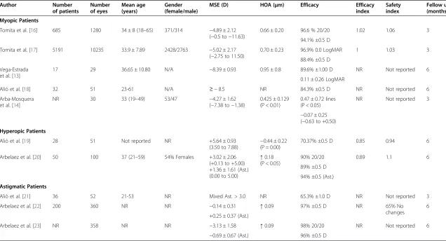
Outcomes of 6thgeneration lasers
Our studies focused on the promised safety and accur-acy of the new platforms (Table 4) [13,14,16-23] in my-opic correction through two separate reports of patients with high myopia of−8.50 D or more. In the first study, we studied 29 eyes of 17 patients, with a mean age of 36.65 ± 10.80 years, and a mean spherical equivalent (MSE) of−8.39 ± 0.93 D, after 6 months follow up. We found the efficacy of the treatment to be 89.6% within ±1.00 D of tar-get refraction, and post operative high order aberrations (HOA) to be 0.95 ± 0.80μm [13]. We confirmed the results with a larger sample in a second study, to show an efficacy of 84.3% within ±0.5 D of target refraction [18]. At the same time, Arba-Mosquera et al. published results of the 3 month follow up of 30 eyes with a mean spherical equivalent (MSE) of −4.27 ± 1.62 D showing a mean re-sidual spherical equivalent of−0.07 ± 0.25 D and postoper-ative HOA of 0.425 ± 0.129 μm [14]. Later on, Tomita et al. used a larger sample of 10235 eyes of 5191 patients. In this study, the MSE was−5.02 ± 2.17 D and follow up results up to 3 months showed an efficacy index of 1.00; 88.4% within ±0.50 D of target refraction and a safety index of 1.03; 96.9% achived 0.0 LogMAR or better, and postop-erative HOA were 0.70 ± 0.23μm [17]. The previous stud-ies were conducted on the AMARIS excimer laser platform of SCHWIND eye-tech-solutions, Kleinostheim, Germany. Kanellopoulos et al. reported his results with the
Alcon-WaveLight EX500 excimer laser by (Alcon Labora-tories, Fort Worth, TX), where he followed 58 patients who underwent bilateral surgery for 12 months. The pre-operative MSE was−7.67 ± 1.55 D, and his results showed 100% were within 1.0 D defocus equivalent with a kerato-metric stability of 0.22 D [24].
The small spot hyperopic laser in situ keratomileusis (H-LASIK) ablation at the periphery of the cornea pro-ducing certain degrees of steepness. This treatment mo-dality had several limitations as decentration, decrease in best corrected visual acuity (BCVA), high frequency for the need of retreatments, residual refractive errors and induction of astigmatism due to the high levels of cor-neal aberrations as negative spherical aberration, all cause loss of the efficiency of treatments and changes in biomechanics of the cornea [20]. Our studies included a sample of 51 eyes in 28 patients of a MSE of +5.64 ± 0.93 D followed up for 6 months, we reported an efficacy of 70.37% within ±0.5 D of target refraction, the HOA postoperatively was −0.44 ± 0.22 μm, with a efficacy and safety index of 0.85 and 0.94 respectively [19]. Another 6 months retrospective follow up of 51 eyes with a spherical equivalent of more than 5.5 D showed signifi-cant increase in corneal root mean square (RMS) HOA, RMS spherical aberration (SA) and RMS coma were also observed six months after surgery (p < 0.01). Corneal asphericity for the 4.5 mm (Q45) and 8 mm (Q8) of cor-neal diameter also changed significantly during the post-operative period (p < 0.01). Strehl ratio change was not statistically significant (p = 0.77) [25]. Arbelaez et al. published a study with MSE +3.02 ± 2.06 D (astigmatism was +1.36 ± 1.61 D), they reported 6 months follow up with a mean postoperative increase in HOA of 0.18 μm (P < 0.05), 89% were within ±0.5 D of the target refrac-tion and 94% were ±0.5 D of target astigmatism [20]. Kanellopoulos used a larger sample of 202 eyes with a longer follow up of 2 years to demonstrate the safety and efficacy of topography guided ablation using a 400 Hz WaveLight excimer laser by (Alcon Laboratories, Fort Worth, TX). In his study the MSE was +3.04 ± 1.75 D, the results showed that the mean refraction spherical equivalent was ±0.50 D of target refraction in over 80% of cases, with an increase in the RMS of 15% [26].
Table 4 Outcomes of the AMARIS-SCHWIND 6thgeneration excimer laser
Author Number
of patients
Number of eyes
Mean age (years)
Gender (female/male)
MSE (D) HOA (μm) Efficacy Efficacy
index
Safety index
Fellow up (months)
Myopic Patients
Tomita et al. [16] 685 1280 34 ± 8 (18–65) 371/314 −4.89 ± 2.12
(−0.5 to−11.63)
0.66 ± 0.20 96.6 % 20/20 1.02 1.06 3
94.1% ±0.5 D
Tomita et al. [17] 5191 10235 33.9 ± 7.89 2428/2763 −5.02 ± 2.17
(−2.75 to 11.50)
0.70 ± 0.23 96.9% 0.0 LogMAR 1 1.03 3
88.4% ±0.5 D
Vega-Estrada et al. [13]
17 29 36.65 ± 10.80 N/A −8.39 ± 0.93 0.95 ± 0.8 89.6% ±1.00 D NR Not reported 6
0.11 ± 0.26 LogMAR
Alió et al. [18] 32 51 23-61 N/A ≥ −8.5 NR 84.3% ±0.5 D NR Not reported 6
Arba-Mosquera et al. [14]
NR 30 33 (19–49) 53/47 −4.27 ± 1.62
(−7.38 to−1.38)
0.425 ± 0.129 (P < 0.01)
0.47 ± 0.72 lines (P < 0.05)
NR Not reported 3
−0.07 ± 0.25 (−0.63 to +0.50)
Hyperopic Patients
Alió et al. [19] 28 51 Not reported NR +5.64 ± 0.93
(3.50 to 7.88) −
0.44 ± 0.22 (P = 0.00)
70.37% ±0.5 D 0.85 0.94 6
Arbelaez et al. [20] 50 100 37 (21–59) 54% Females +3.02 ± 2.06
(+0.13 to +5.00) +1.36 ± 1.61 (Ast.) (0.00 to 5.00)
↑0.18 (P < 0.05)
90% 20/20 0.89 1.1 6
89% ±0.5 D
94% ±0.5 (Ast.)
Astigmatic Patients
Alió et al. [21] 36 52 21-53 NR Mixed Ast. > 3.0 NR 65.3% ±1.0 D NR Not reported 3
Arbelaez et al. [22] 200 360 NR NR −0.14 ± 0.31 ↑0.09 97% ±0.5 D NR 65% No
changes
6
+0.25 ± 0.37 (Ast.)
Arbelaez et al. [23] NR 358 NR NR −3.13 ± 1.58 ↑0.09 98% 20/20 NR Not reported 6
−0.69 ± 0.67 (Ast.) 96% ±0.5 D
MSE: mean spherical equivalent, HOA: high order aberrations, Ast.: astigmatism, NR: not reported, N/A: not available.
↑: increase in high order aberrations.
Alió
Eye
and
Vision
(2015) 2:6
Page
6
of
refraction [21]. Earlier, Arbelaez et al. published a num-ber of reports, with an average of 1.26 ± 3.29 D of astig-matism, and with 6 months follow up found an increase of 0.09μm with a residual astigmatism 0.50 ± 0.26 D (P < 0.0001) [12,22,23]. The mean decrease of astigmatic magnitude in these reports was 93%, indicating a slight under correction of the preoperative astigmatism, but with marked improvements from the 36% to 91% re-ported with the use of older excimer laser platforms [27,28]. Also, recent reports showed a 100% efficacy within ±0.25 D after 12 months [24]. Topography-guided hyperopic astigmatism correction showed a correction of a mean preoperative cylinder value of−1.24 ± 1.41 D to the respective postoperative value of−0.35 ± 0.25 D [26].
New platforms had shown similar satisfactory results with PRK. Aslanides et al. in his 2 years follow up of 80 eyes with mild to high myopia and myopic astigmatism found 91% to be within ±0.5 D of target refraction, but re-ported a statistically significant increase in postoperative coma (+0.12 μm) and spherical aberration (+0.14 μm) compared to preoperative values (P< 0.001, both cases) when performed using the SCHWIND AMARIS [29,30].
Challenges facing excimer laser refractive surgery today In spite of the astonishing progress in the field of laser refractive surgery, a number of challenges are still facing the technology. One of the most critical is the limita-tions in the visual and optical outcomes in patients with high hyperopic refractive errors, as most authors re-ported a significant induction of the corneal HOA, most significantly in the RMS coma [19,20].
The efficacy of the excimer laser technology even with latest platforms in the treatment of presbyopia is still the subject of great debate. PresbyLASIK has been described in three different approaches to create multifocality: the transitional multifocality, the central presbyLASIK and the peripheral presbyLASIK. Both central and peripheral techniques reportedly obtained adequate spectacle inde-pendence for near and for far with a degree of neuroa-daptation process needed in peripheral techniques. An intentional increase in coma aberrations was noticed in transitional multifocality giving it a range of very limited use and outcomes. The level of scientific evidence we already have is enough to consider presbyLASIK as a useful tool in the correction of presbyopia [31]. Epstein et al. reported a 4 years follow up of 103 patients who underwent peripheral presbyLASIK, 89% of hyperopes and 92% of myopes was completely spectacle independ-ent, with distance unaided visual acuity of 20/20 in 67.9% of hyperopes and 70.7% in myopes. They also re-ported that the overall increase in the HOA was mani-fested to a greater extent in hyperopic cases [32]. Other programs as The Supracor presbyopia procedure showed good near visual acuity outcomes over 6 months follow
up, but loss of corrected distance visual acuity (CDVA) occurred in 39.1% of eyes, also reported in PresbyMAX [33]. At 1 year, 70% of patients achieved uncorrected distance visual acuity (UDVA) 0.1 logMAR or better, 84% patients obtained uncorrected near visual acuity (UNVA) 0.1 logRAD or better, and 83% of eyes were within 0.75 D of defocus [34].
Ablation centration is a major issue in the excimer laser development, the decentration of ablation can lead to under correction and irregular astigmatism, which is most important in hyperopic patients [35,36], who tend to have a larger angle kappa values [35]. There are four main methods of centration in laser refractive surgery that has been suggested in literature; center of the pupil, coaxially sighted corneal light reflex (CSCLR), corneal vertex normal and between the pupillary and visual axis [37]. Many reports had demonstrated that pupil-centered and vertex-centered treatments provide similar visual and optical out-comes. However, in eyes showing large temporal pupil decentration, pupil-centered ablation seemed to produce a lower amount of coma and consequently, a reduced loss of BCVA compared with vertex-centered patients [26,38,39].
Patient satisfaction with the results of treatment with the recent excimer laser was remarkable. Kyprianou et al. reported a 100% satisfaction in 32 patients, with average age of 31.9 years and a preoperative MSE of −3.05 D. This was evaluated by a question-naire consisting of 21 questions. Patients were most satisfied in questions concerning quality of vision, distance vision, when watching TV and driving dur-ing daytime and durdur-ing the night [43].
Conclusion
In summary, the latest generation of excimer laser plat-forms had introduced a large number of features as faster laser, smaller spot size, a high speed tracker, pupil monitoring and online pachymetry, all of which provided superior treatment with significant improvement of in-duced post operative HOA and control of thermal dam-age. This technology is still facing major limitations in terms of high hyperopic, presbyopic treatments, along with difficulties in laser centration along with the limita-tion of the customized treatments, generated by the bio-mechanical patterns of wound healing [44].
Competing interests
The authors declare that they have no competing interests.
Authors’contributions
Both authors set the framework of the article, ME was responsible for writing the draft manuscript and with literature review, JA, was responsible for scientific proofing and several adjustments of the draft manuscript. Both authors read and approved the final manuscript.
Acknowledgements
This study has been supported in part by a grant from the Spanish Ministry of Health, InstitutoCarlos III, Red Temática de Investigación Cooperativa en Salud“Patología ocular del envejecimiento, calidad visual y calidad de vida”, Subproyecto de Calidad Visual (RD07/0062) and a grant from the Spanish Ministry of Economy and Competitiveness, Instituto Carlos III, Red Temática de Investigación Cooperativa en Salud (RETICS)“Prevención, detección precoz y tratamiento de la patología ocular prevalente, degenerativa y crónica”. Subprograma“dioptrio ocular y patologías frecuentes” (RD12/0034/0007).
Author details 1
Vissum Corporación Alicante, Alicante, Spain.2Division of Ophthalmology, Universidad Miguel Hernández, Alicante, Spain.3Clinical research fellow in
Vissum Corporación Alicante, Universidad Miguel Hernández, Alicante, Spain.
4Avda de Denia s/n, Edificio Vissum, 03016 Alicante, Spain.
Received: 20 June 2014 Accepted: 11 February 2015
References
1. Krueger RR, Rabinowitz YS, Binder PS. The 25th anniversary of excimer lasers in refractive surgery: historical review. J Refract Surg. 2010;26(10):749–60. 2. AAO Refractive Management/Intervention PPP Panel, Hoskins Center for Quality
Eye Care. Refractive Errors & Refractive Surgery PPP–2013. 2013. http://one.aao. org/preferred-practice-pattern/refractive-errors—surgery-ppp-2013.
3. AAO Quality of Care Secretariat, Hoskins Center for Quality Eye Care. Summary Recommendations for Keratorefractive Laser Surgery–2013. 2013. http://one. aao.org/clinical-statement/summary-recommendations-lasik—january-2008. 4. Anschütz T. Laser Correction of Hyperopia and Presbyopia. Int Ophthalmol
Clin. 1994;34(4):107–37.
5. Müller B, Boeck T, Hartmann C. Effect of excimer laser beam delivery and beam shaping on corneal sphericity in photorefractive keratectomy. J Cataract Refract Surg. 2004;30(2):464–70.
6. Poirier L, Coulon P, Williamson W, Verin P. Energy fluctuations in an excimer laser during photorefractive keratectomy. J Refract Corneal Surg. 1994;10 Suppl 2:S258–61.
7. Azar DT, Chang JH, Han KY. Wound healing after keratorefractive surgery: review of biological and optical considerations. Cornea. 2012;31 Suppl 1:S9–19.
8. Kruger RR, Saedy NF, McDonell PJ. Clinical analysis of steep central islands after excimer laser photorefractive keratectomy. Arch Ophthalmol. 1996;114:377–81. 9. SCHWIND eye-tech-solutions. The SCHWIND AMARIS family. http://www.
schwind-amaris.com/en/home. Accessed 1 May 2014.
10. ALCON surgical. WaveLight® Allegretto Wave® Eye-Q Laser. http://www.alcon-surgical.com/wavelight-allegretto-wave-eye-q-laser.aspx. Accessed 1 May 2014. 11. NIDEK CO., LTD. NIDEK advanced vision excimer laser system NAVEX Quest. http://www.nidek-intl.com/products/ref_surgical/navex-quest.html. Accessed 1 May 2014.
12. Arbelaez MC, Vidal C, Arba-Mosquera S. Excimer laser correction of moderate to high astigmatism with a non-wavefront-guided aberration-free ablation profile: Six-month results. J Cataract Refract Surg. 2009;35(10):1789–98. 13. Vega-Estrada A, Alió JL, Arba Mosquera S, Moreno LJ. Corneal higher order
aberrations after LASIK for high myopia with a fast repetition rate excimer laser, optimized ablation profile, and femtosecond laser-assisted flap. J Refract Surg. 2012;28(10):689–96.
14. Arba-Mosquera S, Arbelaez MC. Three-month clinical outcomes with static and dynamic cyclotorsion correction using the SCHWIND AMARIS. Cornea. 2011;30(9):951–7.
15. De Ortueta D, Magnago T, Triefenbach N, Arba Mosquera S, Sauer U, Brunsmann U. In vivo measurements of thermal load during ablation in high-speed laser corneal refractive surgery. J Refract Surg. 2012;28(1):53–8. 16. Tomita M, Watabe M, Yukawa S, Nakamura N, Nakamura T, Magnago T.
Safety, efficacy, and predictability of laser in situ keratomileusis to correct myopia or myopic astigmatism with a 750 Hz scanning-spot laser system. J Cataract Refract Surg. 2014;40(2):251–8.
17. Tomita M, Waring 4th GO, Magnago T, Watabe M. Clinical results of using a high-repetition-rate excimer laser with an optimized ablation profile for myopic correction in 10 235 eyes. J Cataract Refract Surg. 2013;39(10):1543–9. 18. Alio JL, Vega-Estrada A, Piñero DP. Laser-assisted in situ keratomileusis in high
levels of myopia with the amaris excimer laser using optimized aspherical profiles. Am J Ophthalmol. 2011;152(6):954–63.
19. Alió JL, El Aswad A, Vega-Estrada A, Javaloy J. Laser in situ keratomileusis for high hyperopia (>5.0 diopters) using optimized aspheric profiles: efficacy and safety. J Cataract Refract Surg. 2013;39(4):519–27.
20. Arbelaez MC, Vidal C, Arba MS. Six-month clinical outcomes after hyperopic cor-rection with the SCHWIND AMARIS Total-Tech laser. J Optom. 2010;3:198–205. 21. Alio JL, El Aswad A, Plaza-Puche AB. Laser-Assisted In Situ Keratomileusis in
High Mixed Astigmatism With Optimized, Fast-Repetition and Cyclotorsion Control Excimer Laser. Am J Ophthalmol. 2013;155(5):829–36.
22. Arbelaez MC, Arba MS. The SCWIND AMARIS Total-Tech Laser as An All-Rounder in Refractive Surgery. Middle East Afr J Ophthalmol. 2009;16(1):46–53. 23. Arbelaez MC, Aslanides IM, Barraquer C, Carones F, Feuermannova A,
Neuhann T, et al. LASIK for myopia and astigmatism using the SCWIND AMARIS excimer laser: an international multicenter trial. J Refract Surg. 2010;26(2):88–98.
24. Kanellopoulos AJ, Asimellis G. Refractive and keratometric stability in high myopic LASIK with high-frequency femtosecond and excimer lasers. J Refract Surg. 2013;29(12):832–7.
25. El Aswad A, Vega A, Arba Samuel, Alio JL, Plaza-Puche A, Wróbel-Dudzińska D. Anterior corneal surface aberrations following excimer laser correction of high hyperopia. J Refract Surg. In Press.
26. Kanellopoulos AJ. Topography-guided hyperopic and hyperopic astigmatism femtosecond laser-assisted LASIK: long-term experience with the 400 Hz eye-Q excimer platform. Clin Ophthalmol. 2012;6:895–901.
27. Barraquer CC, Gutie’rrez MAM. Results of laser in situ keratomileusis in hyperopic compound astigmatism. J Cataract Refract Surg. 1999;25:1198–204.
28. Payvar S, Hashemi H. Laser in situ keratomileusis for myopic astigmatism with the Nidek EC-5000 laser. J Refract Surg. 2002;18:225–33.
30. Moshirfar M, Churgin D, Betts B, Hsu M, Sikder S, Neuffer M, et al. Prospective, randomized, fellow eye comparison of WaveLight® Allegretto Wave® Eye-Q versus VISX CustomVue TM STAR S4 IRTM in photorefractive keratectomy: analysis of visual outcomes and higher-order aberrations. Clin Ophthalmol. 2011;5:1185–93.
31. Alió JL, Amparo F, Ortiz D, Moreno L. Corneal multifocality with excimer laser for presbyopia correction. Curr Opin Ophthalmol. 2009;20(4):264–71. 32. Epstein RL, Gurgos MA. Presbyopia treatment by monocular peripheral
presbyLASIK. J Refract Surg. 2009;25(6):516–23.
33. Cosar CB, Sener AB. Supracor hyperopia and presbyopia correction: 6-month results. Eur J Ophthalmol. 2014;24(3):325–9.
34. Luger MH, Ewering T, Arba-Mosquera S. One-year experience in presbyopia correction with biaspheric multifocal central presbyopia laser in situ keratomileusis. Cornea. 2013;32(5):644–52.
35. Basmak H, Sahin A, Yildirim N, Papakostas TD, Kanellopoulos AJ. Measurement of angle kappa with synoptophore and Orbscan II in a normal population. J Refract Surg. 2007;23(5):456–60.
36. Pande M, Hillman JS. Optical zone centration in keratorefractive surgery. Entrance pupil center, visual axis, coaxially sighted corneal reflex, or geometric corneal center? Ophthalmology. 1993;100(8):1230–7.
37. Moshirfar M, Hoggan RN, Muthappan V. Angle Kappa and its importance in refractive surgery. Oman J Ophthalmol. 2013;6(3):151–8.
38. De Ortueta D, Schreyger FD. Centration on the cornea vertex normal during hyperopic refractive photoablation using videokeratoscopy. J Refract Surg. 2007;23(2):198–200.
39. Soler V, Benito A, Soler P, Triozon C, Arné JL, Madariaga V, et al. A randomized comparison of pupil-centered versus vertex-centered ablation in LASIK correction of hyperopia. Am J Ophthalmol. 2011;152(4):591–9. 40. Dupps Jr WJ, Wilson SE. Biomechanics and wound healing in the cornea.
Exp Eye Res. 2006;83(4):709–20.
41. Ortiz D, Piñero D, Shabayek MH, Arnalich-Montiel F, Alió JL. Corneal biomechanical properties in normal, post-laser in situ keratomileusis, and keratoconiceyes. J Cataract Refract Surg. 2007;33(8):1371–5.
42. Yu CQ, Manche EE. Comparison of 2 wavefront-guided excimer lasers for myopic laser in situ keratomileusis: one-year results. J Cataract Refract Surg. 2014;40(3):412–2.
43. Kyprianou G, Machácková M, Feuermannová A, Rozsíval P, Langrová H. Subjective visual perception after laser treatment of myopia on two types of lasers. Cesk Slov Oftalmol. 2010;66(5):213–9.
44. Alio JL, Rosman M, Arba MS. Minimally Invasive Refractive Surgery. In: Howard Fibe I, Mojon DS, editors. Minimally Invasive Ophthalamic Surgery. NewYork: Springer-Verlag; 2010. p. 100–1.
Submit your next manuscript to BioMed Central and take full advantage of:
• Convenient online submission
• Thorough peer review
• No space constraints or color figure charges
• Immediate publication on acceptance
• Inclusion in PubMed, CAS, Scopus and Google Scholar
• Research which is freely available for redistribution
![Table 1 FDA Indications for LASIK and PRK: [2]](https://thumb-us.123doks.com/thumbv2/123dok_us/9460113.1929076/2.595.57.291.528.725/table-fda-indications-lasik-prk.webp)