Comparative Effects of Copper, Iron, Vanadium and Titanium on
Low Density Lipoprotein Oxidation in vitro
Mohsen Ani
*1, Ali Asghar Moshtaghie
1and Hassan Ahmadvand
21
Dept. of Clinical Biochemistry, School of Pharmacy, Isfahan University of Medical Sciences, Isfahan; 2Dept. of Clinical Biochemistry, School of Medicine, Lorestan University of Medical Sciences, Khoramabad, Iran
Received 9 May 2006; revised 19 September 2006; accepted 2 October 2006
ABSTRACT
Introduction: Oxidation of low density lipoprotein (LDL) has been strongly implicated in the phathogenesis of atherosclerosis. The use of oxidants in dietary food stuff may lead to the production of oxidized LDL and may increase both the development and the progression of atherosclerosis. The present work investigated the effects of some elements including: copper (Cu), iron (Fe), vanadium (V) and titanium (Ti) on in vitro LDL oxidation quantitatively. Methods: The first LDL fraction was isolated from fresh plasma by single vertical discontinuous density gradient ultracentrifugation. The formation of conjugated dienes and thiobarbituric acid reactive substances and increase in electrophoretic mobility of LDL were monitored as markers of the oxidation of LDL. Results: It was demonstrated that Cu, Fe, V and Ti exhibited strong oxidant activity in this respect (P<0.001). Oxidation of LDL in the presence of Cu was more and appeared to be in this order Cu>Fe> V>Ti. Discussion: Cu, Fe, V and Ti are redox-active transition metals that may cause oxidative damage to lipids, proteins and DNA molecules. We suggest that these elements may also influence the oxidation of LDL
in vivo, which could increase both the development and progression of atherosclerosis. Iran. Biomed. J. 11 (2): 113-118, 2007
Keywords: Copper (Cu), Iron (Fe), Vanadium (V), Titanium (Ti), Low-density lipoprotein (LDL) oxidation
INTRODUCTION
ardiovascular diseases are the leading causes of mortality in the world. Although an increased concentration of plasma low density lipoprotein (LDL) is believed to constitute a major risk factor for atherosclerosis, the underlying mechanisms for this effect remain unclear. To date, considerable evidence support a role for oxidatively modified LDL in the pathogenesis of atherosclerosis [1]. The uptake of oxidized LDL (Ox-LDL) by macrophages results in the formation of foam cells and cholesterol accumulated in vascular endothelial cells could promote the development of the characteristic fatty streaks found in atherosclerotic lesions. Ox-LDL is cytotoxic to arterial wall cells stimulates haemostatic and thrombotic process and secretion of cytokines and growth factors from cells of the arterial wall [1-4]. Ox-LDL as well as lipid peroxidation products of LDL have been detected in human and animal atherosclerotic lesions, and this
finding adds support to the contributory role of Ox-LDL in clinical condition. Finally, Ox-Ox-LDL has been reported to compromise endothelial integrity, a salient feature of atherosclerosis [4, 5].
Copper (Cu) and iron (Fe) are redox-active metals that can participate in electron transfer reactions with the consequent production of oxidant species capable of oxidizing cell components. Cu and Fe can catalyze the formation of the highly reactive hydroxyl radicals from hydrogen peroxide (H2O2)
via the Haber-Weiss reaction and decompose lipid peroxides to peroxyl and alkoxyl radicals, which favor the propagation of lipid oxidation [6]. Oxidants such as Cu and Fe have been shown to increase formation of atherosclerotic lesions in animal models and epidemiological data suggest that a direct relationship exists between the intake of oxidant and the risk of coronary artery disease. Cu, Fe, vanadium (V) and titanium (Ti) are redox-active transition metal that may cause oxidative damage to lipids, proteins and DNA molecules. Reactive
C
oxygen species (ROS) so formed initiate lipid oxidation involved in atherogenesis [7-11]. In this study, we investigated the effects of Cu, Fe, V and Ti on LDL oxidation in vitro by monitoring the formation of conjugated dienes, the formation of thiobarbituric acid reactive substances (TBARS) and measurements of electrophoretic mobility (REM) of LDL.
MATERIALS AND METHODS
Chemicals. CuSO4, Fe2(SO4)3, V3O5, TiCl3,
disodium ethylene diamine tetraacetate, Potassium bromide (KBr), sodium chloride (NaCl), disodium hydrogen phosphate (Na2HPO4) and oil red O were
purchased from Sigma Chemical Co.(USA).
Blood sampling. Fasting blood samples after an overnight fasting were collected in EDTA containing tubes (1.6 mg EDTA/ml blood). To obtain plasma, the centrifugation (2000 ×g, at 4°C, for 10 min) was started within 1 min following venous puncture to avoid auto-oxidation of the sample. To minimize oxidation in vitro, sodium azide (0.06% wt/vol) was added to plasma immediately after collection.
LDL isolation. The LDL fraction was isolated from fresh plasma by single vertical discontinuous density gradient ultracentrifugation [12]. The density of the plasma was adjusted to 1.21 g/ml by the addition of solid KBr (0.365 g/ml). Centrifuge tubes were loaded by layering 1.5 ml of density-adjusted plasma under 3.5 ml of 0.154 mol/L NaCl, and centrifuged in a Beckman L7-55 ultracentrifuge (100,000 ×g) at 10°C for 2.5 hours. The yellow LDL band located in the upper middle portion of the tube was collected into a syringe by puncturing the tube. The isolated LDL was dialyzed at 4°C for 48 h against three changes of deoxygenated-PBS (0.01mol/L Na2HPO4, 0.16 mol/L NaCl, pH 7.4)
containing 0.01% NaN3 and 0.01% EDTA.
LDL oxidation. continuous monitoring of formation of conjugated dienes in LDL. After isolation of total LDL, the protein content of LDL was measured [13]. LDL was adjusted to 150 µg/ml of LDL protein with 10 mM PBS, pH 7.4. The oxidative modification of LDL was initiated by addition of freshly prepared 10 µM CuSO4 solution
in water bath at 37°C for 5 h.
The kinetics of LDL oxidation was monitored every 10 minutes by removing an aliquot, measuring its absorbance at 234 nm, and then returning it to the LDL sample. The lag phase was calculated from the oxidation profile of each LDL preparation by drawing a tangent to the slope of the propagation phase and extrapolation into intercept the initial-absorbance axis.
The lag phase represented the length of the
antioxidant-protected phase during LDL oxidation by 10 µM Cu, Fe, V and Ti solution in vitro. The lag time was measured as the time period until the conjugated dienes began to increase [14]. The formation of conjugated dienes was calculated as conjugated dienes equivalent content (nmol/mg-protein) at 5 h. The conjugated dienes concentration was calculated by using the extinction coefficient for diene conjugates at 234 nm (29,500/mol./L.cm).
Assay of the formation of TBARS. Lipid peroxidation end product was determined as TBARS according to the method of Sheu and Chen(16). After initiating the oxidation process with CuSO4,
the sample mixtures were incubated at 37°C for 5 h in a water bath and the reaction terminated by adding EDTA (2 mM). TBARS formation was measured in a spectrophotometer at 532 nm. The results were recorded as malondialdehyde (MDA) equivalent content (nmol/mg LDL-protein) [15, 16].
Electrophoretic mobility of LDL. The electro-phoretic mobility of n-LDL and Ox-LDL was determined by agarose gel electrophoresis (1% agarose and 0.1 mol/L barbital buffer) under the condition of 200 V, 80 mA in 50 mM barbital buffer, then the gel was fixed in ethanol, acetic acid and water 6:1:3 for 15 min, oven dried (80°C for 1 h), and then stained with oil-red for 16 h. After staining, the gel was washed by bleaching solution (ethanol and water 5:3), until the background adjacent to protein or lipoprotein bands was cleared [1, 17].
Statistical analysis. Each data value is presented as the mean +S.D. The variables used to describe the difference between the oxidation curves were lag time, conjugated dienes, MDA and REM. These parameters were obtained using student's t-test for independent data and differences were considered significant when P<0.05.
Fig. 1. The effects of Cu, Fe, V and Ti on LDL oxidation in l0 mM PBS, pH 7.4 at 37°C for 5 h. Each point represents the mean of three experiments.
RESULTS
The effects of Cu, Fe, V and Ti on LDL oxidation are shown in Figure 1. It clearly shows that CuSO4
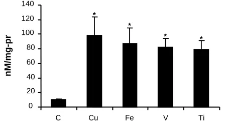
dramatically increased oxidation of LDL. The formation of conjugated dienes, a marker of LDL oxidation, was also increased by Fe, V and Ti. The levels of the conjugated dienes at 5 h and lag time of all experiment groups are shown in Figures 2 and 3.
Fig. 2. Oxidant effects on lag time of Cu, Fe, V and Ti induced LDL oxidation. Each point represents the means of five experiments. *, significance compared to ox-LDL (n-LDL+ Cu) by student's t-test.
Thus, CuSO4 increased the level of the conjugated
dienes by about three folds and was significantly different from control (430.4 + 11.46 nmol/mg LDL-protein vs 146.2 + 6.21 nmol/mg LDL-LDL-protein,
P<0.05 by student's t-test). Fe, V, Ti increased final levels of conjugated dienes in the medium (413.3 + 11.6, 387.47 + 8.6 and 374.13 + 10.1), respectively. The oxidative effect of Cu, Fe, V, and Ti on LDL
was determined and expressed by measurement of MDA equivalent content. The levels of the MDA at 5 h of incubation in all experiment groups are showed in Figure 4. The addition of CuSO4
increased TBARS formation about 11 folds that was compared statistically with control (109.06 + 25.31 nmol/mg LDL-protein VS 9.11 + 1.34 nmol/mg LDL-protein, P<0.001 by student's t-test). Fe, V and Ti also significantly increased TBARS formation about 8, 7 and 5.7 fold in comparison with control LDL.
Fig. 3. The effects of Cu, Fe, V and Ti on the formation of conjugated dienes of LDL oxidation. (C) n-LDL; (Cu) n-LDL + Fe n-LDL + Fe(V) n-LDL + V, (Ti) n-LDL + Ti. Each point represents the means of five experiments. *, significance compared to n-LDL by student's t-test.
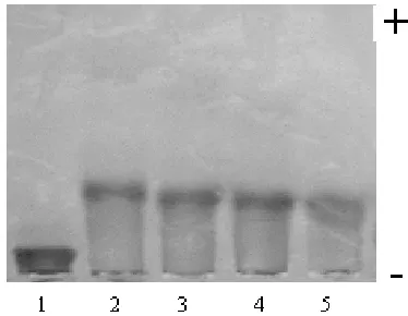
Effect of Cu, Fe, V and Ti on electrophoretic mobility of LDL. The effects of Cu, Fe, V and Ti on the electrophoretic mobility of LDL are shown in Figure 5 and 6. The electrophoretic mobility of LDL in all experimental groups is shown Figure 7. After LDL oxidation by CuSO4 the Ox-LDL moved faster
than non-Ox-LDL on agarose gel electrophoresis. CuSO4 and Fe, V and Ti significantly increased the
electrophoretic mobility of LDL.
Fig 4. The effects of Cu, Fe, V and Ti on the formation of MDA. (C) n-LDL; (Cu) n-LDL + Fe n-LDL + Fe(V) n-LDL + V, (Ti) n-LDL + Ti. Each Point represents the mean of five experiments.*, significance compared to n-LDL by student's t-test.
0 20 40 60 80 100 120 140
C Cu Fe V Ti
nM/mg-pr
*
*
* *
0 100 200 300 400 500
C Cu Fe V Ti
nM/mg-pr
*
*
* *
0.2 0.4 0.6 0.8 1.0 1.2 1.4 1.6 1.8
2.0
0 50 100 150 200 250
min
A23
4
C Cu Fe V Ti
0 20 40 60 80 100 120
Cu Fe V Ti
min
*
Fig. 5. The effects of Cu, Fe, V and Ti on electrophoretic mobility of LDL. (1) n-LDL; (2) n-LDL + Cu; (3) n-LDL + Fe; (4) n-LDL + V; (5) n-LDL + Ti.
DISCUSSION
Lipid peroxidation in micelles, liposomes and LDL can be initiated simply by adding redox-active transition metal, which has been assumed to participate in some metabolic complication [18]. The peroxyl (LOO-) and alkoxyl (LOo) radicals generated in these reactions are believed to be responsible for initiating oxidation of the parent polyunsaturated fatty acid (PUFA) through hydrogen-atom abstraction. Trace metals have been shown to oxidize LDL even in the absence of the cells. This oxidation is associated with free radical-mediated peroxidation of some lipids within the LDL particles, resulting in recognition by cell surface receptor and internalization of the LDL by macrophages. The oxidative modification of LDL by some transition metal ions such as Cu or Fe has
Fig. 6. The effects of Cu, Fe, V and Ti on electrophoretic mobility of LDL. (1) n-LDL; (2) n-LDL + Cu; (3) n-LDL + Fe; (4) n-LDL+V; (5) n-LDL + Ti.
already been reported and their importance in the progression of atherosclerosis has been emphasized [19]. Transition metals such as Cu and Fe have also been extensively studied as catalysts for LDL oxidation in vitro. There is evidence for the presence of redox-active metals in atheromatous plaques, and physiological forms of these metals such as hemin and ceruloplasmin can promote LDL oxidation in vitro [20, 21]. The oxidative modification of LDL (OX-LDL) is the major factor that stimulates the development of atherosclerosis [5]. Therefore, the major aim of this study was to determine and compare the oxidant effects of Cu, Fe, V and Ti using the in vitro model of LDL oxidation. The oxidative modification of LDL induced by Cu ion is related to free radical reaction. However, the
Fig. 7. The effects of Cu, Fe, V and Ti on rate the electrophoretic mobility of LDL oxidation. (C) LDL; (Cu) n-LDL + Fe n-n-LDL + Fe(V) n-n-LDL + V, (Ti) n-n-LDL + Ti. Each Point represents the means of three experiments. *, significance compared to n-LDL by STUDENT'S t-test.
mechanism of the oxidant production has not been elucidated yet. LDL oxidation may require the generation of superoxide anion and probably the ultimate generation of hydroxyl radicals by the Fenton reaction. After the oxidation by Cu ion, PUFA of LDL were oxidized which resulted in an elevation of lipid peroxides and depletion of vitamins in ox-LDL [1]. Vanadium can induce the formation of reactive oxidizing substances (ROS) in biological systems through: (1) Fenton-like reaction and 2)Vanadate bioreduction mediated by reduced glutathione oxidases with ROS as a by-product [22]. Vanadium is a heavy metal with increased environmental circulation.In high doses,it may have various anthropogenic activities which are great public health concerns due to its toxicity and accumulative behavior at specific target organs, such as the liver and kidney, including oxidative damage,
0 1 2 3 4 5 6
C Cu Fe V Ti
cm
*
*
*
*
lipid peroxidation and changes in hematological, reproductive and respiratory systems. Formation of ROS induced by V in biological systems may involve Fenton-like reactions, vanadate bioreduction mediated by reduced glutathione (GSH), flavoenzymes or nicotinamide adenine dinucleotide, reduced form and nicotinamide adenine dinucleotide phosphate, reduced form oxidation or interaction with mitochondria [22-26]. Ti is another element with oxidation potential. Ti salts are widely used in industry for ceramic painting in pharmacy for tablet coating and making chemical sunscreens and in medicine as photocatalysts with bacteriocidal activity. This may address the idea that the exposure to these salts could play a role in the pathogenesis of atherosclerosis [27-29]. Our results clearly showed that Cu increased conjugated dienes and TBARS and led to increase in electrophoretic mobility of LDL in vitro. In this study, the oxidation effects of V and Ti on LDL oxidation showed to be less than Fe or Cu, probably because the redox potential of Ti and V is less than Cu and/or Fe.
REFERENCES
1. Joseph, L.W. and Steinberg, D. (1991) Role of oxidized low density lipoprotein in atherogenesis. J. Clin. Invest. 88: 1785-1792.
2. Yamamoto, A., Tembal, H., Horibe, H., Mabuchi, H., Saito, Y., Matsuzawa, Y., Kita, T. and Nakamura, H. (1990) Influence of life style and excess body weight on HDL-cholesterol and other lipid parameters in men. J. Atheroscler. Thromb. 10 (3): 165-175.
3. Koizumi, J., Shimizu, M., Miyamoto, S., Hideki Origasa, H. and Mabuchi, H. (2002) Effect of pravastatin-induced LDL-cholesterol reduction on coronary heart disease and cerebrovascular disease in Japanease: Hokuriku lipid coronary heart disease study-pravastatin. Atherosclerosis trial (Holicos-PAT). J. Atheroscler. Thromb. 9 (5): 251-259. 4. Yu, L.H., Liu, G.T., Sun, Y.M. and Zhang, H.Y.
(2004) Antioxidative effect of schisanhenol on human low density lipoprotein and its quantum chemical calculation. Acta Pharmacol. Sci. 25 (8): 1038-1044.
5. Aviram, M. and Rosenblat, M. (1994) Macrophage-mediated oxidation of extracellular low density lipoprotein requires an initial binding of the lipoprotein to its receptor. J. Lipid Res. 35: 385-398.
6. Paola Zago, M. and Oteiza, P.I. (2001) The antioxidant properties of zinc: Interaction with iron and antioxidant. Free Radic. Biol. Med. 31 (2): 266-274.
7. Zacharski, L.R., Chow, B., Lavori, P.W. and Howes, P.S. (2000) The iron (Fe) and atherosclerosis study (FeAST): A pilot study of reduction of body iron stores in atherosclerotic peripheral vascular disease.
Am. Heart J. 139 (2): 337-345.
8. Gaetke, L.M. and Chow, C.K. (2003) Copper toxicity, oxidative stress, and antioxidant nutrients.
Toxicology 189: 147-163.
9. Lynch, S.M. and Frei, B. (1993) Mechanisms of copper- and iron-dependent oxidative modification of human low density lipoprotein. J. Lipid Res. 34:
1745-1753.
10. Gieseg, S.P. and Esterbauer, H. (1994) Low density lipoprotein is saturable by pro-oxidant copper.
FEBS Lett. 343: 188-194.
11. Van Reyk, D.M., Jessup, W. and Dean, R.T. (1999) Prooxidant and antioxidant activities of macrophages in metal-mediated LDL oxidation.
Vasc. Biol. 19: 1119-1124.
12. Richard, S.C., Sunder, R.M., Robert, R.H. and Alan, C. (1999) Inhibition of LDL oxidation in vitro but not ex vivo by troglitazone. Diabetes 48: 783-790. 13. Lowry, O.H., Rosenbrough, N.J., Farr, A.L. and
Randall, R.J. (1951) Protein measurement with the folin-phenol reagent. J. Biol. Chem. 193: 256-275. 14. Khursheed, P.N., Enrique, B., Maria, A.L. and
Charles, S.L. (1999) Oxidation of LDL in baboons is increased by alcohol and attenuated by poly-enolphosphatidylcholine. J. Lipid Res. 40:
983-987.
15. Seven, A., Civelek, S., Inci, E., Inci, F., Korkut, N. and Burccak, G. (1997) Biochemical evaluation of oxidative stress in patients with laryngeal carcinoma. Turk ORL Arflivi. 35 (3-4): 88-92. 16. Sheu, L.U., Chen, P.H., Tseng, W.C., Chen, C.Y.,
Tsai, L.Y. and Huang, Y.L. (2003) Spectrophotometric determination of a thiobarbituric acid-reactive substance in human hair. Anal. Sci.
19: 958-960.
17. Noble, R.P. (1968) Electrophoretic separation of plasma lipoproteins in agarose gel. J. Lipid Res. 9: 693-700.
18. Morgan, J. and Leake, D.S. (1995) Oxidation of low density lipoprotein by iron or copper at acidic pH. J.
Lipid Res. 36: 2504-2512.
19. Burkitt, M.J. (2001) A critical overview of chemistry of copper-dependent low density lipoprotein oxidation: Roles of lipid hydroperoxides, a-tocopherol, thiols, and ceruloplasmin. Arch. Biochem. Biophys. 394 (1): 117-135.
20. Tribble, D.L., Chu, B.M., Levine, G.A., Krauss, R.M. and Gong, E.L. (1996) Selective resistance of LDL core lipids to iron-mediated oxidation.
Arterioscl. Thromb. Vasc. Biol. 16:1580-1587
.
21. Hippeli, S. and Elstner, E.F. (1999) Transition metal ion-catalyzed oxygen activation during pathogenic processes. FEBS letters. 443: 1-7.
22. Cortizo, A.M., Bruzzone, L., Molinuevo, S. and Etcheverry, S.B. (2000) A possible role of oxidative stress in the vanadium-induced cytotoxicity in the MC3T3E1 osteoblast and UMR106 osteosarcoma cell lines. Toxicology 147: 89-99.
23. Vanadate oligomers (2005) In vivo effects in hepatic vanadium accumulation and stress markers. J. Inorg. Biochem. 99: 1238-1244.
24. Aureliano, M. and Gandara, R.M.C. (2005) Decavanadate effects in biological systems. J. Inorg. Biochem. 99: 979-985.
25. Reul, B.A., Amin, S.S., Buchet, J.P., Ongemba, L.N., Crans, D.C. and Brichard, S.M. (1999) Effects of vanadium complexes with organic ligands on
glucose metabolism: a comparison study in diabetic rats. Br. J. Pharmacol. 126: 467-477.
26. Mukherjee, B., Patra, B., Mahapatra, S., Banerjee, P., Tiwari, A. and Chatterjee, M. (2004) Vanadium an element of atypical biological significance.
Toxicol. Lett. 150: 135-143.
27. Habashi, F. (1997) Titanium. In: Handbook of Extractive Metallurgy, Wiley-VCH, Weinham (GmbH), pp. 1129-1180.
28. Vanzillotta, P.S., Sader, M.S., Bastos, I.N. and Almeida Soares, G.D. (2005) Improvement of in vitro bioactivity by three different surface treatments. Dent. Mater. 1-8.
29. Cotton, S.A. (1995) Titanium, Zirconium and Hofnium Inorg. Chem. 92: 147-156.