ISSN 0015–5659 www.fm.viamedica.pl
The influence of post-fixation on
visualising vimentin in the retina using
immunofluorescence method
B. Baykal
1, C. Korkmaz
1, N. Kocabiyik
2, O.M. Ceylan
31Department of Histology and Embryology, Gulhane Faculty of Medicine, University of Health Sciences, Ankara, Turkey 2Department of Anatomy, Gulhane Faculty of Medicine, University of Health Sciences, Ankara, Turkey
3Department of Ophthalmology, Diskapi Yildirim Beyazit Training and Research Hospital, University of Health Sciences, Ankara, Turkey
[Received: 9 June 2017; Accepted: 24 July 2017]
Background: Post-fixation of sections is especially required for cryostat sections of fresh frozen tissues. Vimentin is an intermediate filament in both fibrillary and non-fibrillary form, expressed in Müller’s cells and astrocytes of the retina. Our aim was to determine the best post-fixation method for visualising vimentin in archival mouse eyes.
Materials and methods: We used an archival mouse eye, slightly pre-fixed with paraformaldehyde and stored at –80°C for 4 years. We tried three fixatives (pa-raformaldehyde [PFA], alcohol/acetic acid [AAA] and methanol) for post-fixation of eye sections.
Results: We showed that post-fixation alters the labelling properties of vimentin expressed in the retina. In the sections with no post-fixation, vimentin positivity was observed in and around the nuclei in non-fibrillary form. In PFA post-fixed sections, the vimentin in the retina was not observed as fibrils. Positivity was observed in the nuclei and in perinuclear regions of the cells. In AAA post-fixed sections, positive labelling was observed around the nuclei as fibrils. In methanol post-fixed sections, labelling was observed around the nuclei as fibrils.
Conclusions: We conclude that post-fixation with AAA is more convenient for immunofluorescent labelling of vimentin in the retina for slightly PFA pre-fixed and long-term stored retina. (Folia Morphol 2018; 77, 2: 246–252)
Key words: vimentin, post-fixation, paraformaldehyde, alcohol/acetic acid, methanol
INTRODUCTION
Vimentin is a 57 kDa intermediate filament ex -pressed in cells of mesenchymal origin [5]. It is located both in the cell cytoplasm and nucleus [10]. It can
be present in fibrillary and non-fibrillary forms [11].
Vimentin is the major cell skeleton component in the immature glial cells of the rat brain [4]. In the adults,
it is expressed in Müller’s cells and astrocytes of the
retina [3, 16, 19].
The eyeball consists of three layers: corneoscleral layer (includes sclera and cornea), vascular layer (uvea) and retina. Retina is arranged into ten layers. These layers are retinal pigment epithelium (RPE), rod and cone cells layer (RCL), outer limiting membrane (OLM),
B. Baykal et al., Post-fixation alters vimentin visualisation
outer nuclear layer (ONL), outer plexiform layer (OPL), inner nuclear layer (INL), inner plexiform layer (IPL), ganglion cell layer (GCL), nerve fibre layer (NFL) and
inner limiting membrane (ILM). The cell bodies of
Müller’s cells are localised in the INL and their pro
-cesses extend to OLM and ILM. Thus, the pro-cesses of Müller’s cells traverse almost the entire retina [17]. Astrocytes are localised in NFL and GCL [16].
The expression of vimentin increases after a me -chanical damage like retinal detachment [15]. In ad-dition, the vimentin positivity in the processes of
Müller’s cells, which extend through the both borders
of retina, reaches the deep layers of retina after retinal
detachment [14]. Together with the vimentin expres -sion in cell bodies and processes of the astrocytes,
vimentin expression in the retina extends from the NFL to ONL under pathologic conditions [16].
Post-fixation is a method, which is performed by
immersion of sections of fresh frozen tissues into
a fixative especially after being sectioned by a cry -ostat. An increase in staining intensity has been
re-ported after this method [18]. Perfusion fixation with glutaraldehyde and post-fixation with osmium tetrox -ide is a method used for electron microscopy [21].
During a study on vimentin expression in slightly paraformaldehyde (PFA) pre-fixed and long-term cryo-preserved archival retina, we encountered difficulty
in visualisation of vimentin positivity. Thinking that
it may be due to the fixative used for post-fixation, we planned this study in which we tried post-fixation with three fixatives.
MATERIALS AND METHODS
The source of archival tissues
The eye used in this study is an archival tissue from
a male Swiss-Webster mice (100 days old), which un
-dergone acute carbon monoxide (CO) poisoning in a previous study, which was approved by the local ethi -cal committee (July 06, 2005; approval number: 05/68; Animal Research Ethics Committee of Gulhane Military
Medical Faculty). Also, this study on archival tissues was
approved by the local ethical committee (January 21, 2011; approval number: 11/1; Animal Research Ethics
Committee of Gulhane Military Medical Faculty). Acute carbon monoxide poisoning was performed using the method described by Gilmer et al. [7]. Briefly, the animal first inhaled 1000 ppm. CO for 40 min and then 50,000 ppm. CO until the loss of consciousness. This animal was perfusion fixed by first infusion of physiological saline solution and then a 4% solution of PFA in phosphate
buffered saline (PBS) under general anaesthesia with xylazine/ketamine. After an in-situ waiting time of 30 min, its brain was used and the eyes were preserved at –80°C
for 4 years after an overnight incubation in 30% sucrose
solution in PBS at 4°C.
Sectioning
The eye was first kept inside the cryostat (Shandon, Thermo Electron Corporation) at –25°C for 30 min for
thermal equilibration and then serial sections at a
thick-ness of 25 µm were taken on 4 slides in a systematically random manner. Each of the 4 slides included 8–10 sections of the same eye. Thus, at least 5–6 sections were analysed for each of the post-fixation method.
Post-fixation
Three different post-fixation fixatives were applied to the sections on three slides. For control purposes post-fixation was not applied to fourth slide. PFA was used on
one of the slides, the other one received alcohol/acetic acid (AAA) (2/1, v/v) and the other one received methanol
for post-fixation. Post-fixation with PFA was performed at room temperature for 5 min. Post-fixation with AAA and methanol was performed at –20°C for 5 min.
Labelling
The sections were washed with PBS for 3 × 5 min
and were incubated in blocking solution (5% bovine
serum albumin and 1% Triton X-100 in PBS) for 2 h.
After that, the sections were incubated overnight
(17 h) in blocking solution containing Cy3 conjugated anti-vimentin antibody (Sigma-Aldrich, Cat# C9080) at
a dilution of 1/20 at 4°C. After washing with PBS for
3 × 5 min, counterstaining was performed by incu -bating in PBS containing Hoechst 33258 at a dilution
of 1/500 for 5 min. The sections were coverslipped with PBS containing 50% glycerine and sealed with
nail polish.
Imaging
Imaging was performed under a fluorescent mi
-croscope (DMI6000B, Leica, Germany) which was equipped with appropriate filter cubes for Cy3 and Hoechst 33258. Densitometric analysis was performed using the same imaging software (Leica Application Suite Advanced Fluorescence, Leica, Germany).
RESULTS
sections of retina, were analysed for each one of post-fixation method.
1. In the sections that were not post-fixed, vimentin positivity was observed in and around the nuclei of GCL and INL of the retina. No fibrillary organisation was observed inside the retina. The vimentin in the
sclera and the connective tissue surrounding the
eyeball was observed as fibrils (Fig. 1).
2. In the sections post-fixed with PFA, the vimentin in the retina was not observed as fibrils. Positiv
-ity was observed in the nuclei and in perinuclear regions of the cells of GCL and INL (Fig. 2). In the surrounding connective tissue vimentin was observed as fibrils. Also positive regions were observed in striated muscles (Fig. 3).
3. In the sections post-fixed with AAA, positive label
-ling was observed around the nuclei of GCL and vimentin was observed as fibrils extending from NFL to INL (Fig. 4). In the surrounding connective tissue vimentin was also observed as fibrils (Fig. 5). 4. In the sections post-fixed with methanol, labelling
was observed around the nuclei and vimentin was observed as fibrils extending to INL. In addition to that, intensive positivity was observed in INL and OPL (Fig. 6). Vimentin is observed as fibrils in the sclera (Fig. 7).
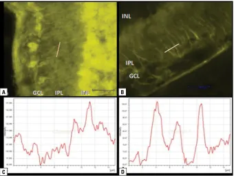
5. According to densitometric analysis of images of
AAA and methanol post-fixed sections, the peaks
are more regular and the background noise is
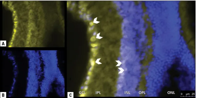
lower in AAA post-fixed sections (Fig. 8). Figure 1. A section with no post-fixation. A. Vimentin labelled with Cy3; B. Nuclei stained with Hoechst; C. Overlay of panels A and B. Nuclear and perinuclear positive labelling is observed in ganglion cell layer (GCL) and inner nuclear layer (INL) of the retina (arrow heads). Positive labelling in fibrillary form is observed in the sclera (arrow). IPL — inner plexiform layer; ONL — outer nuclear layer; OPL — outer plexiform layer.
Figure 2. Retina in a paraformaldehyde post-fixed section. A. Vimentin labelled with Cy3; B. Nuclei stained with Hoechst; C. Overlay of a pan-els A and B. Nuclear and perinuclear positive labelling is observed in ganglion cell layer (GCL) and inner nuclear layer (INL) of the retina (arrow heads); IPL — inner plexiform layer; ONL — outer nuclear layer; OPL — outer plexiform layer.
A
B C
A
B. Baykal et al., Post-fixation alters vimentin visualisation
Figure 3. Connective and muscle tissues in a section post-fixed with paraformaldehyde. A. Vimentin labelled with Cy3; B. Nuclei stained with Hoechst; C. Overlay of a panels A and B. Fibrils in the connective tissue (arrow) and non-fibrillary vimentin in the striated muscle tissue can be observed (arrow head).
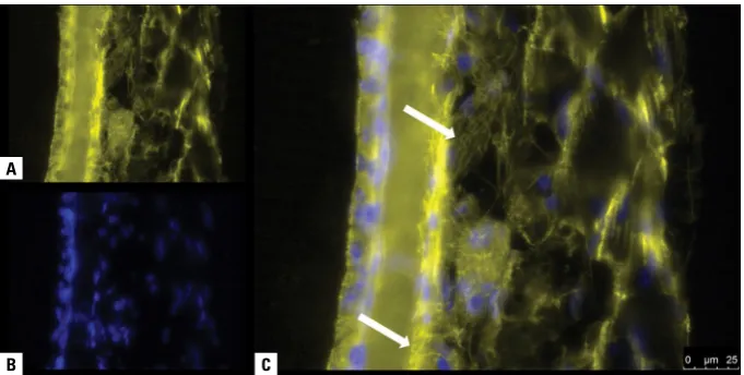
Figure 4. Retina in a section post-fixed withalcohol/acetic acid. A. Vimentin labelled with Cy3; B. Nuclei stained with Hoechst; C. Overlay of a panels A and B. Vimentin isextending from ganglion cell layer (GCL) to inner nuclear layer (INL) as fibrils (arrows). IPL — inner plexiform layer; ONL — outer nuclear layer; OPL — outer plexiform layer.
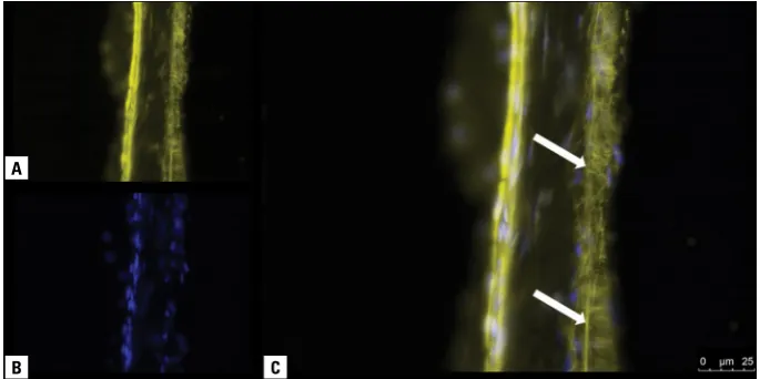
Figure 5. Sclera and connective tissue in a section post-fixed with alcohol/acetic acid. A. Vimentin labelled with Cy3; B. Nuclei stained with Hoechst; C. Overlay of panels A and B. Vimentin positive labelling is observed in the form of fibrils (arrows).
A
B C
A
B
C
A
DISCUSSION
In this study, immunofluorescent vimentin label -ling is performed on sections taken from an archival
eye, which was slightly fixed via perfusion-fixation with PFA and preserved at –80°C for 4 years. Although no statement regarding interaction with mouse tis -sues takes place in the user manual of the antibody
used, the producer has stated a cross-reaction with mouse vimentin had been detected in the first lot of product. During trial labellings, because post-fixation was performed with PFA, fibrillary labelling has not been observed in the retina. However, positive label -ling in the surrounding tissue and positive label-ling in
the retina after appropriate post-fixation has proved that the antibody works on mouse tissues. We used sections from a single eye and therefore we kept only the post-fixation step as a variable and all other
conditions constant.
Fresh frozen sections tend to break. In order to
prevent this, incubation in sucrose is required for taking good sections and good images. But, incuba-tion in sucrose can only be performed if the tissue
is pre-fixed, because, for sucrose to infiltrate the
tissue, an overnight incubation is required and this
time period causes unfixed tissue to be degraded by its own lysosomal enzymes. The 30 min pre-fixation Figure 6. Retina in a section post-fixed with methanol. A. Vimentin labelled with Cy3; B. Nuclei stained with Hoechst; C. Overlay of panels A and B. Nuclear and perinuclear positive labelling (arrow heads) is observed in ganglion cell layer (GCL) and inner nuclear layer (INL). Positive labelling in fibrillary form extending from GCL to INL (arrows) is also observed. Increased intensity is observed in outer plexiform layer (OPL); IPL — inner plexiform layer; ONL — outer nuclear layer.
Figure 7. Sclera in a section post-fixed with methanol. A. Vimentin labelled with Cy3; B. Nuclei stained with Hoechst; C. Overlay of panels A and B. Positive labelling in fibrillary form is observed (arrows).
A
B C
A
B. Baykal et al., Post-fixation alters vimentin visualisation
is a slight fixation because, for a good fixation with PFA, the tissues are usually offered to be immersed
in it at least overnight.
Because fixation changes the chemical properties
of tissue components and the three-dimensional con-formation of proteins, it has important effects on the
affinity and selectivity of the antibodies [6]. For this
reason, optimum conditions must be detected for every molecule of interest. Alves et al. (1992) [1] have
stated that different fixatives may be required for
different epitops and has reported better interaction
for vimentin in sections fixed with ethanol than those fixed with formalin. Helfand et al. (2011) [11] have suggested that methanol fixation is optimal for visu
-alisation of vimentin. However, methanol is reported to cause protein coagulation, which results in decreased
antigenicity and loss of cell architecture [12]. Boon and
Kok [2] have reported that post-fixation with formalin
causes false-negative immune labelling for vimentin.
An interesting finding in our study is that, positive
labelling for vimentin has been observed after
fixation with all three fixatives and also without post-fixation, but variations have been observed in vimentin labelling in the retina according to the post-fixation
method. We observed positive regions for vimentin in the striated muscles, which was also reported by
Granger and Lazarides [8].
Fixation with PFA before labelling vimentin in the
retina has been used in several studies [13, 14, 20]. In these studies the specimens have directly undergone labelling processes. Guidry et al. (2002) [9], on a study
on human retina, have used PFA pre-fixation for 6 h
and after sectioning the retina, they have stored the
slides at –20°C. Before immunofluorescence experi
-ments, they have warmed the slides to room tempera
-ture and post-fixed them with acetone for 3 min. According to our findings, it is obvious that the pre-fixation and storing at –80°C, has provided enough
preservation for vimentin and it is also obvious that
the fixative used for post-fixation has an effect on visualisation of vimentin as fibrils. Researchers who have difficulty in visualisation of vimentin should try pre- and post-fixation methods mentioned here and
in other studies.
CONCLUSIONS
In this study we tried three fixatives for post-fixation of archival slightly PFA pre-fixed eye sections Figure 8. Densitometric analysis of fibrils positively labelled after methanol (A, C) andalcohol/acetic acid (AAA) (B, D) post-fixation. Intensity values of pixels on the lines are shown as histograms (C, D). The peaks in section post-fixed with AAA (D) are more regular and background noise is observed to be lower. GCL — ganglion cell layer; INL — inner nuclear layer; IPL — inner plexiform layer; ONL — outer nuclear layer; OPL — outer plexiform layer.
A B
for labelling vimentin in the retina. In sections which were not post-fixed and those post-fixed with PFA, vimentin was not visible as fibrils. In the sections post-fixed with AAA or methanol, vimentin was visible as fibrils. Densitometric analysis of the images from AAA and methanol post-fixed sections revealed that AAA post-fixation yields more regular peaks and less background noise. In conclusion, for studies in which vimentin expression in the retina has to be shown extending to the outer layers of the retina in a fibril
-lary manner, AAA post-fixation is more convenient. For antibodies which are thought not to work, trials must be performed with combinations of variables
in every step. It should also be noted that vimentin
could be visualised using immunofluorescence even after storing of tissues perfusion-fixed with PFA at –80°C for 4 years.
Acknowledgements
This study has been supported by The Scientific
and Technological Research Council of Turkey (TUBI-TAK) (Grant No: 110S308).
REFERENCES
1. Alves VA, Wakamatsu A, Kanamura CT, et al. [The importance of fixation in immunohistochemistry: distribution of vimentin and cytokeratins in samples fixed in alcohol and formol]. Rev Hosp Clin Fac Med Sao Paulo. 1992; 47(1): 19–24, indexed in Pubmed: 1284893.
2. Boon ME, Kok LP. Formalin is deleterious to cytoskeleton proteins: do we need to replace it by formalin-free Kryofix? Eur J Morphol. 1991; 29(3): 173–180, indexed in Pub
-med: 1726665.
3. Dahl D, Bignami A, Weber K, et al. Filament proteins in rat optic nerves undergoing Wallerian degeneration: localiza
-tion of vimentin, the fibroblastic 100-A filament protein, in normal and reactive astrocytes. Exp Neurol. 1981; 73(2): 496–506, indexed in Pubmed: 7021171.
4. Dahl D, Rueger DC, Bignami A, et al. Vimentin, the 57 000
molecular weight protein of fibroblast filaments, is the
major cytoskeletal component in immature glia. Eur J Cell
Biol. 1981; 24(2): 191–196, indexed in Pubmed: 7285936. 5. Franke WW, Schmid E, Osborn M, et al. Different intermedi
-ate-sized filaments distinguished by immunofluorescence mi
-croscopy. Proc Natl Acad Sci U S A. 1978; 75(10): 5034–5038, doi: 10.1073/pnas.75.10.5034.
6. Fritschy JM. Is my antibody-staining specific? How to deal with pitfalls of immunohistochemistry. Eur J Neu
-rosci. 2008; 28(12): 2365–2370, doi: 10.1111/j.1460-9568.2008.06552.x, indexed in Pubmed: 19087167. 7. Gilmer B, Kilkenny J, Tomaszewski C, et al. Hyperbaric oxygen
does not prevent neurologic sequelae after carbon monoxide poisoning. Acad Emerg Med. 2002; 9(1): 1–8, indexed in Pubmed: 11772662.
8. Granger BL, Lazarides E. Desmin and vimentin coexist at the periphery of the myofibril Z disc. Cell. 1979; 18(4): 1053–1063, doi: 10.1016/0092-8674(79)90218-6, indexed in Pubmed: 391403.
9. Guidry C, Medeiros NE, Curcio CA. Phenotypic variation of retinal pigment epithelium in age-related macular
degen-eration. Invest Ophthalmol Vis Sci. 2002; 43(1): 267–273, indexed in Pubmed: 11773041.
10. Hartig R, Shoeman RL, Janetzko A, et al. DNA-mediated
transport of the intermediate filament protein vimentin into
the nucleus of cultured cells. J Cell Sci. 1998; 111 (Pt 24):
3573–3584, indexed in Pubmed: 9819349.
11. Helfand BT, Mendez MG, Murthy SN, et al. Vimentin organi-zation modulates the formation of lamellipodia. Mol Biol Cell.
2011; 22(8): 1274–1289, doi: 10.1091/mbc.E10-08-0699,
indexed in Pubmed: 21346197.
12. Hoetelmans RW, Prins FA, Cornelese-ten Velde I, et al. Ef -fects of acetone, methanol, or paraformaldehyde on cellular
structure, visualized by reflection contrast microscopy and
transmission and scanning electron microscopy. Appl
Im-munohistochem Mol Morphol. 2001; 9(4): 346–351, indexed in Pubmed: 11759062.
13. Johansson UE, Eftekhari S, Warfvinge K. A battery of cell- and structure-specific markers for the adult porcine retina. J Histochem Cytochem. 2010; 58(4): 377–389, doi: 10.1369/ jhc.2009.954933, indexed in Pubmed: 20086234.
14. Lewis GP, Fisher SK. Müller cell outgrowth after retinal detachment: association with cone photoreceptors. Invest Ophthalmol Vis Sci. 2000; 41(6): 1542–1545, indexed in Pubmed: 10798674.
15. Nakazawa T, Takeda M, Lewis GP, et al. Attenuated glial reactions and photoreceptor degeneration after retinal
detachment in mice deficient in glial fibrillary acidic pro -tein and vimentin. Invest Ophthalmol Vis Sci. 2007; 48(6):
2760–2768, doi: 10.1167/iovs.06-1398, indexed in Pub
-med: 17525210.
16. Rehak M, Hollborn M, Iandiev I, et al. Retinal gene expression and Müller cell responses after branch retinal vein occlusion in the rat. Invest Ophthalmol Vis Sci. 2009; 50(5): 2359–2367, doi: 10.1167/iovs.08-2332, indexed in Pubmed: 18806298. 17. Ross M, Pawlina W. Eye. In: Histology: A Text and Atlas with
Correlated Cell and Molecular Biology. Lippincott Williams & Wilkins, China. 2011: 896–908.
18. Rustad OJ, Kaye V, Cerio R, et al. Postfixation of cryostat sections improves tumor definition in Mohs surgery. J Der
-matol Surg Oncol. 1989; 15(12): 1262–1267, indexed in Pubmed: 2480374.
19. Schnitzer J. Chapter 7 Astrocytes in mammalian retina.
Prog Retinal Res. 1988; 7: 209–231, doi: 10.1016/0278-4327(88)90009-0.
20. Sethi CS, Lewis GP, Fisher SK, et al. Glial remodeling and neural plasticity in human retinal detachment with prolif -erative vitreoretinopathy. Invest Ophthalmol Vis Sci. 2005;
46(1): 329–342, doi: 10.1167/iovs.03-0518, indexed in Pubmed: 15623793.
21. van Harreveld A, Khattab FI. Perfusion fixation with glutaral